the 2007 Abstract Presentations - Wound Healing Society
the 2007 Abstract Presentations - Wound Healing Society
the 2007 Abstract Presentations - Wound Healing Society
Create successful ePaper yourself
Turn your PDF publications into a flip-book with our unique Google optimized e-Paper software.
<strong>Abstract</strong>s<br />
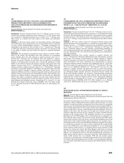
100<br />
COMPARISON OF CELL VITALITY AND CHEMOKINE<br />
PRODUCTION BETWEEN CELLS EXPERIENCING<br />
NEGATIVE PRESSURE MANIFOLDED WITH DIFFERENT<br />
DRESSINGS<br />
Amy K. McNulty, Marisa Schmidt, Teri Feeley, Kris Kieswetter<br />
Kinetic Concepts, Inc.<br />
Introduction: Vacuum Assisted Closure (V.A.C. s ) Therapy creates an environment<br />
that promotes granulation tissue formation (1–3). The current study<br />
was conducted to determine potential ways in which V.A.C. s Therapy may<br />
affect tissues at a cellular level with respect to cell vitality and chemokine<br />
expression.<br />
Methods: A 3D tissue culture system was developed whereby adult human<br />
dermal fibroblasts were encapsulated in a provisional fibrin matrix, simulating<br />
an acute wound. Subatmospheric pressure ( 125 mmHg, continuous) was<br />
manifolded to this matrix for 48 h via ei<strong>the</strong>r V.A.C. s GranuFoam s Dressing<br />
or USP Type VII gauze. Following treatment, cell vitality was assessed as well<br />
as <strong>the</strong> ability of media from <strong>the</strong> treated cell cultures to stimulate fibroblast<br />
migration.<br />
Discussion: The production of granulation tissue requires an influx of fibroblasts<br />
to <strong>the</strong> wound. The fibroblasts must <strong>the</strong>n syn<strong>the</strong>size an extracellular<br />
matrix. Results show that cells exposed to negative pressure and V.A.C. s<br />
GranuFoam s Dressing were less dendritic than cells exposed to negative<br />
pressure and gauze. Changes in cell shape may be reflective of associated<br />
changes in cell physiology. Cell death was a statistically significant 2.4 fold<br />
higher in cell cultures treated with negative pressure using gauze than with<br />
negative pressure using V.A.C. s GranuFoam s Dressing. Media from cell<br />
cultures exposed to negative pressure using V.A.C. s GranuFoam s Dressing<br />
stimulated a statistically significant 3 fold higher cell migration than did media<br />
from cell cultures exposed to negative pressure and gauze. There was no<br />
significant difference in ability to stimulate migration between cells treated with<br />
negative pressure using gauze and unconditioned media. This indicates that<br />
exposure to negative pressure using gauze does not provide any additional<br />
benefit with respect to migration. Results from this study indicate that <strong>the</strong><br />
dressing choice interfacing <strong>the</strong> wound and negative pressure source dramatically<br />
influences <strong>the</strong> biological outcome. Armstrong, D. G., Lavery, L. A., and<br />
Diabetic Foot Study Consortium. Lancet 366[9498], 1704–1710. 2005. Joseph<br />
E, et al., <strong>Wound</strong>s 12(3), 60–67. 2000. Mooney JF, III, et al., Clin Orthop. 376,<br />
26–31. 2000.<br />
101<br />
COMPARISON OF CELL ENERGETICS BETWEEN CELLS<br />
EXPERIENCING NEGATIVE PRESSURE MANIFOLDED<br />
WITH V.A.C. s GRANUFOAM s DRESSING VS. GAUZE<br />
Amy K. McNulty, Marisa Schmidt, Teri Feeley, Kris Kieswetter<br />
Kinetic Concepts, Inc.<br />
Introduction: Vacuum Assisted Closure s (V.A.C. s ) Therapy creates an environment<br />
that promotes granulation tissue formation (1–3) and prepares wounds<br />
for closure. <strong>Wound</strong> healing and <strong>the</strong> production of granulation tissue are highly<br />
energetic processes. The following study was conducted to assess whe<strong>the</strong>r or<br />
not V.A.C. s Therapy using GranuFoam s Dressing positively affects cellular<br />
energetics.<br />
Methods: A 3D tissue culture system was developed whereby human adult<br />
dermal fibroblasts were encapsulated in a syn<strong>the</strong>tic provisional fibrin matrix.<br />
Negative pressure ( 125 mmHg, continuous) was manifolded to this matrix<br />
for 48 hours via ei<strong>the</strong>r V.A.C. s GranuFoam s Dressing or USP Type VII<br />
Gauze. Following negative pressure treatment, mitochondria exhibiting an<br />
active membrane potential were visualized using MitoCapture TM stain (BioVision,<br />
Inc., Mountain View, CA). The cell areas occupied by active mitochondria<br />
were <strong>the</strong>n measured.<br />
Discussion: Fibroblasts from cultures exposed to negative pressure and<br />
V.A.C. s GranuFoam s Dressing exhibited large rings of perinuclear, punctate<br />
fluorescence. The staining of mitochondria in cultures exposed to negative<br />
pressure using gauze was much less, and similar to that exhibited by cells in a<br />
static environment. Cell areas containing active mitochondria for cultures<br />
exposed to negative pressure and gauze were significantly less (p o 0.05) than<br />
for cultures exposed to negative pressure and V.A.C. s GranuFoam s Dressing.<br />
There was no significant difference (p 4 0.05) between cell areas for cells<br />
exposed to negative pressure and gauze and cells in a static environment.<br />
Increases in numbers of active mitochondria per cell typically occur when cells<br />
are undergoing highly energetic processes. Energetic processes include cell<br />
division and protein syn<strong>the</strong>sis, two processes occurring during granulation<br />
tissue formation. Results from this study indicate that <strong>the</strong> dressing choice<br />
interfacing <strong>the</strong> wound and negative pressure source dramatically influences<br />
cellular energetics. Armstrong, D. G., Lavery, L. A., and Diabetic Foot Study<br />
Consortium. Lancet 366[9498], 1704–1710. 2005. Joseph E, et al., <strong>Wound</strong>s<br />
12(3), 60–67. 2000. Mooney JF, III, et al., Clin Orthop. 376, 26–31. 2000.<br />
102<br />
WOUND HEALING AFTER PHOTOCHEMICAL TISSUE<br />
BONDING<br />
Min Yao, Kenneth Bujold, Robert Redmond, Irene Kochevar<br />
Wellman Center for Photomedicine, Massachusetts General Hospital, Harvard<br />
Medical School, Boston, MA 02114<br />
Successful wound healing is <strong>the</strong> result of a complex cellular and tissue structure<br />
restoration, which is influenced by many systemic and local factors. As a local<br />
regulator, wound closure plays an important role in this process by reattaching<br />
injured tissue and initiating healing. Photochemical tissue bonding (PTB) is a<br />
new sutureless technology for <strong>the</strong> closing wounds and reattaching tissues that<br />
utilizes visible light to activate a photochemical dye (Rose Bengal). The dye is<br />
applied to <strong>the</strong> tissue, <strong>the</strong>n <strong>the</strong> tissues are brought into intimate contact and<br />
exposed to light that activates <strong>the</strong> dye, leading to covalent crosslinks between<br />
proteins on <strong>the</strong> wound surfaces. The immediate formation of a continuous,<br />
fluid-tight seal with PTB suggests that wound healing may be enhanced over<br />
that obtained using sutures. Our previous studies showing that PTB repairs<br />
incision and excisions in skin and cornea, and reattaches peripheral nerves and<br />
small arteries with excellent healing support this notion. For example, equivalent<br />
healing of skin excisions was produced by PTB and standard suture repair<br />
at 6 weeks after treatment. In order to better understand <strong>the</strong> wound healing<br />
process after PTB and to more efficiently manipulate it, we are studying <strong>the</strong><br />
tissue responses. Phototoxicity is a potential adverse tissue response to PTB<br />
because our studies have shown that Rose Bengal is phototoxic to cultured<br />
cells. We evaluated this possibility by treating full-thickness skin incisions with<br />
PTB ex vivo, culturing <strong>the</strong> skin for 24 h and quantifying <strong>the</strong> cytotoxicity on<br />
H&E-stained sections relative to untreated control skin. Tight bonding with a<br />
barely visible incision line was obtained. PTB did not produce greater<br />
cytotoxicity to keratinocytes or fibroblasts than found in <strong>the</strong> control samples.<br />
To fur<strong>the</strong>r explore <strong>the</strong> healing process in PTB, we are investigating inflammation,<br />
activation of fibroblast and scar formation after PTB treatment of in skin<br />
excisions.<br />
<strong>Wound</strong> Rep Reg (<strong>2007</strong>) 15 A14–A54 c <strong>2007</strong> by <strong>the</strong> <strong>Wound</strong> <strong>Healing</strong> <strong>Society</strong><br />
A41






![2010 Abstracts-pah[2] - Wound Healing Society](https://img.yumpu.com/3748463/1/190x245/2010-abstracts-pah2-wound-healing-society.jpg?quality=85)