May-June 2013 Issue - American College of Phlebology
May-June 2013 Issue - American College of Phlebology
May-June 2013 Issue - American College of Phlebology

Create successful ePaper yourself
Turn your PDF publications into a flip-book with our unique Google optimized e-Paper software.
COMMENTARY<br />
The clinical phlebologist is well aware that<br />
sclerosants act at limited areas <strong>of</strong> clinical activity<br />
from the point where they are injected. In the past,<br />
it was felt that this clinical observation was related<br />
to the dilution <strong>of</strong> the sclerosant and its subsequent<br />
decreased potency in this regard, based solely upon<br />
concentration/sclerosant endothelial interaction<br />
kinetically.<br />
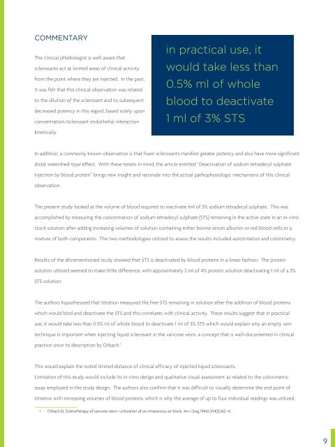
in practical use, it<br />
would take less than<br />
0.5% ml <strong>of</strong> whole<br />
blood to deactivate<br />
1 ml <strong>of</strong> 3% STS<br />
In addition, a commonly known observation is that foam sclerosants manifest greater potency and also have more significant<br />
distal watershed-type effect. With these tenets in mind, the article entitled “Deactivation <strong>of</strong> sodium tetradecyl sulphate<br />
injection by blood protein” brings new insight and rationale into the actual pathophysiologic mechanisms <strong>of</strong> this clinical<br />
observation.<br />
The present study looked at the volume <strong>of</strong> blood required to inactivate 1ml <strong>of</strong> 3% sodium tetradecyl sulphate. This was<br />
accomplished by measuring the concentration <strong>of</strong> sodium tetradecyl sulphate (STS) remaining in the active state in an in-vitro<br />
stock solution after adding increasing volumes <strong>of</strong> solution containing either bovine serum albumin or red blood cells or a<br />
mixture <strong>of</strong> both components. The two methodologies utilized to assess the results included autotritation and colorimetry.<br />
Results <strong>of</strong> the aforementioned study showed that STS is deactivated by blood proteins in a linear fashion. The protein<br />
solution utilized seemed to make little difference, with approximately 2 ml <strong>of</strong> 4% protein solution deactivating 1 ml <strong>of</strong> a 3%<br />
STS solution.<br />
The authors hypothesized that titration measured the free STS remaining in solution after the addition <strong>of</strong> blood proteins<br />
which would bind and deactivate the STS and this correlates with clinical activity. These results suggest that in practical<br />
use, it would take less than 0.5% ml <strong>of</strong> whole blood to deactivate 1 ml <strong>of</strong> 3% STS which would explain why an empty vein<br />
technique is important when injecting liquid sclerosant in the varicose veins, a concept that is well-documented in clinical<br />
practice since its description by Orbach. 1<br />
This would explain the noted limited distance <strong>of</strong> clinical efficacy <strong>of</strong> injected liquid sclerosants.<br />
Limitation <strong>of</strong> this study would include its in-vitro design and qualitative visual assessment as related to the colorimetric<br />
assay employed in the study design. The authors also confirm that it was difficult to visually determine the end point <strong>of</strong><br />
titration with increasing volumes <strong>of</strong> blood proteins, which is why the average <strong>of</strong> up to four individual readings was utilized.<br />
1 Orbach EJ. Sclerotherapy <strong>of</strong> varicose veins—utilization <strong>of</strong> an intravenous air block. Am J Surg 1944;LXVI(3):362–6.<br />
9