Cloning, Functional Expression and Characterization of L ...
Cloning, Functional Expression and Characterization of L ...
Cloning, Functional Expression and Characterization of L ...

Create successful ePaper yourself
Turn your PDF publications into a flip-book with our unique Google optimized e-Paper software.
J. VIDYA et al.: L-Asparaginase II from E. coli MTCC 739, Food Technol. Biotechnol. 49 (3) 286–290 (2011)<br />
287<br />
by replacement <strong>of</strong> certain amino acid residues by site-<br />
-directed mutagenesis (8), attachment <strong>of</strong> some chemical<br />
moieties to the purified protein (9) or immobilization <strong>of</strong><br />
the asparaginase in nanoparticles (10) or microparticles<br />
<strong>of</strong> natural silk sericin protein (11).<br />
The aim <strong>of</strong> the current study is to construct an expression<br />
cassette <strong>of</strong> L-asparaginase II gene from E. coli<br />
MTCC 739 bearing a C-terminal His 6 tag, in frame with<br />
pelB leader sequence <strong>of</strong> pET20b under the control <strong>of</strong> a<br />
T7 inducible promoter. The ansB gene from E. coli MTCC<br />
739 was cloned <strong>and</strong> expressed in E. coli DE3 cells. The<br />
expressed protein was purified <strong>and</strong> studied for operational<br />
properties.<br />
Materials <strong>and</strong> Methods<br />
Bacterial strains <strong>and</strong> plasmids<br />
E. coli MTCC 739 was used as a source <strong>of</strong> the L-asparaginase<br />
II gene. E. coli strains BL21(DE3) <strong>and</strong> DH5a,<br />
<strong>and</strong> pET-20b plasmid as an expression vector for ansB<br />
gene were obtained from Novagen (Milan, Italy).<br />
Construction <strong>of</strong> recombinant plasmid <strong>and</strong> cloning <strong>of</strong><br />
ansB gene<br />
Genomic DNA <strong>of</strong> E. coli MTCC 739 (NII08131) was<br />
used as template for polymerase chain reaction (PCR)<br />
amplification <strong>of</strong> L-asparaginase II (ansB) genebyexcluding<br />
the native signal sequence using AnsBF (forward, 5’-<br />
-GCGGAATTCGTTACCCAATATCACCA-3’ <strong>and</strong> AnsBR<br />
(reverse, 5’-GGCGAAGCTTGTACTGATTGAAGA-3’) with<br />
EcoRI <strong>and</strong> HindIII restriction sites underlined respectively.<br />
The PCR conditions were as follows: initial denaturationat94°Cfor4min,denaturationat95°Cfor40<br />
s, annealing at 57.4 °C for 30 s, extension at 72 °C for 90<br />
s for 30 cycles, <strong>and</strong> a final extension at 72 °C for 10 min.<br />
The 981-bp amplicons, digested by EcoRI <strong>and</strong> HindIII, were<br />
cloned in pET20b vector having the same restriction termini.<br />
The recombinant construct thus obtained (pET20b-<br />
-M2His) bearing an N-terminal pelB leader <strong>and</strong> a C-<br />
-terminal His 6 tag was then transformed into chemically<br />
competent E. coli DH5a cells. The clones were analyzed<br />
by a restriction digestion for an insert release <strong>of</strong> the recombinant<br />
plasmid isolated from the confirmed clones.<br />
The plasmid DNA was isolated from the confirmed clone<br />
<strong>of</strong> E. coli DH5a cells <strong>and</strong> used for transformation <strong>of</strong> E.<br />
coli DE3 competent cells for protein expression.<br />
Soluble expression <strong>and</strong> purification <strong>of</strong> the recombinant<br />
enzyme<br />
To optimize the expression <strong>of</strong> L-asparaginase gene<br />
in E. coli, the effect <strong>of</strong> various isopropyl b-D-1-thiogalactopyranoside<br />
(IPTG) concentrations was studied. E. coli<br />
DE3 cells harbouring the expression plasmid pET20b-<br />
-M2His was grown in Luria-Bertani broth containing 0.5 %<br />
glucose <strong>and</strong> 50 mg/mL <strong>of</strong> ampicillin <strong>and</strong> induced with<br />
different IPTG concentrations starting from 10 to 400 mM<br />
at the A 600 nm <strong>of</strong> 0.6. The culture was then allowed to<br />
grow for a postinduction period <strong>of</strong> 3hat37°C<strong>and</strong>200<br />
rpm. After the harvest, absorbance (at 600 nm) was measured<br />
<strong>and</strong> all the samples were diluted to an absorbance<br />
<strong>of</strong> 1 for a normalised SDS-PAGE <strong>and</strong> enzymatic assays.<br />
Periplasmic <strong>and</strong> cytosolic fractions were collected <strong>and</strong> analysed<br />
to establish the location <strong>of</strong> expressed recombinant<br />
protein in the cell compartments. For the preparation <strong>of</strong><br />
periplasmic fractions, 50-mL culture was pelleted <strong>and</strong><br />
resuspended in Tris-sucrose buffer (30 mM Tris, pH=8,<br />
20 % sucrose <strong>and</strong> 1 mM EDTA) followed by shaking for<br />
10 min <strong>and</strong> pelleting at 8000×g for 20 min. The pellet<br />
was resuspended again in 10 mL <strong>of</strong> 5 mM MgSO 4 , kept<br />
in an ice bath <strong>and</strong> shaken for 10 min, <strong>and</strong> after centrifugation<br />
at 8000×g for 20 min the supernatant was collected<br />
as periplasmic fraction. The cytosolic fraction was<br />
prepared by sonicating the pellet collected from 50 mL<br />
<strong>of</strong> culture in lysis buffer (50 mM NaH 2 PO 4 , 300 mM<br />
NaCl <strong>and</strong> 10 mM imidazole). For recombinant protein<br />
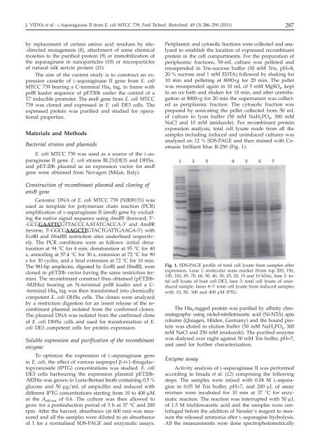
expression analysis, total cell lysate made from all the<br />
samples including induced <strong>and</strong> uninduced cultures was<br />
analysed on 12 % SDS-PAGE <strong>and</strong> then stained with Coomasie<br />
brilliant blue R-250 (Fig. 1).<br />
Fig. 1. SDS-PAGE pr<strong>of</strong>ile <strong>of</strong> total cell lysate from samples after<br />
expression. Lane 1: molecular mass marker (from top 200, 150,<br />
120, 100, 85, 70, 60, 50, 40, 30, 25, 20, 15 <strong>and</strong> 10 kDa), lane 2: total<br />
cell lysate <strong>of</strong> host cell DE3, lane 3: total cell lysate <strong>of</strong> uninduced<br />
sample, lanes 4–7: total cell lysate from induced samples<br />
with 10, 50, 100 <strong>and</strong> 400 mM IPTG<br />
The His 6 -tagged protein was purified by affinity chromatography<br />
using nickel-nitrilotriacetic acid (Ni-NTA) spin<br />
column (Quiagen, Hilden, Germany) <strong>and</strong> the bound protein<br />
was eluted in elution buffer (50 mM NaH 2 PO 4 , 300<br />
mM NaCl <strong>and</strong> 250 mM imidazole). The purified enzyme<br />
was dialysed over night against 50 mM Tris buffer, pH=7,<br />
<strong>and</strong> used for further characterization.<br />
Enzyme assay<br />
Activity analysis <strong>of</strong> L-asparaginase II was performed<br />
according to Imada et al. (12) comprising the following<br />
steps. The samples were mixed with 0.04 M L-asparagine<br />
in 0.05 M Tris buffer, pH=7, <strong>and</strong> 200 mL <strong>of</strong> assay<br />
mixture were incubated for 10 min at 37 °C for enzymatic<br />
reaction. The reaction was interrupted with 50 mL<br />
<strong>of</strong> 1.5 M trichloroacetic acid <strong>and</strong> the samples were centrifuged<br />
before the addition <strong>of</strong> Nessler’s reagent to measure<br />
the released ammonia after L-asparagine hydrolysis.<br />
All the measurements were done spectrophotometrically