

MEIOFAUNA MARINA
MEIOFAUNA MARINA
MEIOFAUNA MARINA

You also want an ePaper? Increase the reach of your titles
YUMPU automatically turns print PDFs into web optimized ePapers that Google loves.
90<br />
morphology of species belonging to 21 different<br />
genera from twelve families. At least one species<br />
for each of the eight macrodasyidan families has<br />
been investigated, and we have data on eight<br />
species from the only four chaetonotidan families<br />
with amphigonic reproduction. As the number of<br />
species investigated increased, it became more<br />
and more evident that it is not possible to define a<br />
general model for gastrotrich spermatozoa (compare<br />
Ferraguti & Balsamo 1995 with Balsamo et al.<br />
1999 and Marotta et al. 2005). Among macrodasyidans,<br />
the spermatozoa, besides being all filiform<br />
and flagellated cells (with the exception of Dactylopodola<br />
baltica: Fischer 1996 and Dolichodasys: Ruppert<br />
& Shaw 1977) which however is a character<br />
common to many animal species with internal<br />
fertilization, do not show characters common to<br />
all the species investigated. For example, one of<br />
the most peculiar characters of macrodasyidan<br />
gastrotrich spermatozoa, i. e. the presence of one<br />
or more mitochondria surrounded by the nucleus<br />
is not present in the family Macrodasyidae and<br />
in Xenodasys eknomios (Guidi et al. 2009). Another<br />
macrodasyidan sperm character, the striated cylinder<br />
involving the flagellar axoneme is absent<br />
in the Lepidodasys species examined (Guidi et al.<br />
2004), in one of the two Urodasys species (Guidi<br />
et al. 2007), in two Crasiella species (Guidi et al.<br />
2010), in Xenodasys eknomios (Guidi et al. 2009) and<br />
in the three Turbanellidae studied (Balsamo et al.<br />
2002), but is present in all the remaining twelve<br />
species examined.<br />
Thaumastodermatidae is the most speciose<br />
family within Macrodasyida, with near 150<br />
species (Hochberg 2001, Hummon & Todaro<br />
2010, Hummon 2011) distributed in eight genera<br />
and two recognized subfamilies: Acanthodasys<br />
and Diplodasys (Diplodasyinae) vs. Hemidasys,<br />
Oregodasys (= Platydasys), Pseudostomella, Ptychostomella,<br />
Tetranchyroderma and Thaumastoderma<br />
(Thaumastodermatinae). It is worth to notice<br />
that recently Hemidasys has been considered<br />
extinct (Hummon & Todaro 2010). Phylogenetic<br />
relationships within the family are debated, with<br />
contrasting scenarios hypothesized on gross<br />
anatomy level data (e. g. Hochberg & Litvaitis<br />
2000 vs. Hochberg & Litvaitis 2001 vs. Kieneke<br />
et al. 2008).<br />
We have spermatological data on species<br />
of four out of the eight genera: Acanthodasys<br />
aculeatus (Guidi et al. 2003), Diplodasys ankeli,<br />
Pseudostomella etrusca, Tetranchyroderma (three<br />
species) (Ferraguti & Balsamo 1995). It appears<br />
that the general structure of the spermatozoa in<br />
the family Thaumastodermatidae is pretty constant.<br />
In particular all sperm models have a more<br />
or less complex, helical acrosome containing a<br />
tubular structure made of piled hollow cylinders,<br />
a nuclear-mitochondrial region in which one or<br />
more mitochondria are surrounded by a helical<br />
nucleus, and a tail in which the flagellar axoneme<br />
is surrounded by a striated cylinder (sensu Ferraguti<br />
& Balsamo 1994). To assess to what extent<br />
this general model is valid for other members of<br />
the family, and in search for possible character<br />
ground pattern we have studied the spermatozoa<br />
of Thaumastoderma moebjergi Clausen, 2004. Within<br />
this framework the study bears supplementary<br />
relevance as a recent phylogenetic investigation<br />
based on molecular traits found Thaumastoderma<br />
to be one of the most basal taxon along the Thaumastodermatinae<br />
evolutionary line (Todaro et al.<br />
2011).<br />
Materials and methods<br />
Marine sediment containing Thaumastoderma<br />
moebjergi Clausen, 2004 was collected in September<br />
2007 during a two-week workshop held<br />
at the Sven Lovén Centre for Marine Sciences on<br />
Tjärnö, an island on the Swedish west coast (for<br />
details see Willems et al. 2009). Gastrotrichs were<br />
extracted from the sediment by the narcotizationdecantation<br />
technique, using an isosmotic (7 %)<br />
magnesium chloride solution (Todaro & Hummon<br />
2008). The fauna-containing supernatant was then<br />
poured directly into a 5-cm diameter Petri dish<br />
and scanned for specimens under a Wild M3 dissecting<br />
microscope set at 50 × magnification. For<br />
optical microscopy, the gastrotrichs were removed<br />
with a micropipette from the Petri dish, fresh-<br />
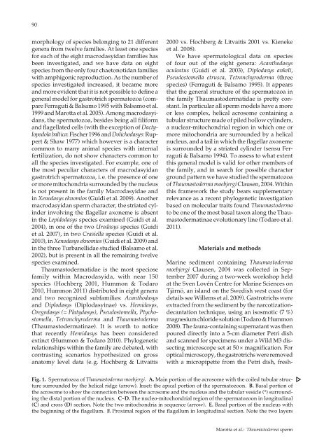
Fig. 1. Spermatozoa of Thaumastoderma moebjergi. A. Main portion of the acrosome with the coiled tubular structure<br />
surrounded by the helical ridge (arrow). Inset: the apical portion of the spermatozoon. B. Basal portion of<br />
the acrosome to show the connection between the acrosome and the nucleus and the tubular vesicle (*) surrounding<br />
the distal portion of the nucleus. C-D. The nucleo-mitochondrial region of the spermatozoon in longitudinal<br />
(C) and cross (D) section. Note the two mitochondria in sequence (arrow). E. Basal portion of the nucleus with<br />
the beginning of the flagellum. F. Proximal region of the flagellum in longitudinal section. Note the two layers<br />
Marotta et al.: Thaumastoderma sperm