MEEK Micrografting - AFS Medical
MEEK Micrografting - AFS Medical
MEEK Micrografting - AFS Medical

Create successful ePaper yourself
Turn your PDF publications into a flip-book with our unique Google optimized e-Paper software.
The <strong>MEEK</strong> technique<br />
In 1958 a remarkable technique<br />
for expanding autografts was<br />
described by the American<br />
surgeon C.P. Meek 1,2) . With a<br />
so-called ‘<strong>MEEK</strong>-WALL’<br />
dermatome postage stamp<br />
autografts were obtained and<br />
expanded with a ratio 1:9 using<br />
double pleated gauzes. The<br />
method however required too<br />
much skill and it became<br />
eclipsed by the introduction of<br />
mesh skin grafts by Tanner in<br />
1964. Eventually the <strong>MEEK</strong><br />
technique was discontinued.<br />
However, lack of autograft<br />
donorsites is still encountered as<br />
a limiting factor in achieving<br />
wound closure in case of<br />
extensive burns. The meshgraft<br />
technique requires donorsites of<br />
suitable size and shape and<br />
epithelialization may be delayed<br />
in case of expansion ratios<br />
greater than 1:4.<br />
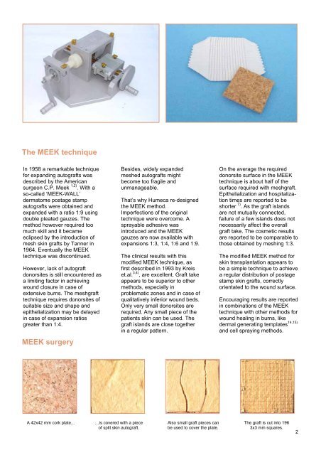
<strong>MEEK</strong> surgery<br />
Besides, widely expanded<br />
meshed autografts might<br />
become too fragile and<br />
unmanageable.<br />
That’s why Humeca re-designed<br />
the <strong>MEEK</strong> method.<br />
Imperfections of the original<br />
technique were overcome. A<br />
sprayable adhesive was<br />
introduced and the <strong>MEEK</strong><br />
gauzes are now available with<br />
expansions 1:3, 1:4, 1:6 and 1:9.<br />
The clinical results with this<br />
modified <strong>MEEK</strong> technique, as<br />
first described in 1993 by Kreis<br />
et.al. 3,4) , are excellent. Graft take<br />
appears to be superior to other<br />
methods, especially in<br />
problematic zones and in case of<br />
qualitatively inferior wound beds.<br />
Only very small donorsites are<br />
required. Any small piece of the<br />
patients skin can be used. The<br />
graft islands are close together<br />
in a regular pattern.<br />
On the average the required<br />
donorsite surface in the <strong>MEEK</strong><br />
technique is about half of the<br />
surface required with meshgraft.<br />
Epithelialization and hospitalization<br />
times are reported to be<br />
shorter 7) . As the graft islands<br />
are not mutually connected,<br />
failure of a few islands does not<br />
necessarily affect the overall<br />
graft take. The cosmetic results<br />
are reported to be comparable to<br />
those obtained by meshing 1:3.<br />
The modified <strong>MEEK</strong> method for<br />
skin transplantation appears to<br />
be a simple technique to achieve<br />
a regular distribution of postage<br />
stamp skin grafts, correctly<br />
orientated to the wound surface.<br />
Encouraging results are reported<br />
in combinations of the <strong>MEEK</strong><br />
technique with other methods for<br />
wound healing in burns, like<br />
dermal generating templates 14,15)<br />
and cell spraying methods.<br />
A 42x42 mm cork plate... …is covered with a piece Also small graft pieces can The graft is cut into 196<br />
of split skin autograft. be used to cover the plate. 3x3 mm squares.<br />
2