The Long QT Syndrome - SADS Foundation
The Long QT Syndrome - SADS Foundation
The Long QT Syndrome - SADS Foundation

Create successful ePaper yourself
Turn your PDF publications into a flip-book with our unique Google optimized e-Paper software.
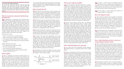
A note from the <strong>SADS</strong> <strong>Foundation</strong>.<br />
We provide this information with the hope that informing<br />
physicians, other health care providers, and the public will<br />
encourage early and correct diagnosis and proper therapy,<br />
resulting in the reduction and ultimately elimination of cardiac<br />
arrest and sudden death from inherited <strong>Long</strong> <strong>QT</strong> <strong>Syndrome</strong><br />
(L<strong>QT</strong>S).<br />
Why do I need to know about the Inherited <strong>Long</strong><br />
<strong>QT</strong> <strong>Syndrome</strong><br />
We estimate that 1 in 2500 people in the United States have<br />
L<strong>QT</strong>S. L<strong>QT</strong>S-precipitated sudden deaths continue to claim<br />
otherwise healthy infants, children, adolescents, and adults at an<br />
unacceptably high rate. However, with increased awareness,<br />
genetic testing, and effective treatment options, L<strong>QT</strong>S can be<br />
diagnosed early and sudden death prevented. Still, this condition is<br />
often undetected prior to death and not recognized as the cause of<br />
death. Family members of individuals with unexplained death<br />
should be tested for L<strong>QT</strong>S and other genetic arrhythmias. L<strong>QT</strong>S is<br />
a highly treatable disorder and, with correct diagnosis and<br />
common treatments, most deaths are preventable.<br />
Physicians need to know:<br />
• When to consider L<strong>QT</strong>S as a possible diagnosis.<br />
• When to refer patients for diagnosis & treatment.<br />
• About genetic testing for L<strong>QT</strong>S and other <strong>SADS</strong> conditions.<br />
• How to develop a family pedigree and screen family members<br />
for L<strong>QT</strong>S.<br />
Patients and Parents need to know:<br />
• <strong>The</strong> warning signs and symptoms of L<strong>QT</strong>S.<br />
• Who to see for proper testing.<br />
• How to protect their children and themselves.<br />
• How to expand their family pedigree contact other family<br />
members who may be at risk.<br />
What is L<strong>QT</strong>S<br />
L<strong>QT</strong>S is a disturbance of the heart’s electrical system. It is<br />
caused by abnormalities of microscopic pores in the heart cells<br />
called ion channels. Ions such as potassium, sodium, calcium and<br />
chloride pass back and forth across the cell membrane through ion<br />
channels. As they do, they generate the electrical activity<br />
(depolarization and repolarization) that controls the heart’s<br />
beating. Our window to this electrical activity of the heart is<br />
through an electrocardiogram (EKG or ECG). Potassium and<br />
sodium ion channels are two of the major sites affected in L<strong>QT</strong>S.<br />
<strong>The</strong> abnormal channels prolong the repolarization (“recharging”)<br />
process and the <strong>QT</strong> interval, thus predisposing patients to certain<br />
cardiac arrhythmias. Thus, L<strong>QT</strong>S is a glitch in the electrical<br />
recharging phase of the heart.<br />
What is the <strong>QT</strong> interval<br />
<strong>The</strong> <strong>QT</strong> interval is a time interval on the ECG. It represents the<br />
time from the electrical stimulation (depolarization) of the<br />
heart’s pumping chambers (ventricles) to the end of the recharging<br />
of the electrical system (repolarization). It is measured in<br />
milliseconds and closely approximates the time from the<br />
beginning of the ventricles’ contraction until the end of relaxation.<br />
<strong>The</strong> <strong>QT</strong> interval varies in each person and between persons—<br />
like most physiologic parameters, such as blood pressure or<br />
heart rate. In particular, the <strong>QT</strong> varies with the heart rate. It shortens<br />
as the rate increases and lengthens as the rate decreases. <strong>The</strong>refore,<br />
there is a range of normal or healthy <strong>QT</strong> intervals. In contrast, the<br />
long <strong>QT</strong> heart often recharges sluggishly or inefficiently as<br />
evidenced by a “prolonged <strong>QT</strong> interval” on the ECG.<br />
To determine if a given <strong>QT</strong> is normal for a given heart rate, the<br />
<strong>QT</strong> is corrected for the heart rate using a simple mathematical<br />
formula, and the resultant quantity is called the <strong>QT</strong>c. <strong>The</strong> <strong>QT</strong>c is<br />
the value that doctors generally use when assessing for L<strong>QT</strong>S.<br />
<strong>The</strong> “normal” <strong>QT</strong>c varies from approximately 350 to 480<br />
milliseconds. About 90% of people have a value between 380<br />
and 440 ms, which is the range doctors generally consider as the<br />
“normal” range. However, this “normal” range is affected by age<br />
and gender. For example, women tend to have slightly longer <strong>QT</strong><br />
intervals than men and while a <strong>QT</strong>c of 440 ms represents the top<br />
2.5 percentile in a 4-day-old infant, that same value represents the<br />
top 15-20th percentile for a 40-year-old female. <strong>The</strong> diagram<br />
below provides an example of a normal and a prolonged <strong>QT</strong>c<br />
interval. <strong>The</strong> RR interval determines the heart rate.<br />
In this diagram, since the heart rate (RR) is the same for both<br />
examples but the <strong>QT</strong> interval is longer in the lower panel, the<br />
<strong>QT</strong>c is longer in the lower panel example.<br />
What are the symptoms of L<strong>QT</strong>S<br />
Nearly half of patients with L<strong>QT</strong>S NEVER have a symptom.<br />
However, if/when the L<strong>QT</strong>S heart “spins electrically out of<br />
control” into its trademark cardiac arrhythmia called torsade de<br />
pointes, sudden, temporary, loss of consciousness (syncope) is the<br />
most common event. <strong>The</strong>se events usually occur without warning<br />
and are often triggered by exertion or auditory stimuli. Typically,<br />
the onset of symptoms is earlier in boys than in girls. Statistically,<br />
the greatest risk window includes the first three decades of life (the<br />
first two decades in males and the second and third decades in<br />
females. However, there are tragic exceptions to these trends and<br />
L<strong>QT</strong>S-related events can continue during the 40’s and 50’s. When<br />
presenting later, a second hit, due to a medication that further<br />
aggravates the <strong>QT</strong> interval or low potassium, is often present.<br />
In patients who experience syncope only, the potentially<br />
dangerous rhythm spontaneously returns to normal, usually<br />
within about one minute, and the patient quickly regains<br />
consciousness without disorientation or confusion. Some patients<br />
may experience slight fatigue afterwards; others feel fine and<br />
resume their regular activities. If this bad L<strong>QT</strong>S rhythm persists<br />
longer, patients may then manifest a generalized seizure. In fact,<br />
some patients with L<strong>QT</strong>S have been misdiagnosed initially with<br />
epilepsy and even treated with anti-epileptic medication. In both<br />
the syncope and seizure presentations, the long <strong>QT</strong> heart<br />
eventually caught itself, reverted back to normal sinus rhythm, and<br />
the “spell” ended. On the other hand, in a minority of patients, the<br />
torsade rhythm persists, then degenerates into the heart rhythm<br />
known as ventricular fibrillation, which rarely reverts back to a<br />
normal rhythm without medical intervention. If the ventricular<br />
fibrillation is not converted, usually by electrical defibrillation, the<br />
outcome is sudden cardiac death or sudden cardiac arrest.<br />
When should the diagnosis be suspected<br />
In any young person with unexplained syncope (fainting),<br />
unexplained seizures, or unexplained cardiac arrest or sudden<br />
death.<br />
Usually, a careful history of the events surrounding the syncope<br />
differentiates L<strong>QT</strong>S induced syncope from the common faint,<br />
known as vasovagal or neurocardiogenic syncope. <strong>The</strong> L<strong>QT</strong>S<br />
syncope is usually precipitous and without warning. It often occurs<br />
during or just after physical exertion, emotional excitement or<br />
sudden auditory arousal (such as a doorbell or alarm clock), but<br />
may occur during sleep or at rest. Conversely, in vasovagal<br />
syncope, most times there are warning symptoms, such as<br />
dizziness, blurring or blackening of vision, tingling or sweating,<br />
for seconds to even minutes prior to the syncope. Also, a<br />
precipitating event is usually present, commonly pain, injury,<br />
nausea, or an unpleasant or stressful experience.<br />
When there is a family history of unexplained syncope,<br />
unexplained seizures, or sudden death in young people. As<br />
noted above, at least one-third to one-half of individuals never<br />
exhibit symptoms, and, therefore, the lack of prior symptoms does<br />
not exclude a person or family from having L<strong>QT</strong>S.<br />
When the autopsy is normal following the sudden and<br />
unexpected death of a young person.\<br />
How is the diagnosis made<br />
L<strong>QT</strong>S is diagnosed primarily upon recognition of a prolonged<br />
<strong>QT</strong>interval on the ECG. A <strong>QT</strong>c of 470 milliseconds (ms) in<br />
males and 480 milliseconds in females is generally considered<br />
strongly suspicious for L<strong>QT</strong>S, in the absence of medications,<br />
which prolong the <strong>QT</strong> interval or other forms of heart disease. A<br />
<strong>QT</strong>c of less than 400 ms in males and 410 ms in females makes the<br />
diagnosis extremely unlikely. <strong>The</strong> computer generated <strong>QT</strong>c may be<br />
incorrect, so when the diagnosis of L<strong>QT</strong>S is considered, the<br />
physician should verify the computer measurement.<br />
Not all L<strong>QT</strong>S patients have a prolonged <strong>QT</strong>c on the initial ECG,<br />
however. In fact, about 30 – 50% have a <strong>QT</strong>c that overlaps<br />
with the normal range and over 10% have a <strong>QT</strong>c < 440 ms. <strong>QT</strong>c’s<br />
in this range are inconclusive, therefore, and must be clarified by<br />
additional testing such as exercise stress testing, the epinephrine<br />
<strong>QT</strong> stress test, and genetic testing.<br />
How is L<strong>QT</strong>S inherited, and who in a known or<br />
suspected family should be tested<br />
L<strong>QT</strong>S is usually inherited by autosomal dominant transmission.<br />
This means that it generally affects boys and girls equally, and<br />
that each child of an affected parent has a 50% chance of inheriting<br />
the genetic abnormality. In a really large family, close to 50% of<br />
the children would inherit the disease-causing gene. In average<br />
size families, it can range from all to none as each child has an<br />
independent 50/50 chance of inheriting the “genetic defect.” Once<br />
a family member is identified with L<strong>QT</strong>S, it is extremely<br />
important that other family members be tested for the syndrome. It<br />
is especially important to know which parent and grandparent has<br />
the abnormality, since brothers and sisters, aunts, uncles, nephews,<br />
nieces, and cousins on the affected side are potentially at risk.<br />
T<br />
his prospective screening, by ECG, is extremely important so<br />
that all affected family members are identified and treated<br />
early in order to prevent the tragic and unnecessary sudden deaths<br />
that may occur.