peer reviewed - Irish Veterinary Journal
peer reviewed - Irish Veterinary Journal
peer reviewed - Irish Veterinary Journal
Create successful ePaper yourself
Turn your PDF publications into a flip-book with our unique Google optimized e-Paper software.
Volume 57 (1) : January, 2004<br />
<strong>Irish</strong> <strong>Veterinary</strong> <strong>Journal</strong><br />
<strong>peer</strong> <strong>reviewed</strong><br />
<strong>peer</strong> <strong>reviewed</strong><br />
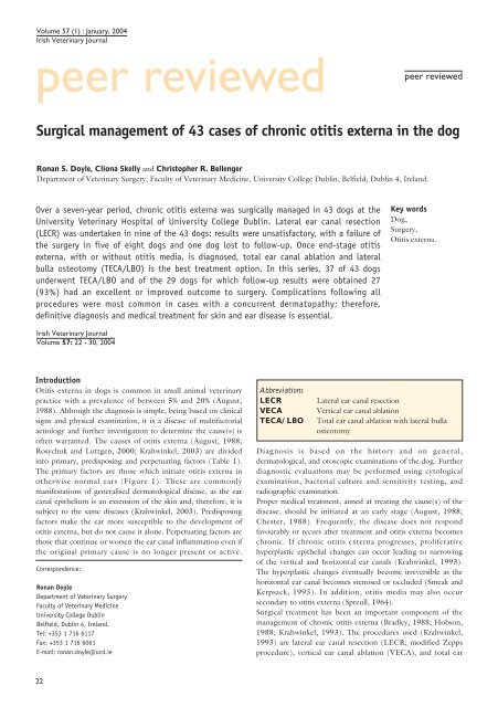
Surgical management of 43 cases of chronic otitis externa in the dog<br />
Ronan S. Doyle, Cliona Skelly and Christopher R. Bellenger<br />
Department of <strong>Veterinary</strong> Surgery, Faculty of <strong>Veterinary</strong> Medicine, University College Dublin, Belfield, Dublin 4, Ireland.<br />
Over a seven-year period, chronic otitis externa was surgically managed in 43 dogs at the<br />
University <strong>Veterinary</strong> Hospital of University College Dublin. Lateral ear canal resection<br />
(LECR) was undertaken in nine of the 43 dogs: results were unsatisfactory, with a failure of<br />
the surgery in five of eight dogs and one dog lost to follow-up. Once end-stage otitis<br />
externa, with or without otitis media, is diagnosed, total ear canal ablation and lateral<br />
bulla osteotomy (TECA/LBO) is the best treatment option. In this series, 37 of 43 dogs<br />
underwent TECA/LBO and of the 29 dogs for which follow-up results were obtained 27<br />
(93%) had an excellent or improved outcome to surgery. Complications following all<br />
procedures were most common in cases with a concurrent dermatopathy; therefore,<br />
definitive diagnosis and medical treatment for skin and ear disease is essential.<br />
Key words<br />
Dog,<br />
Surgery,<br />
Otitis externa.<br />
<strong>Irish</strong> <strong>Veterinary</strong> <strong>Journal</strong><br />
Volume 57: 22 - 30, 2004<br />
Introduction<br />
Otitis externa in dogs is common in small animal veterinary<br />
practice with a prevalence of between 5% and 20% (August,<br />
1988). Although the diagnosis is simple, being based on clinical<br />
signs and physical examination, it is a disease of multifactorial<br />
aetiology and further investigation to determine the cause(s) is<br />
often warranted. The causes of otitis externa (August, 1988;<br />
Rosychuk and Luttgen, 2000; Krahwinkel, 2003) are divided<br />
into primary, predisposing and perpetuating factors (Table 1).<br />
The primary factors are those which initiate otitis externa in<br />
otherwise normal ears (Figure 1). These are commonly<br />
manifestations of generalised dermatological disease, as the ear<br />
canal epithelium is an extension of the skin and, therefore, it is<br />
subject to the same diseases (Krahwinkel, 2003). Predisposing<br />
factors make the ear more susceptible to the development of<br />
otitis externa, but do not cause it alone. Perpetuating factors are<br />
those that continue or worsen the ear canal inflammation even if<br />
the original primary cause is no longer present or active.<br />
Correspondence:<br />
Ronan Doyle<br />
Department of <strong>Veterinary</strong> Surgery<br />
Faculty of <strong>Veterinary</strong> Medicine<br />
University College Dublin<br />
Belfield, Dublin 4, Ireland.<br />
Tel: +353 1 716 6117<br />
Fax: +353 1 716 6061<br />
E-mail: ronan.doyle@ucd.ie<br />
Abbreviations<br />
LECR<br />
VECA<br />
TECA/LBO<br />
Lateral ear canal resection<br />
Vertical ear canal ablation<br />
Total ear canal ablation with lateral bulla<br />
osteotomy<br />
Diagnosis is based on the history and on general,<br />
dermatological, and otoscopic examinations of the dog. Further<br />
diagnostic evaluations may be performed using cytological<br />
examination, bacterial culture and sensitivity testing, and<br />
radiographic examination.<br />
Proper medical treatment, aimed at treating the cause(s) of the<br />
disease, should be initiated at an early stage (August, 1988;<br />
Chester, 1988). Frequently, the disease does not respond<br />
favourably or recurs after treatment and otitis externa becomes<br />
chronic. If chronic otitis externa progresses, proliferative<br />
hyperplastic epithelial changes can occur leading to narrowing<br />
of the vertical and horizontal ear canals (Krahwinkel, 1993).<br />
The hyperplastic changes eventually become irreversible as the<br />
horizontal ear canal becomes stenosed or occluded (Smeak and<br />
Kerpsack, 1993). In addition, otitis media may also occur<br />
secondary to otitis externa (Spreull, 1964).<br />
Surgical treatment has been an important component of the<br />
management of chronic otitis externa (Bradley, 1988; Hobson,<br />
1988; Krahwinkel, 1993). The procedures used (Krahwinkel,<br />
1993) are lateral ear canal resection (LECR; modified Zepps<br />
procedure), vertical ear canal ablation (VECA), and total ear<br />
22
Volume 57 (1) : January, 2004<br />
<strong>Irish</strong> <strong>Veterinary</strong> <strong>Journal</strong><br />
<strong>peer</strong> <strong>reviewed</strong><br />
TABLE 1: Causes of otitis externa<br />
Primary factors Predisposing factors Perpetuating factors<br />
Hypersensitivity diseases Ear canal conformation Bacteria<br />
External parasites Temperature and humidity Yeasts<br />
Foreign bodies Obstructive ear disease Contact allergy and irritants<br />
Disorders of keratinisation Ear canal maceration Proliferative changes<br />
Autoimmune disease Systemic disease Otitis media<br />
Juvenile cellulitis<br />
Inappropriate treatment<br />
canal ablation with lateral bulla osteotomy (TECA/LBO). The<br />
correct procedure for an individual case depends on the accurate<br />
assessment of the extent of the disease within the external ear<br />
canal and tympanic bulla.<br />
In this paper, we evaluate and compare the indications, clinical<br />
and surgical findings, complications and long-term outcome of<br />
the surgical management of chronic otitis externa in dogs at<br />
University College Dublin and emphasise clinically relevant<br />
aspects of case management.<br />
Materials and methods<br />
We <strong>reviewed</strong> the case records of 43 dogs (61 surgical<br />
procedures) referred between August 1995 and March 2002 to<br />
the University <strong>Veterinary</strong> Hospital, University College Dublin.<br />
All 43 dogs presented with chronic otitis externa in one or both<br />
ears. There were six West Highland White terriers, five<br />
crossbred terriers, four Labrador retrievers, four Cocker<br />
spaniels, four German shepherds, and three Springer spaniels,<br />
with no other breed represented more than twice. Ages ranged<br />
from three to 14 years, with a median age of seven years. There<br />
were 24 males and 19 females.<br />
Pre-operative evaluation included history, general physical<br />
examination, clinical signs, duration of clinical signs and<br />
response to previous medication. Haematological and serum<br />
biochemical examinations were performed prior to anaesthesia<br />
in all dogs greater than five years of age or in dogs with<br />
suspected concomitant disease. Otoscopic examination was<br />
performed in all cases with the dog under sedation or general<br />
anaesthesia. Skull radiography was performed in certain cases to<br />
determine the extent of changes within the horizontal ear canal<br />
and tympanic bulla. Standard views included dorso-ventral,<br />
lateral, lateral oblique and rostro-caudal (open-mouth: Figure<br />
2) projections. A board-certified radiologist assessed all<br />
radiographs. In the majority of cases, specimens for<br />
microbiological examination (smears for Gram stain,<br />
bacteriological culture and susceptibility to antibiotics) were<br />
taken at surgery from either the horizontal canal (LECR) or the<br />
tympanic bulla (TECA/LBO). Histopathology was performed<br />
on excised tissue where there was a suspicion of neoplasia (such<br />
as abnormal masses within the ear canal). Concurrent<br />
dermatopathy was defined as the presence of dermatological<br />
lesions not affecting the ear canal.<br />
Chronic otitis externa was defined as medically non-responsive<br />
FIGURE 1: Cross-sectional diagram of the external ear canal<br />
and middle ear.<br />
FIGURE 2:<br />
Rostro-caudal<br />
skull radiograph of<br />
Case 20. Arrow<br />
indicates<br />
thickening of right<br />
tympanic bulla<br />
wall suggestive of<br />
otitis media.<br />
Arrowhead<br />
indicates normal<br />
left tympanic bulla.<br />
23
Volume 57 (1) : January, 2004<br />
<strong>Irish</strong> <strong>Veterinary</strong> <strong>Journal</strong><br />
<strong>peer</strong> <strong>reviewed</strong><br />
or recurrent otitis externa. End-stage otitis externa was defined<br />
as chronic otitis externa with marked stenosis and/or<br />
calcification of the horizontal ear canal, as determined by<br />
otoscopic examination and skull radiography. A tentative<br />
diagnosis of otitis media was made if the tympanic membrane<br />
was perforated or absent on otoscopic examination or if there<br />
was evidence of radiological changes within the bulla. Otitis<br />
media was confirmed on surgical exploration of the bulla.<br />
All surgical procedures were performed under general<br />
anaesthesia using a standard surgical technique (Krahwinkel,<br />
1993) by staff surgeons or by surgical residents. Post-operative<br />
analgesia was provided using a combination of opioids, nonsteroidal<br />
anti-inflammatory drugs and bupivicaine ‘splash block’<br />
(Buback et al., 1996). Post-operative complications were<br />
defined as those occurring up to four months after surgery.<br />
Results of treatment were obtained by either physical<br />
examination of the dogs or telephone follow-up four months or<br />
more after surgery.<br />
For LECR and VECA, the results of surgery were evaluated,<br />
using the criteria of Gregory and Vasseur (1983), as either:<br />
• excellent – clinical signs were resolved with minimal or no<br />
care required by the owner;<br />
• improved – occasional recurrence of clinical signs requiring<br />
professional attention;<br />
• poor – no improvement.<br />
For the TECA/LBO procedures, results were evaluated, using<br />
the criteria of Mason et al. (1988), as either:<br />
• excellent – resolution of clinical signs of ear disease without<br />
long-term complication;<br />
• improved – improvement of clinical signs after surgery but<br />
continued disease of the remaining medial wall of the pinna<br />
requiring treatment, or facial nerve paralysis not requiring<br />
treatment;<br />
• poor – continuing ear canal or middle ear disease present, or<br />
permanent facial nerve paralysis requiring continued medical<br />
treatment.<br />
FIGURE 3: Postoperative<br />
failed<br />
lateral ear canal<br />
resection. Arrow<br />
indicates the<br />
hyperplastic medial<br />
wall. Arrowhead<br />
indicates occluded<br />
horizontal ear canal.<br />
Results<br />
The duration of ear disease ranged from one to 84 months, with<br />
a median of 12 months. Previous medical treatments such as<br />
topical and systemic antibiotics and corticosteroids had been<br />
used in all cases. Nineteen dogs had dermatological lesions not<br />
involving the ears: disorders of keratinisation (‘seborrhoea’) in<br />
three dogs, pyoderma in two dogs, confirmed atopy in one dog<br />
and suspected hypersensitivity skin disease (atopy, food allergy,<br />
flea-bite allergic dermatitis, contact allergic dermatitis) in 13<br />
dogs. A decision on the appropriate surgical management for<br />
each case was made based on the clinical evaluation of the<br />
extent of the ear disease and after discussion with the owner.<br />
Thirteen ears were treated with LECR (Table 2): one ear in<br />
each of five dogs and both ears in four dogs. One dog (Case no.<br />
9, Table 2) had concurrent bilateral otitis media, which was<br />
responding to medical treatment at the time of surgery. Followup<br />
results were obtained in eight dogs from four to 50 months<br />
after surgery. Results were excellent in one dog, improved in<br />
two dogs, and poor in five dogs. An excellent result did not<br />
occur in any dog that had concurrent dermatological lesions.<br />
Two of the dogs that had a poor result following LECR (Case<br />
nos. 2 and 8; Table 2) subsequently had TECA/LBO surgery<br />
on the affected ear.<br />
One dog, which had chronic otitis externa with ulceration of<br />
the medial wall of the vertical ear canal, was treated with VECA.<br />
Results were excellent in this dog. VECA is rarely indicated<br />
because irreversible epithelial changes in chronic otitis externa<br />
are rarely confined to the vertical ear canal.<br />
TECA/LBO was performed in 37 dogs (47 ears); ten dogs had<br />
bilateral TECA/LBO. All these dogs had chronic otitis externa,<br />
except for one, which presented with para-aural fistulation as a<br />
complication of previous TECA without LBO for chronic otitis<br />
externa (Case no. 41, Table 2). Prior to TECA/LBO, twelve<br />
dogs (14 ears) had undergone previous surgical treatment for<br />
chronic otitis externa: LECR (Figure 3) in 11 ears (two of<br />
which are reported above: Case nos. 2 and 8; Table 2); VECA<br />
in two ears; and TECA in one ear.<br />
Chronic otitis externa had progressed to end-stage otitis externa<br />
characterised by irreversible narrowing of the horizontal ear<br />
canal in 41 of 47 ears. Otitis media was present in 32 of 47 ears,<br />
diagnosed either before TECA/LBO surgery or confirmed at<br />
the time of surgery. One dog had sustained a traumatic ear<br />
canal separation, which led to chronic otitis externa and otitis<br />
media (Case no. 41, previously reported in Connery et al.,<br />
2001).<br />
Pre-operative skull radiography had permitted evaluation of 27<br />
of the 32 ears with otitis media. However, nine of 27 ears were<br />
negative radiographically for otitis media, representing a false-<br />
24
Volume 57 (1) : January, 2004<br />
<strong>Irish</strong> <strong>Veterinary</strong> <strong>Journal</strong><br />
<strong>peer</strong> <strong>reviewed</strong><br />
TABLE 2: Case records of 43 dogs presented with chronic otitis externa<br />
Case Signalment Ear Duration (months) Clinical Surgical Concurrent Postoperative Follow-up result<br />
number (breed, age, of ear disease to time and procedure dermatopathy complications (months post-op.)<br />
sex) of surgery and previous surgical (TECA/LBO<br />
surgery findings only)<br />
1 Sp.sp., 3y, M R 24m COE LECR None ------- Excellent (4m)<br />
L 24m (same time as R ear) COE LECR -------<br />
2 York. T., 3y, M R 24m COE LECR Pyoderma ------- Poor (50m) - needs<br />
TECA/LBO<br />
L 24m (same time as R ear) COE LECR ------- Poor (4m)<br />
28m (4m after LECR) ESOE TECA/LBO None Improved (46m)<br />
3 WHFT, 6y, M R 12m COE LECR Ker. disorder ------- Poor (25m) - euthanasia<br />
due to ear disease<br />
L 12m (same time as R ear) COE LECR<br />
4 WHWT, 7y, F L 4m COE LECR Suspect ------- Poor (4m)<br />
hypersensitivity<br />
5 Lab., 3y, M L 8m COE LECR None ------- Lost to follow-up<br />
6 St. Ber., 3y, M R 10m COE LECR Suspect ------- Poor (4m)<br />
hypersensitivity<br />
L 10m (same time as R ear) COE LECR<br />
7 Lab., 8y, M L 84m COE LECR Suspect ------- Improved (6m)<br />
hypersensitivity<br />
R 78m ESOE TECA/LBO Minor wound Improved (12m)<br />
dehiscence<br />
8 St. Ber., 4y, F L 4m COE LECR None ------- Poor (4m)<br />
8m (4m after LECR) ESOE TECA/LBO None None Excellent (6m)<br />
9 B. Collie, 5y, F R 6m COE, OM LECR None ------- Improved (4m)<br />
L 6m (same time as R ear) COE, OM TECA/LBO None None Excellent (4m)<br />
10 C.Sp., 6y, M R 18m COE - VECA None ------- Excellent (11m)<br />
ulceration of<br />
medial wall<br />
of VEC<br />
11 GSD, 4y, F R 36m; LECR COE, OM TECA/LBO Atopy None Lost to follow-up<br />
12 Pom., 7y, F L 12m ESOE, OM TECA/LBO None None Lost to follow-up<br />
R 14m (2m after L ear) ESOE, OM TECA/LBO None<br />
13 Rott., 9y, M R 6m; LECR ESOE TECA/LBO None None Lost to follow-up<br />
14 Cairn T., 6y, F L 14m; LECR ESOE TECA/LBO Ker. disorder None Excellent (42m)<br />
15 Boxer, 10y, F R 60m ESOE, OM TECA/LBO Suspect Para-aural fistula, Poor - euthanasia (6m)<br />
hypersensitivity vestibular<br />
problem<br />
16 Old Eng., 6y, F R 30m; LECR ESOE, OM TECA/LBO Pyoderma None Excellent (34m)<br />
17 WHWT, 12y, M R 60m ESOE TECA/LBO None Drooped ear Excellent (32m)<br />
carriage<br />
18 WHWT, 7y, F R 6m; LECR ESOE TECA/LBO Suspect None Excellent (33m)<br />
hypersensitivity<br />
L 13m (7m after R ear); ESOE, OM, TECA/LBO None Excellent (26m)<br />
LECR<br />
para-aural<br />
abscess<br />
19 TerrierX, 3y, M R 12m; VECA ESOE, OM TECA/LBO Suspect None Excellent (32m)<br />
hypersensitivity<br />
L 13m (1m after R ear); ESOE, OM TECA/LBO Temporary facial Excellent (31m)<br />
nerve paralysis<br />
Table 2: continued overleaf>>><br />
25
Volume 57 (1) : January, 2004<br />
<strong>Irish</strong> <strong>Veterinary</strong> <strong>Journal</strong><br />
<strong>peer</strong> <strong>reviewed</strong><br />
Volume 57 (1) : January, 2004<br />
<strong>Irish</strong> <strong>Veterinary</strong> <strong>Journal</strong><br />
<strong>peer</strong> <strong>reviewed</strong><br />
Case Signalment Ear Duration (months) Clinical Surgical Concurrent Postoperative Follow-up result<br />
number (breed, age, of ear disease to time and procedure dermatopathy complications (months post-op.)<br />
sex) of surgery and previous surgical (TECA/LBO<br />
surgery findings only)<br />
35 C. Sp., 12y, F L 6m ESOE, OM TECA/LBO None None Excellent (12m)<br />
36 CKCS, 5y, M L 36m; LECR ESOE, OM, TECA/LBO None Continued head Excellent (12m)<br />
pre-op. head<br />
tilt and facial<br />
tilt, facial<br />
nerve paralysis.<br />
nerve paralysis<br />
Resolved by<br />
12m post-op.<br />
37 C. Sp., 7y, F L 6m ESOE, OM TECA/LBO None Permanent facial Improved (10m)<br />
nerve paralysis<br />
38 R. collie, 6y, M R 36m ESOE, OM TECA/LBO None Minor wound Lost to follow-up<br />
dehiscence<br />
L 36m (same time as R ear) ESOE, OM TECA/LBO None<br />
39 TerrierX, 3y, F L 12m, LECR ESOE, OM TECA/LBO Suspect None Excellent (10m)<br />
hypersensitivity<br />
R 18m, LECR ESOE, OM TECA/LBO None Excellent (4m)<br />
40 Bulldog, 5y, M L 1m COE, OM TECA/LBO None Temporary facial Excellent (18m)<br />
nerve paralysis<br />
41 Sp.sp., 8y, M R 24m Traumatic ear TECA/LBO None Temporary Excellent (12m)<br />
canal separation,<br />
vestibular disease<br />
para-aural<br />
abscess, OM<br />
42 WHWT, 11y, M L 36m ESOE, OM TECA/LBO Suspect None Excellent (4m)<br />
hypersensitivity<br />
43 WHWT, 6y, M L 12m ESOE, OM TECA/LBO Suspect None Excellent (6m)<br />
hypersensitivity<br />
COE = chronic otitis externa, ESOE = end-stage otitis externa, LECR = lateral ear canal resection, OM = otitis media, TECA/LBO = total ear canal ablation with<br />
lateral bulla osteotomy, VEC = vertical ear canal, VECA = vertical ear canal ablation.<br />
F = female, L= left, M = male, R = right.<br />
B. Collie = Border collie, Cairn T. = Cairn terrier, CKCS = Cavalier King Charles spaniel, C. Sp. = Cocker spaniel, GSD = German shepherd, Lab. = Labrador retriever,<br />
Old Eng. = Old English sheepdog, Pom. = Pomeranian, Rott. = Rottweiler, R. Collie = Rough collie, Sp.sp.= Springer spaniel, St. Ber. = Saint Bernard, TerrierX =<br />
crossbred terrier, WHFT = Wire Haired Fox terrier, WHWT = West Highland White terrier, York. T. = Yorkshire terrier.<br />
Ker. disorder = disorder of Keratinisation<br />
ear), and loss of ear carriage (three ears). Three ears had<br />
multiple post-operative complications. Facial nerve paralysis was<br />
temporary in two dogs and permanent in two dogs, with the<br />
one dog lost to follow-up. One of the cases of temporary facial<br />
nerve paralysis, a bulldog (Case 40, Table 2), was treated with<br />
synthetic tear solution (Liquifilm; Allergan) until recovery of the<br />
palpebral reflex.<br />
Follow-up results four or more months after surgery were<br />
obtained in 29 of 37 dogs after TECA/LBO, with the<br />
remaining eight dogs lost to follow-up. Results were excellent in<br />
19 dogs, improved in eight dogs, and poor in two dogs. Of the<br />
poor cases, one developed a para-aural fistula in the postoperative<br />
period and the other developed deep aural pain of<br />
unknown origin ten months after surgery. Both owners elected<br />
euthanasia without further investigation for these dogs. Of the<br />
improved cases, six had continued dermatological problems of<br />
the pinna requiring intermittent treatment, and two had<br />
permanent facial nerve paralysis, which did not require<br />
treatment.<br />
Owners observed post-operative hearing loss after TECA/LBO<br />
in some dogs, but they did not think this problem significant<br />
when weighed against the improvement of other signs after<br />
surgery, except in one dog, where hearing loss was thought to<br />
have contributed to the dog being injured by a motor vehicle.<br />
27
Volume 57 (1) : January, 2004<br />
<strong>Irish</strong> <strong>Veterinary</strong> <strong>Journal</strong><br />
<strong>peer</strong> <strong>reviewed</strong><br />
Discussion<br />
Surgical treatment has been an important part of the proper<br />
management of chronic otitis externa, especially after medical<br />
treatment has failed and any underlying systemic disease, which<br />
could predispose to otitis externa, has been cured or controlled.<br />
LECR and VECA have been used to improve the environment<br />
within the horizontal ear canal (Grono, 1970), to permit<br />
drainage of the ear canal and to facilitate further examination,<br />
cleaning and medication of the ear canal. However, for these<br />
procedures to be successful, they must be completed before<br />
there is irreversible narrowing of the horizontal ear canal and<br />
they must be followed with continued medical treatment of the<br />
ear disease (Krahwinkel, 2003). In cases of chronic irreversible<br />
otitis externa, TECA/LBO, a salvage procedure, is considered<br />
the best treatment option (Mason et al., 1988; Beckman et al.,<br />
1990; Matthiesen and Scavelli, 1990; White and Pomeroy,<br />
1990; Devitt et al., 1997; Krahwinkel, 2003) with the principal<br />
aim of making the animal more comfortable by removing the<br />
infected tissue (Smeak and DeHoff, 1986).<br />
All dogs in this report presented with chronic otitis externa, but<br />
usually with a long duration of disease (median 12 months).<br />
LECR was undertaken in nine of the 43 dogs. The follow-up<br />
results for this group of dogs were unsatisfactory, with a<br />
complete failure of the surgery in five of eight dogs. This<br />
compares with previously reported poor responses to surgery of<br />
34.9% (Tufvesson, 1955), 47% (Gregory and Vasseur, 1983)<br />
and 55% (Sylvestre, 1998). Otitis externa is a complex disease<br />
with multiple causes, not all of which respond favourably to<br />
lateral ear canal resection (Gregory and Vasseur, 1983).<br />
Unsatisfactory results can be expected if there is an underlying<br />
otitis media present at the time of surgery (Lane and Little,<br />
1981). Otitis media can occur secondary to otitis externa and<br />
has been reported in 16% of cases of early otitis externa (Spreull,<br />
1964) and between 52% and 83% of dogs with chronic otitis<br />
externa (Spreull, 1964; Cole et al., 1998). It is important to<br />
remember that otitis media can be difficult to diagnose as it has<br />
been reported that the tympanic membrane is intact in 71% of<br />
cases with otitis media (Cole et al., 1998).<br />
An excellent result was not obtained in any dog in this series<br />
that had a concurrent dermatopathy; therefore, definitive<br />
diagnosis and appropriate treatment for the skin and ear disease<br />
is essential. TECA/LBO is indicated over LECR if owners are<br />
unable or unwilling to treat skin or ear disease appropriately<br />
(Smeak and Kerpsack, 1993). TECA/LBO is also indicated if<br />
previous surgical management (LECR, VECA, or TECA alone)<br />
of otitis externa has failed (Figure 3). Case selection for LECR<br />
is critical. Better results are expected with: early surgical<br />
intervention for correctly selected cases; appropriate diagnosis<br />
and treatment of the primary cause of the otitis externa;<br />
appropriate medical treatment of concurrent otitis media if<br />
present and commitment by owners to ongoing post-operative<br />
medical management.<br />
Once end-stage otitis externa, with or without otitis media, is<br />
diagnosed, TECA/LBO is considered the best treatment option<br />
(Mason et al., 1988; Beckman et al., 1990; Matthiesen and<br />
Scavelli, 1990; White and Pomeroy, 1990; Devitt et al, 1997;<br />
Krahwinkel, 2003; White, 2003). Total ear canal ablation alone<br />
is contraindicated due to the high risks of a concurrent otitis<br />
media (Spreull, 1964; Cole et al., 1998) leading to postoperative<br />
para-aural fistulation (Smeak and DeHoff, 1986).<br />
Combining TECA with lateral bulla osteotomy (LBO) gives<br />
access to the tympanic bulla. This allows not only the removal<br />
of any infected tissue and exudate, but also encourages growth<br />
of granulation tissue into the bulla, a result that is believed to<br />
prevent abscess formation (McAnulty et al., 1995).<br />
Of the 37 dogs in which TECA/LBO was performed, 17 dogs<br />
(46%) had generalised skin disease, a finding that compares with<br />
previously reported figures of between 64% and 80% (Mason et<br />
al., 1988; White and Pomeroy, 1990). An underlying<br />
dermatopathy is often the primary cause of the otitis externa<br />
(August, 1988) and the reason that initial surgery often fails<br />
unless this is adequately treated (Lane and Little, 1986).<br />
Ongoing disease of the remaining medial wall of the pinna was<br />
the cause of continuing problems following TECA/LBO in six<br />
of the eight improved cases in the present series. The early<br />
treatment of skin disorders affecting the ear can prevent the<br />
progression of disease, but treatment must also be continued<br />
after ear canal surgery.<br />
Radiography is useful in diagnosing otitis media and in revealing<br />
changes within the ear canal such as stenosis and calcification of<br />
cartilage. However, it is not a highly sensitive tool in the<br />
diagnosis of otitis media. The false-negative rate - the<br />
probability of negative radiographic findings in the presence of<br />
otitis media - in this series was 33%, which compares with<br />
previously reported false-negative rates of 25% (Remedios et al.,<br />
1991) and 14% (Devitt et al., 1997). Negative radiographic<br />
findings do not rule out otitis media and should not discourage<br />
surgical exploration if clinical signs suggest the presence of<br />
disease (Remedios et al., 1991). Positive radiographic findings<br />
of otitis media and narrowing or calcification of the horizontal<br />
ear canal were used in this series as an indication to perform<br />
TECA/LBO.<br />
The bacteriological culture results in this series were similar to<br />
those previously reported (August, 1988; Beckman et al., 1990;<br />
Matthieson and Scavelli, 1990; Devitt et al., 1997; Vogel et al.,<br />
1999). During TECA/LBO surgery, all specimens were<br />
collected from the tympanic bulla. This is important as<br />
differences in total microbiological isolates and antibiotic<br />
susceptibility patterns have been found between the horizontal<br />
ear canal and middle ear in up to 90% of ears with chronic otitis<br />
externa (Cole et al., 1998). Broad-spectrum antibiotics were<br />
administered in all cases in the post-operative period; however,<br />
antibiotic susceptibility testing of cultured pathogens is still<br />
important, to verify efficacy of the selected antibiotic.<br />
TECA/LBO is a technically difficult procedure and a high<br />
complication rate has been reported (Mason et al., 1988;<br />
28
Volume 57 (1) : January, 2004<br />
<strong>Irish</strong> <strong>Veterinary</strong> <strong>Journal</strong><br />
<strong>peer</strong> <strong>reviewed</strong><br />
Beckman et al., 1990; Matthiesen and Scavelli, 1990; White and<br />
Pomeroy, 1990; Devitt et al., 1997). There is potential for<br />
iatrogenic damage to the vital structures surrounding the<br />
external ear canal and tympanic bulla such as the facial nerve,<br />
inner ear, superficial temporal and great auricular vessels,<br />
retroarticular vein, and branches of the external carotid artery.<br />
Facial nerve injury is a common surgical complication,<br />
characterised most commonly by palpebral reflex deficit and<br />
drooping of the ipsilateral muscles of facial expression. In our<br />
case series, temporary or permanent facial nerve deficits were<br />
observed in five of 37 ears (14%). Devitt et al. (1997) combined<br />
data from previous studies (Smeak and DeHoff, 1986; Mason et<br />
al., 1988; Beckman et al., 1990; White and Pomeroy, 1990;<br />
Matthieson and Scavelli, 1990; Devitt et al., 1997) and found<br />
that facial nerve deficits occurred in approximately 24% of dogs<br />
undergoing TECA/LBO. They also found that the facial nerve<br />
deficits were permanent in 10% of dogs; however, this rarely<br />
caused long-term complications for the dogs (White and<br />
Pomeroy, 1990).<br />
TECA/LBO must deal with the presence of infected tissue and<br />
debris within the bulla or the horizontal canal. Careful removal<br />
of all pus, exudate and potentially infective material, vigorous<br />
flushing of the surgical site with sterile saline, and appropriate<br />
antibiotic administration are necessary to prevent wound<br />
dehiscence and post-operative para-aural abscessation (Vogel et<br />
al., 1999). Para-aural abscessation and fistulation is a serious<br />
complication, which can be more difficult to treat than the<br />
original problem (Smeak and Kerpsack, 1993; Smeak et al.,<br />
1996). Recent reports document para-aural abscessation and<br />
fistulation occurring in less than 10% of dogs after TECA/LBO<br />
(Smeak and DeHoff, 1986; Mason et al., 1988; Beckman et al.,<br />
1990; White and Pomeroy, 1990; Matthieson and Scavelli,<br />
1990; Devitt et al., 1997). This complication led to the<br />
euthanasia of one dog in the present study.<br />
The tympanic membrane has an epithelial surface and should be<br />
removed during surgery, as it can become a nidus for infection<br />
and may be associated with abscessation (McAnulty et al.,<br />
1995). Hearing is effectively lost after TECA/LBO (McAnulty<br />
et al., 1995), although it has already been lost pre-operatively in<br />
many dogs with end stage otitis externa as the ear canal and<br />
bulla are not patent. Dermatitis at the surgical site is the most<br />
common complication (Devitt et al., 1997). This problem was<br />
seen in six of the 10 dogs that had an improved or poor<br />
outcome in this series. Therefore, in those cases with concurrent<br />
skin disease, ongoing treatment of the dermatopathy is required<br />
following TECA/LBO.<br />
In this series, 27 of 29 dogs (93%) undergoing TECA/LBO for<br />
which follow-up results were obtained had an excellent or<br />
improved outcome to surgery. This compares favourably with<br />
previous reports that have documented that TECA/LBO has<br />
resolved the original ear disease in 76% to 95% of dogs (Mason<br />
et al., 1988; Beckman et al., 1990; White and Pomeroy, 1990;<br />
Matthieson and Scavelli, 1990).<br />
Conclusion<br />
Otitis externa is a common disease which, although easy to<br />
diagnose, requires correct identification and proper medical<br />
treatment of its cause(s) at an early stage. Surgical management<br />
is indicated if medical treatment fails to correct the cause(s) or if<br />
episodes of otitis externa are recurrent. The correct selection of<br />
a surgical procedure for an individual case is important and is<br />
completely dependent on an accurate assessment of the ear canal<br />
and tympanic bulla using otoscopy, cytology, microbiology and<br />
radiography. If the ear canal is normal or there are early<br />
reversible changes, then lateral ear canal resection (LECR) is<br />
indicated. In the unusual situation where irreversible changes<br />
are confined to the vertical ear canal, then vertical ear canal<br />
ablation (VECA) is indicated. However, either technique alone<br />
is not a cure for otitis externa and, to provide a reasonable<br />
prognosis, they must be carried out early in the disease before<br />
horizontal ear canal changes and otitis media occur and they<br />
must be followed with continued medical treatment. Once there<br />
are irreversible changes within the horizontal ear canal, with or<br />
without otitis media, total ear canal ablation and lateral bulla<br />
osteotomy (TECA/LBO) is the treatment of choice. This is a<br />
technically demanding surgery with a potentially high<br />
complication rate for the inexperienced surgeon; however, in<br />
this case series an excellent result was recorded in the majority<br />
of cases.<br />
Acknowledgements<br />
The authors thank the veterinary surgeons who referred the<br />
cases for management, J.M.L. Hughes and Professor Boyd<br />
Jones for reading the manuscript, and the clinicians, surgeons,<br />
anaesthetists, technicians, nursing staff and final year veterinary<br />
students of the University <strong>Veterinary</strong> Hospital, University<br />
College Dublin, who assisted in the management of these cases.<br />
References<br />
August, J.R. (1988). Otitis externa: A disease of multifactorial etiology.<br />
<strong>Veterinary</strong> Clinics of North America: Small Animal Practice 18:<br />
731-742.<br />
Beckman, S.L., Henry, W.B. and Cechner, P. (1990). Total ear canal<br />
ablation combining bulla osteotomy and curettage in dogs with<br />
chronic otitis externa and media. <strong>Journal</strong> of the American <strong>Veterinary</strong><br />
Medical Association 196: 84-90.<br />
Bradley, R.L. (1988). Surgical management of otitis externa.<br />
<strong>Veterinary</strong> Clinics of North America: Small Animal Practice 18: 813-<br />
819.<br />
Buback, J.L., Boothe, H.W., Carroll, G.L. and Green, R.W. (1996).<br />
Comparison of three methods for relief of pain after ear canal<br />
ablation in dogs. <strong>Veterinary</strong> Surgery 25: 380-385.<br />
Chester, D.K. (1988). Medical management of otitis externa.<br />
<strong>Veterinary</strong> Clinics of North America: Small Animal Practice 18: 799-<br />
812.<br />
Cole, L.K., Kwochka K.W., Kowalski, J.J. and Hillier, A. (1998).<br />
Microbial flora and antimicrobial susceptibility patterns of isolated<br />
29
Volume 57 (1) : January, 2004<br />
<strong>Irish</strong> <strong>Veterinary</strong> <strong>Journal</strong><br />
<strong>peer</strong> <strong>reviewed</strong><br />
pathogens from the horizontal ear canal and middle ear in dogs with<br />
otitis media. <strong>Journal</strong> of the American <strong>Veterinary</strong> Medical Association<br />
212: 534-538.<br />
Connery, N.A., McAllister, H. and Hay, C.W. (2001). Para-aural<br />
abscessation following traumatic ear canal separation in a dog.<br />
<strong>Journal</strong> of Small Animal Practice 42: 253-256.<br />
Devitt, C.M., Seim, H.B., Willer, R., McPherron, M. and Neely, M.<br />
(1997). Passive drainage versus primary closure after total ear canal<br />
ablation – lateral bulla osteotomy in dogs: 59 dogs (1985-1995).<br />
<strong>Veterinary</strong> Surgery 26: 210-216.<br />
Gregory, C.R. and Vasseur, P.B. (1983). Clinical results of lateral ear<br />
resection in dogs. <strong>Journal</strong> of the American <strong>Veterinary</strong> Medical<br />
Association 182: 1087-1090.<br />
Grono, L.R. (1970). Studies of the microclimate in the external<br />
auditory canal in the dog. Research in <strong>Veterinary</strong> Science 11: 307-<br />
319<br />
Hayes, H.M. and Pickle, L.W. (1987). Effects of ear type and weather<br />
on the hospital prevalence of canine otitis externa. Research in<br />
<strong>Veterinary</strong> Science 42: 294-298.<br />
Hobson, H.P. (1988). Surgical management of advanced ear disease.<br />
<strong>Veterinary</strong> Clinics of North America: Small Animal Practice 18: 821-<br />
844.<br />
Krahwinkel, D.J. (1993). External ear canal. In: Textbook of Small<br />
Animal Surgery, Volume II. Second Edition. Edited by D. Slatter.<br />
Philadelphia: Saunders. pp 1560-1567.<br />
Krahwinkel, D.J. (2003). External ear canal. In: Textbook of Small<br />
Animal Surgery, Volume II. Third Edition. Edited by D. Slatter.<br />
Philadelphia: Saunders. pp 1746-1757.<br />
Lane, J.G. and Little, C.J.L. (1986). Surgery of the canine external<br />
auditory meatus: a review of failures. <strong>Journal</strong> of Small Animal<br />
Practice 27: 247-254.<br />
McAnulty, J.F., Hattel, A. and Harvey, C.E. (1995). Wound healing<br />
and brain stem auditory evoked potentials after experimental total<br />
ear canal ablation with lateral tympanic bulla osteotomy in dogs.<br />
<strong>Veterinary</strong> Surgery 24: 1-8.<br />
Mason, L.K., Harvey, C.E. and Orsher, R.J. (1988). Total ear canal<br />
ablation combined with lateral bulla osteotomy for end-stage otitis in<br />
dogs. <strong>Veterinary</strong> Surgery 17: 263-268.<br />
Matthieson, D.T. and Scavelli, T. (1990). Total ear canal ablation and<br />
lateral bulla osteotomy in 38 dogs. <strong>Journal</strong> of the American Animal<br />
Hospital Association 26: 257-267.<br />
Remedios, A.M., Fowler, J.D. and Pharr, J.W. (1991). A comparison<br />
of radiographic versus surgical diagnosis of otitis media. <strong>Journal</strong> of<br />
the American Animal Hospital Association 27: 183-188.<br />
Rosychuk, R.A. and Luttgen, P. (2000). Diseases of the ear. In:<br />
Textbook of <strong>Veterinary</strong> Internal Medicine: diseases of the dog and cat,<br />
Volume II. Fifth Edition. Edited by S.J. Ettinger and D.J. Feldman.<br />
Philadelphia: Saunders. pp 986 – 1002.<br />
Smeak, D.D., Crocker, C.B. and Birchard, S.J. (1996). Treatment of<br />
recurrent otitis media after total ear canal ablation and lateral bulla<br />
osteotomy in dogs: nine cases (1986-1994). <strong>Journal</strong> of the American<br />
<strong>Veterinary</strong> Medical Association 209: 937-942.<br />
Smeak, D.D. and DeHoff, W.D. (1986). Total ear canal ablation:<br />
Clinical results in the dog and cat. <strong>Veterinary</strong> Surgery 15: 161-170.<br />
Smeak, D.D. and Kerpsack, S.J. (1993). Total ear canal ablation and<br />
lateral bulla osteotomy for management of end-stage otitis. Seminars<br />
in <strong>Veterinary</strong> Medicine and Surgery (Small Animal) 8: 30-41.<br />
Spreull, J.S.A. (1964). Treatment of otitis media in the dog. <strong>Journal</strong> of<br />
Small Animal Practice 5: 107-152.<br />
Sylvestre, A.M. (1998). Potential factors affecting the outcome of dogs<br />
with a resection of the lateral wall of the vertical ear canal. Canadian<br />
<strong>Veterinary</strong> <strong>Journal</strong> 39: 157-160.<br />
Tufvesson, G. (1955). Operation for otitis externa in dogs according<br />
to Zepp’s method. American <strong>Journal</strong> of <strong>Veterinary</strong> Research 16:<br />
565-570.<br />
Vogel, P.L., Komtebedde, J., Hirsh, D.C. and Kass, P.H. (1999).<br />
Wound contamination and antimicrobial susceptibility of bacteria<br />
cultured during ear canal ablation and lateral bulla osteotomy in<br />
dogs. <strong>Journal</strong> of the American <strong>Veterinary</strong> Medical Association 214:<br />
1641-1643.<br />
White, R.A.S. and Pomeroy, C.J. (1990). Total ear canal ablation and<br />
lateral bulla osteotomy in the dog. <strong>Journal</strong> of Small Animal Practice<br />
31: 547-553.<br />
White R.A.S. (2003). Middle ear. In: Textbook of Small Animal<br />
Surgery, Volume II. Third Edition. Edited by D. Slatter.<br />
Philadelphia: Saunders. pp 1757-1767. ■<br />
30


