Male breast cancer. Case report and brief review - Middle East ...
Male breast cancer. Case report and brief review - Middle East ...
Male breast cancer. Case report and brief review - Middle East ...

You also want an ePaper? Increase the reach of your titles
YUMPU automatically turns print PDFs into web optimized ePapers that Google loves.
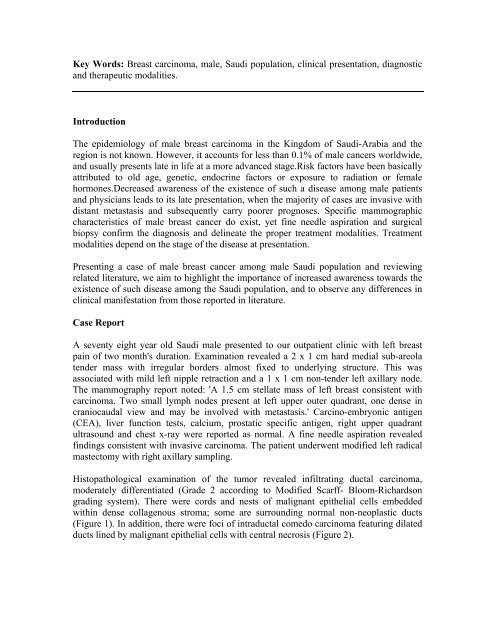
Key Words: Breast carcinoma, male, Saudi population, clinical presentation, diagnostic<br />
<strong>and</strong> therapeutic modalities.<br />
Introduction<br />
The epidemiology of male <strong>breast</strong> carcinoma in the Kingdom of Saudi-Arabia <strong>and</strong> the<br />
region is not known. However, it accounts for less than 0.1% of male <strong>cancer</strong>s worldwide,<br />
<strong>and</strong> usually presents late in life at a more advanced stage.Risk factors have been basically<br />
attributed to old age, genetic, endocrine factors or exposure to radiation or female<br />
hormones.Decreased awareness of the existence of such a disease among male patients<br />
<strong>and</strong> physicians leads to its late presentation, when the majority of cases are invasive with<br />
distant metastasis <strong>and</strong> subsequently carry poorer prognoses. Specific mammographic<br />
characteristics of male <strong>breast</strong> <strong>cancer</strong> do exist, yet fine needle aspiration <strong>and</strong> surgical<br />
biopsy confirm the diagnosis <strong>and</strong> delineate the proper treatment modalities. Treatment<br />
modalities depend on the stage of the disease at presentation.<br />
Presenting a case of male <strong>breast</strong> <strong>cancer</strong> among male Saudi population <strong>and</strong> <strong>review</strong>ing<br />
related literature, we aim to highlight the importance of increased awareness towards the<br />
existence of such disease among the Saudi population, <strong>and</strong> to observe any differences in<br />
clinical manifestation from those <strong>report</strong>ed in literature.<br />
<strong>Case</strong> Report<br />
A seventy eight year old Saudi male presented to our outpatient clinic with left <strong>breast</strong><br />
pain of two month's duration. Examination revealed a 2 x 1 cm hard medial sub-areola<br />
tender mass with irregular borders almost fixed to underlying structure. This was<br />
associated with mild left nipple retraction <strong>and</strong> a 1 x 1 cm non-tender left axillary node.<br />
The mammography <strong>report</strong> noted: 'A 1.5 cm stellate mass of left <strong>breast</strong> consistent with<br />
carcinoma. Two small lymph nodes present at left upper outer quadrant, one dense in<br />
craniocaudal view <strong>and</strong> may be involved with metastasis.' Carcino-embryonic antigen<br />
(CEA), liver function tests, calcium, prostatic specific antigen, right upper quadrant<br />
ultrasound <strong>and</strong> chest x-ray were <strong>report</strong>ed as normal. A fine needle aspiration revealed<br />
findings consistent with invasive carcinoma. The patient underwent modified left radical<br />
mastectomy with right axillary sampling.<br />
Histopathological examination of the tumor revealed infiltrating ductal carcinoma,<br />
moderately differentiated (Grade 2 according to Modified Scarff- Bloom-Richardson<br />
grading system). There were cords <strong>and</strong> nests of malignant epithelial cells embedded<br />
within dense collagenous stroma; some are surrounding normal non-neoplastic ducts<br />
(Figure 1). In addition, there were foci of intraductal comedo carcinoma featuring dilated<br />
ducts lined by malignant epithelial cells with central necrosis (Figure 2).