Download full PDF - UBC Medical Journal
Download full PDF - UBC Medical Journal
Download full PDF - UBC Medical Journal

You also want an ePaper? Increase the reach of your titles
YUMPU automatically turns print PDFs into web optimized ePapers that Google loves.
CASE AND ELECTIVE REPORTS<br />
pulmonary sarcoidosis, systemic sclerosis, Sjogren’s syndrome,<br />
polymyositis, and dermatomyositis. 6,7 This report describes two<br />
cases of HLH in adults in order to illustrate the clinical features<br />
and the importance of prompt diagnosis and therapy.<br />
CASE REPORT 1<br />
A 21–year-old Filipino man presented with a 10–day history of<br />
fever, headache and scleral icterus. He was previously healthy<br />
and taking no regular medications. His family history was<br />
unremarkable. The physical exam revealed a temperature of<br />
40.7 °C and scleral icterus but was otherwise normal. His initial<br />
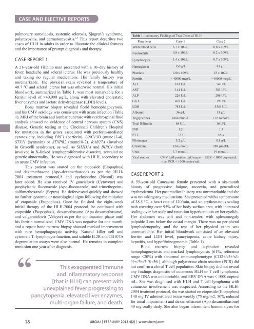
bloodwork, summarized in Table 1, was most remarkable for a<br />
ferritin level of >40,000 µg/L, along with elevated cholestatic<br />
liver enzymes and lactate dehydrogenase (LDH) levels.<br />
Bone marrow biopsy revealed florid hemophagocytosis,<br />
and his CMV serology was consistent with acute infection (Table<br />
1). MRI of the brain and lumbar puncture with cerebrospinal fluid<br />
analysis showed no evidence of central nervous system (CNS)<br />
disease. Genetic testing at the Cincinnati Children’s Hospital<br />
for mutations in the genes associated with perforin-mediated<br />
cytotoxicity, including PRF1 (perforin), UNC13D (munc13-4),<br />
STX11 (syntaxin) or STXPB2 (munc18-2). RAB27A (involved<br />
in Griscelli syndrome), as well as SH2D1A and BIRC4 (both<br />
involved in X-linked lymphoproliferative disorder), revealed no<br />
genetic abnormality. He was diagnosed with HLH, secondary to<br />
an acute CMV infection.<br />
This patient was started on the etoposide (Etopophos)<br />
and dexamethasone (Apo-dexamethasone) as per the HLH-<br />
2004 treatment protocol,8 and cyclosporine (Neoral) was<br />
later added. He also received IV ganciclovir (Cytovene) and<br />
prophylactic fluconazole (Apo-fluconazole) and trimethoprim–<br />
sulfamethoxazole (Septra). He defervesced quickly and showed<br />
no further systemic or neurological signs following the initiation<br />
of etoposide (Etopophos). Once he finished the eight–week<br />
initial therapy of the HLH-2004 protocol, he continued with<br />
etoposide (Etopophos), dexamethasone (Apo-dexamethasone),<br />
and valganciclovir (Valcyte) as per the continuation phase until<br />
his ferritin normalized, CMV DNA was negative for one month,<br />
and a repeat bone marrow biopsy showed marked improvement<br />
with rare hemophagocytic activity. Natural killer cell and<br />
cytotoxic T–lymphocyte function, and soluble IL2R and CD107A<br />
degranulation assays were also normal. He remains in complete<br />
remission one year after diagnosis.<br />
“<br />
This exaggerated immune<br />
and inflammatory response<br />
[that is HLH] can present with<br />
unexplained fever progressing to<br />
pancytopenia, elevated liver enzymes,<br />
multi-organ failure, and death.<br />
Table 1. Laboratory Findings of Two Cases of HLH.<br />
Parameter Case 1 Case 2<br />
White blood cells 6.7 x 10⁹/L 0.8 x 10⁹/L<br />
Neutrophils 4.8 x 10⁹/L 0.2 x 10⁹/L<br />
Lymphocytes 1.4 x 10⁹/L 0.7 x 10⁹/L<br />
Hemoglobin 150 g/L 81 g/L<br />
Platelets 120 x 109/L 23 x 109/L<br />
Ferritin > 40000 mcg/L > 40000 mcg/L<br />
ALT 103 U/L 54 U/L<br />
AST 144 U/L 383 U/L<br />
ALP 226 U/L 206 U/L<br />
GGT 478 U/L 39 U/L<br />
LDH 783 U/L 3546 U/L<br />
Albumin 36 g/L 13 g/L<br />
Triglycerides 4.04 mmol/L 3.18 mmol/L<br />
Total Bilirubin 68 U/L 16 U/L<br />
INR 1.2 1.5<br />
PTT 33 s 65 s<br />
Fibrinogen 3.2 g/L 0.8 g/L<br />
Creatinine 124 µmol/L 288 µmol/L<br />
Urea 3.7 mmol/L 19 mmol/L<br />
Viral studies<br />
CASE REPORT 2<br />
CMV IgM positive, IgG negative,<br />
PCR < 1000 copies/mL<br />
EBV < 1000 copies/mL<br />
A 55-year-old Caucasian female presented with a six-month<br />
history of progressive fatigue, anorexia, and generalized<br />
erythroderma. Her past medical history was unremarkable and she<br />
was not taking any medications. She presented with a temperature<br />
of 38.5 °C, a heart rate of 120/min, and an erythematous scaling<br />
rash covering over 95% of her body surface area, with increased<br />
scaling over her scalp and retention hyperkeratosis on her eyelids.<br />
Her abdomen was soft and non-tender, with splenomegaly<br />
palpable 3 cm below the costal margin. There was no peripheral<br />
lymphadenopathy, and the rest of her physical exam was<br />
unremarkable. Her initial bloodwork consisted of an elevated<br />
ferritin and LDH level, pancytopenia, acute kidney injury,<br />
hepatitis, and hypofibrinogenemia (Table 1).<br />
Bone marrow biopsy and aspiration revealed<br />
hemophagocytosis and marked lymphocytosis (61%, reference<br />
range