PDF download - Translumina
PDF download - Translumina
PDF download - Translumina

You also want an ePaper? Increase the reach of your titles
YUMPU automatically turns print PDFs into web optimized ePapers that Google loves.
JACC Vol. 49, No. 12, 2007 Adriaenssens et al.<br />
March 27, 2007:1265–71 Rapamycin Plus Estradiol-Eluting Stent<br />
nitroglycerin, and the same single worst view projection was<br />
measured at all time points. Qualitative morphologic lesion<br />
characteristics were characterized using standard criteria<br />
(16). Off-line quantitative coronary angiographic analysis<br />
was performed using the automated edge detection system<br />
(QCA-CMS version 6.0, Medis, Medical Imaging Systems,<br />
Leiden, the Netherlands). The contrast-filled non-tapered<br />
catheter tip was used for calibration. The reference diameter<br />
was measured by interpolation. Minimal lumen diameter<br />
was measured within the stent and within the 5-mm<br />
proximal and distal edges of the stent. Quantitative analysis<br />
was performed in the in-stent area (in-stent analysis) and in<br />
the in-segment area including the stented segment as well as<br />
both 5-mm margins proximal and distal to the stent<br />
(in-segment analysis).<br />
The primary end point of the study was in-stent late<br />
lumen loss. Secondary end points were the binary angiographic<br />
restenosis (diameter stenosis of at least 50% based<br />
on in-segment analysis) at follow-up angiography, the need<br />
for target lesion revascularization due to restenosis in the<br />
presence of symptoms or signs of ischemia, the combined<br />
incidence of death and myocardial infarction, and the<br />
incidence of stent thrombosis during the 12-month followup.<br />
The diagnosis of myocardial infarction required the<br />
presence of new Q waves in the electrocardiogram and/or<br />
elevation of creatine kinase or its MB isoenzyme at least 3<br />
times the upper limit of normal in �2 blood samples. Stent<br />
thrombosis was defined as a target lesion occlusion with<br />
Thrombolysis In Myocardial Infarction flow grade 0 or 1<br />
accompanied by angiographically visible thrombus and/or<br />
acute coronary syndrome. Retrospectively, stent thrombosis<br />
was also assessed based on the definitions of the Academic<br />
Research Consortium (ARC) (17).<br />
Statistical methods. The study hypothesis was that ERES<br />
was superior to RES in terms of decreased late lumen loss.<br />
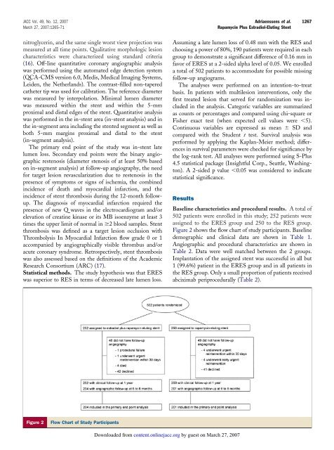
Figure 2 Flow Chart of Study Participants<br />
Downloaded from<br />
content.onlinejacc.org by guest on March 27, 2007<br />
1267<br />
Assuming a late lumen loss of 0.48 mm with the RES and<br />
choosing a power of 80%, 190 patients were required in each<br />
group to demonstrate a significant difference of 0.16 mm in<br />
favor of ERES at a 2-sided alpha level of 0.05. We enrolled<br />
a total of 502 patients to accommodate for possible missing<br />
follow-up angiograms.<br />
The analyses were performed on an intention-to-treat<br />
basis. In patients with multilesion interventions, only the<br />
first treated lesion that served for randomization was included<br />
in the analysis. Categoric variables are summarized<br />
as counts or percentages and compared using chi-square or<br />
Fisher exact test (when expected cell values were �5).<br />
Continuous variables are expressed as mean � SD and<br />
compared with the Student t test. Survival analysis was<br />
performed by applying the Kaplan-Meier method; differences<br />
in survival parameters were checked for significance by<br />
the log-rank test. All analyses were performed using S-Plus<br />
4.5 statistical package (Insightful Corp., Seattle, Washington).<br />
A 2-sided p value �0.05 was considered to indicate<br />
statistical significance.<br />
Results<br />
Baseline characteristics and procedural results. A total of<br />
502 patients were enrolled in this study; 252 patients were<br />
assigned to the ERES group and 250 to the RES group.<br />
Figure 2 shows the flow chart of study participants. Baseline<br />
demographic and clinical data are shown in Table 1.<br />
Angiographic and procedural characteristics are shown in<br />
Table 2. Data were well matched between the 2 groups.<br />
Implantation of the assigned stent was successful in all but<br />
1 (99.6%) patient in the ERES group and in all patients in<br />
the RES group. Only a small proportion of patients received<br />
abciximab periprocedurally (Table 2).