INNOVATIONS FROM THE EDGE - KPIT
INNOVATIONS FROM THE EDGE - KPIT
INNOVATIONS FROM THE EDGE - KPIT

Create successful ePaper yourself
Turn your PDF publications into a flip-book with our unique Google optimized e-Paper software.
on the methods of coding of output information,<br />
which is transmitted out from the brain to the<br />
body organs through the motor neurons and<br />
inter-neurons.<br />
III. Types of Brain Machine Interface<br />
captured, decoded and transmitted to the robot<br />
arm controller. The final robot arm movement is<br />
also visible to the monkey. Scientists at Duke<br />
University are carrying out this experiment [3]. The<br />
figure 4 shows an experimental setup of BMI for<br />
Robot control at Duke University.<br />
Depending upon the placement of electrodes for<br />
picking the neural signals, there are three types of<br />
Brain Machine Interface that are currently under<br />
development.<br />
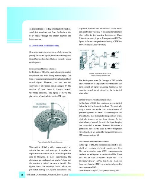
Invasive Brain Machine Interface<br />
In this type of BMI, the electrodes are implanted<br />
deep inside the brain during neurosurgery. This<br />
type of placement produces the highest quality of<br />
neural signals. However, this also has the<br />
drawback of electrodes being damaged by the<br />
reaction of brain tissue to foreign material<br />
(electrode material). The figure 3 shows the<br />
placement of electrode in Invasive BMI type.<br />
Figure 4: Experimental Setup of BMI at<br />
Duke's University (Reference [3])<br />
The development areas for this type of BMI include<br />
the development of implantable electrodes and the<br />
development of signal processing techniques for<br />
decoding neural signals picked by the implanted<br />
electrodes.<br />
Partially Invasive Brain Machine Interface<br />
In this type of BMI, the electrodes are implanted<br />
below the skull and outside the brain. The electrode<br />
array is spread out on the brain surface instead of<br />
penetrating inside the brain. The advantage of this<br />
type of BMI is that it eliminates the possibility of the<br />
electrode damage by the brain tissues. As the<br />
electrode stays beneath the skull, the signal damping<br />
due to the skull is reduced. However, this creates a<br />
permanent hole on the skull. Electrocorticography<br />
(ECoG) methods are utilized for the partially invasive<br />
BMI implementations [4].<br />
Figure 3: Invasive BMI Electrode Placement<br />
This method of BMI is widely experimented on<br />
animals like rats and monkeys. A number of<br />
experiments are carried out for controlling a robot<br />
arm by thoughts. In these experiments, the<br />
electrodes are implanted in a monkey's brain and<br />
the monkey is trained to move a joystick. The<br />
signals from the monkey's brain, which are<br />
generated during the joystick movement, are<br />
Non-Invasive Brain Machine Interface<br />
In this type of BMI, the electrodes are placed on the<br />
s ku l l a t c e r ta i n d e f i n e d p o s i t i o n s . T h e<br />
Electroencephalography (EEG) measurements<br />
methods are widely used in non-invasive BMIs. There<br />
a r e o t h e r n o n - i n v a s i v e m e t h o d s l i k e<br />
Electromyography (EMG), Functional Magnetic<br />
Resonance Imaging (fMRI), etc. which are also used in<br />
the non-invasive BMIs.<br />
In methods utilizing EEG, the signals (neural signals)<br />
14 TechTalk@<strong>KPIT</strong>Cummins, Volume 5, Issue 1, 2012