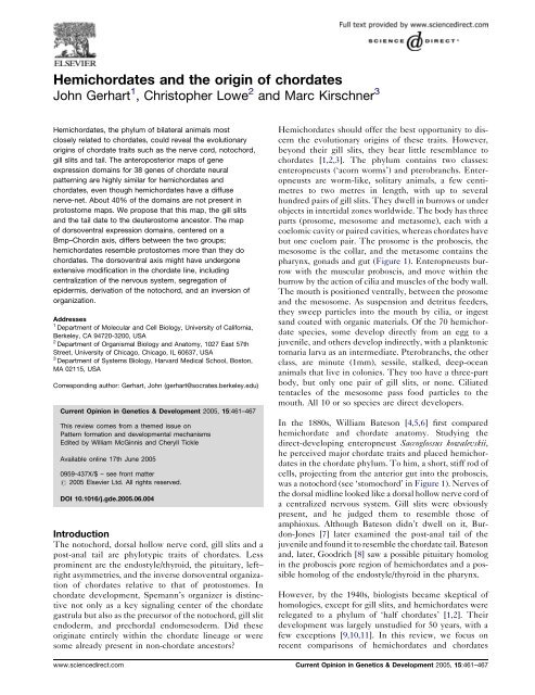
Hemichordates and the origin of chordates
Hemichordates and the origin of chordates
Hemichordates and the origin of chordates

Create successful ePaper yourself
Turn your PDF publications into a flip-book with our unique Google optimized e-Paper software.
<strong>Hemi<strong>chordates</strong></strong> <strong>and</strong> <strong>the</strong> <strong>origin</strong> <strong>of</strong> <strong>chordates</strong><br />
John Gerhart 1 , Christopher Lowe 2 <strong>and</strong> Marc Kirschner 3<br />
<strong>Hemi<strong>chordates</strong></strong>, <strong>the</strong> phylum <strong>of</strong> bilateral animals most<br />
closely related to <strong>chordates</strong>, could reveal <strong>the</strong> evolutionary<br />
<strong>origin</strong>s <strong>of</strong> chordate traits such as <strong>the</strong> nerve cord, notochord,<br />
gill slits <strong>and</strong> tail. The anteroposterior maps <strong>of</strong> gene<br />
expression domains for 38 genes <strong>of</strong> chordate neural<br />
patterning are highly similar for hemi<strong>chordates</strong> <strong>and</strong><br />
<strong>chordates</strong>, even though hemi<strong>chordates</strong> have a diffuse<br />
nerve-net. About 40% <strong>of</strong> <strong>the</strong> domains are not present in<br />
protostome maps. We propose that this map, <strong>the</strong> gill slits<br />
<strong>and</strong> <strong>the</strong> tail date to <strong>the</strong> deuterostome ancestor. The map<br />
<strong>of</strong> dorsoventral expression domains, centered on a<br />
Bmp–Chordin axis, differs between <strong>the</strong> two groups;<br />
hemi<strong>chordates</strong> resemble protostomes more than <strong>the</strong>y do<br />
<strong>chordates</strong>. The dorsoventral axis might have undergone<br />
extensive modification in <strong>the</strong> chordate line, including<br />
centralization <strong>of</strong> <strong>the</strong> nervous system, segregation <strong>of</strong><br />
epidermis, derivation <strong>of</strong> <strong>the</strong> notochord, <strong>and</strong> an inversion <strong>of</strong><br />
organization.<br />
Addresses<br />
1 Department <strong>of</strong> Molecular <strong>and</strong> Cell Biology, University <strong>of</strong> California,<br />
Berkeley, CA 94720-3200, USA<br />
2 Department <strong>of</strong> Organismal Biology <strong>and</strong> Anatomy, 1027 East 57th<br />
Street, University <strong>of</strong> Chicago, Chicago, IL 60637, USA<br />
3 Department <strong>of</strong> Systems Biology, Harvard Medical School, Boston,<br />
MA 02115, USA<br />
Corresponding author: Gerhart, John (gerhart@socrates.berkeley.edu)<br />
Current Opinion in Genetics & Development 2005, 15:461–467<br />
This review comes from a <strong>the</strong>med issue on<br />
Pattern formation <strong>and</strong> developmental mechanisms<br />
Edited by William McGinnis <strong>and</strong> Cheryll Tickle<br />
Available online 17th June 2005<br />
0959-437X/$ – see front matter<br />
# 2005 Elsevier Ltd. All rights reserved.<br />
DOI 10.1016/j.gde.2005.06.004<br />
Introduction<br />
The notochord, dorsal hollow nerve cord, gill slits <strong>and</strong> a<br />
post-anal tail are phylotypic traits <strong>of</strong> <strong>chordates</strong>. Less<br />
prominent are <strong>the</strong> endostyle/thyroid, <strong>the</strong> pituitary, left–<br />
right asymmetries, <strong>and</strong> <strong>the</strong> inverse dorsoventral organization<br />
<strong>of</strong> <strong>chordates</strong> relative to that <strong>of</strong> protostomes. In<br />
chordate development, Spemann’s organizer is distinctive<br />
not only as a key signaling center <strong>of</strong> <strong>the</strong> chordate<br />
gastrula but also as <strong>the</strong> precursor <strong>of</strong> <strong>the</strong> notochord, gill slit<br />
endoderm, <strong>and</strong> prechordal endomesoderm. Did <strong>the</strong>se<br />
<strong>origin</strong>ate entirely within <strong>the</strong> chordate lineage or were<br />
some already present in non-chordate ancestors<br />
<strong>Hemi<strong>chordates</strong></strong> should <strong>of</strong>fer <strong>the</strong> best opportunity to discern<br />
<strong>the</strong> evolutionary <strong>origin</strong>s <strong>of</strong> <strong>the</strong>se traits. However,<br />
beyond <strong>the</strong>ir gill slits, <strong>the</strong>y bear little resemblance to<br />
<strong>chordates</strong> [1,2,3]. The phylum contains two classes:<br />
enteropneusts (‘acorn worms’) <strong>and</strong> pterobranchs. Enteropneusts<br />
are worm-like, solitary animals, a few centimetres<br />
to two metres in length, with up to several<br />
hundred pairs <strong>of</strong> gill slits. They dwell in burrows or under<br />
objects in intertidal zones worldwide. The body has three<br />
parts (prosome, mesosome <strong>and</strong> metasome), each with a<br />
coelomic cavity or paired cavities, whereas <strong>chordates</strong> have<br />
but one coelom pair. The prosome is <strong>the</strong> proboscis, <strong>the</strong><br />
mesosome is <strong>the</strong> collar, <strong>and</strong> <strong>the</strong> metasome contains <strong>the</strong><br />
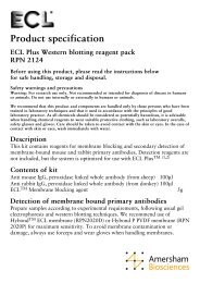
pharynx, gonads <strong>and</strong> gut (Figure 1). Enteropneusts burrow<br />
with <strong>the</strong> muscular proboscis, <strong>and</strong> move within <strong>the</strong><br />
burrow by <strong>the</strong> action <strong>of</strong> cilia <strong>and</strong> muscles <strong>of</strong> <strong>the</strong> body wall.<br />
The mouth is positioned ventrally, between <strong>the</strong> prosome<br />
<strong>and</strong> <strong>the</strong> mesosome. As suspension <strong>and</strong> detritus feeders,<br />
<strong>the</strong>y sweep particles into <strong>the</strong> mouth by cilia, or ingest<br />
s<strong>and</strong> coated with organic materials. Of <strong>the</strong> 70 hemichordate<br />
species, some develop directly from an egg to a<br />
juvenile, <strong>and</strong> o<strong>the</strong>rs develop indirectly, with a planktonic<br />
tornaria larva as an intermediate. Pterobranchs, <strong>the</strong> o<strong>the</strong>r<br />
class, are minute (1mm), sessile, stalked, deep-ocean<br />
animals that live in colonies. They too have a three-part<br />
body, but only one pair <strong>of</strong> gill slits, or none. Ciliated<br />
tentacles <strong>of</strong> <strong>the</strong> mesosome pass food particles to <strong>the</strong><br />
mouth. All 10 or so species are direct developers.<br />
In <strong>the</strong> 1880s, William Bateson [4,5,6] first compared<br />
hemichordate <strong>and</strong> chordate anatomy. Studying <strong>the</strong><br />
direct-developing enteropneust Saccoglossus kowalevskii,<br />
he perceived major chordate traits <strong>and</strong> placed hemi<strong>chordates</strong><br />
in <strong>the</strong> chordate phylum. To him, a short, stiff rod <strong>of</strong><br />
cells, projecting from <strong>the</strong> anterior gut into <strong>the</strong> proboscis,<br />
was a notochord (see ‘stomochord’ in Figure 1). Nerves <strong>of</strong><br />
<strong>the</strong> dorsal midline looked like a dorsal hollow nerve cord <strong>of</strong><br />
a centralized nervous system. Gill slits were obviously<br />
present, <strong>and</strong> he judged <strong>the</strong>m to resemble those <strong>of</strong><br />
amphioxus. Although Bateson didn’t dwell on it, Burdon-Jones<br />
[7] later examined <strong>the</strong> post-anal tail <strong>of</strong> <strong>the</strong><br />
juvenile <strong>and</strong> found it to resemble <strong>the</strong> chordate tail. Bateson<br />
<strong>and</strong>, later, Goodrich [8] saw a possible pituitary homolog<br />
in <strong>the</strong> proboscis pore region <strong>of</strong> hemi<strong>chordates</strong> <strong>and</strong> a possible<br />
homolog <strong>of</strong> <strong>the</strong> endostyle/thyroid in <strong>the</strong> pharynx.<br />
However, by <strong>the</strong> 1940s, biologists became skeptical <strong>of</strong><br />
homologies, except for gill slits, <strong>and</strong> hemi<strong>chordates</strong> were<br />
relegated to a phylum <strong>of</strong> ‘half <strong>chordates</strong>’ [1,2]. Their<br />
development was largely unstudied for 50 years, with a<br />
few exceptions [9,10,11]. In this review, we focus on<br />
recent comparisons <strong>of</strong> hemi<strong>chordates</strong> <strong>and</strong> <strong>chordates</strong><br />
www.sciencedirect.com Current Opinion in Genetics & Development 2005, 15:461–467
462 Pattern formation <strong>and</strong> developmental mechanisms<br />
Figure 1<br />
(a) (b) (c)<br />
Proboscis<br />
Mouth<br />
Collar<br />
Pharynx<br />
Gill<br />
slit<br />
Proboscis<br />
Heart<br />
Kidney<br />
Collar<br />
Tail<br />
Anus<br />
Gut<br />
Stomochord<br />
Mouth<br />
Saccoglossus kowalevskii, a direct-developing enteropneust hemichordate <strong>of</strong> <strong>the</strong> US Atlantic coast. (a) Adult female. Note <strong>the</strong> white proboscis,<br />
orange collar, tan pharynx <strong>and</strong> gut, <strong>and</strong> grey ovaries. Length: 10–18 cm. (b) Juvenile, a week after hatching, two weeks after fertilization <strong>of</strong><br />
<strong>the</strong> egg. Length: 1 mm (c) The ‘notochord’, so-called by Bateson (see text), now called <strong>the</strong> stomochord, located between <strong>the</strong> proboscis <strong>and</strong><br />
collar. Shown in sagittal section. Redrawn from Bateson [5].<br />
regarding <strong>the</strong>ir gene sequences <strong>and</strong> expression domains.<br />
We discuss <strong>the</strong> updated deductions about <strong>the</strong>ir common<br />
ancestor <strong>and</strong>, hence, about <strong>the</strong> <strong>origin</strong> <strong>of</strong> <strong>chordates</strong>.<br />
Modern phylogenies<br />
Recent DNA phylogenies place hemi<strong>chordates</strong> as <strong>the</strong><br />
sister group <strong>of</strong> echinoderms [12–14]. Toge<strong>the</strong>r, <strong>the</strong>se<br />
two are <strong>the</strong> sister group <strong>of</strong> <strong>chordates</strong> (Figure 2). The<br />
three phyla constitute <strong>the</strong> supertaxon <strong>of</strong> deuterostomes<br />
(Xenoturbella might be a fourth [15]). The lineage from<br />
<strong>the</strong> ancestor <strong>of</strong> deuterostomes to <strong>the</strong> ancestor <strong>of</strong> <strong>chordates</strong><br />
bore no branches to extant groups. Paleontology <strong>of</strong> <strong>the</strong><br />
past decade has uncovered a pr<strong>of</strong>usion <strong>of</strong> Cambrian<br />
deuterostomes (e.g. vetulicolians, yunnanozoans) that<br />
has still to be reconciled with this simple phylogeny [16].<br />
O<strong>the</strong>r bilateral animals, comprising approximately 25<br />
phyla, are protostomes. The last common ancestor <strong>of</strong><br />
deuterostomes <strong>and</strong> protostomes is <strong>the</strong> ancestor <strong>of</strong> all<br />
bilateral animals, one node before <strong>the</strong> deuterostome<br />
ancestor.<br />
Four venerable hypo<strong>the</strong>ses <strong>of</strong> chordate<br />
<strong>origin</strong>s<br />
Consistent with <strong>the</strong> modern phylogeny are four hypo<strong>the</strong>ses<br />
for <strong>the</strong> <strong>origin</strong> <strong>of</strong> <strong>chordates</strong> from a deuterostome<br />
ancestor. We present <strong>the</strong>se <strong>and</strong> comment on <strong>the</strong>m in<br />
light <strong>of</strong> recent results.<br />
1. Hemichordate hypo<strong>the</strong>sis: for Bateson [17], <strong>chordates</strong><br />
evolved by <strong>the</strong> exaggeration <strong>of</strong> structures <strong>of</strong> a<br />
hemichordate-like ancestor that had a dorsal central<br />
nervous system. Goodrich [8] proposed that <strong>the</strong> two<br />
anterior coelom pairs shrank to preotic somites in<br />
<strong>chordates</strong>, <strong>and</strong> dorsal anterior structures were displaced<br />
around <strong>the</strong> front end to ventral locations.<br />
2. Auricularian hypo<strong>the</strong>sis: for Garstang [18], <strong>the</strong> chordate<br />
ancestor was a motile, ciliated larva <strong>of</strong> a sessile,<br />
Current Opinion in Genetics & Development 2005, 15:461–467<br />
pterobranch-like adult. Rows <strong>of</strong> cilia, within a diffuse<br />
nervous system, moved dorsally by a series <strong>of</strong><br />
evolutionary intermediates, <strong>and</strong> eventually internalized<br />
at <strong>the</strong> midline as a new, centralized, dorsal nervecord.<br />
From <strong>the</strong> sessile adult, gill slits, a notochord, <strong>and</strong><br />
sexual maturity were transferred to <strong>the</strong> larva by<br />
neoteny, forming a motile protochordate adult.<br />
3. Bilaterial ancestor hypo<strong>the</strong>sis: in <strong>the</strong> 1990s, <strong>chordates</strong><br />
(mice, frogs <strong>and</strong> fish) were found to share many<br />
domains <strong>of</strong> gene expression with protostomes (mainly<br />
Drosophila), such as Hox domains in <strong>the</strong> posterior head<br />
<strong>and</strong> trunk; emx <strong>and</strong> otx domains in <strong>the</strong> anterior head;<br />
pax6 <strong>and</strong> six expression in light receptive organs <strong>of</strong> <strong>the</strong><br />
head; many genes <strong>of</strong> neuron identity <strong>and</strong> differentiation;<br />
nkx2.5 in <strong>the</strong> heart; <strong>and</strong> hh/bmp in <strong>the</strong> gut <strong>and</strong><br />
visceral mesoderm [19,20]. All <strong>the</strong>se domains were<br />
ascribed to <strong>the</strong> ancestor <strong>of</strong> bilateral animals. Given <strong>the</strong><br />
existence <strong>of</strong> such a complex ancestor, <strong>the</strong> evolution <strong>of</strong><br />
deuterostomes <strong>and</strong> <strong>chordates</strong> entailed less innovation<br />
than was previously thought.<br />
4. Inversion hypo<strong>the</strong>sis [21,22]: <strong>the</strong> chordate ancestor<br />
had a protostome-like arrangement <strong>of</strong> organs in <strong>the</strong><br />
dorsoventral dimension: a ventral centralized nervous<br />
system, ventral musculature <strong>and</strong> a dorsal heart. A<br />
descendent in <strong>the</strong> chordate line inverted its body<br />
dorsoventrally <strong>and</strong> evolved a mouth on <strong>the</strong> new ventral<br />
surface, making chordate anatomy <strong>the</strong> inverse to that<br />
<strong>of</strong> protostomes. As recent support, bmp was found to be<br />
expressed dorsally in protostome embryos but ventrally<br />
in chordate embryos, whereas expression <strong>of</strong><br />
chordin, which encodes a Bmp antagonist, is <strong>the</strong><br />
reverse. This Bmp–Chordin axis, which underlies<br />
dorsoventral patterning in all bilateral animals, is<br />
inversely oriented. Motoneurons <strong>and</strong> interneurons<br />
develop in <strong>the</strong> Bmp-free ectoderm, <strong>and</strong> sensory<br />
neurons develop in <strong>the</strong> Bmp area. Although not<br />
logically required, <strong>the</strong> hypo<strong>the</strong>sis usually starts with an<br />
ancestor having a centralized nervous system. Hemi-<br />
www.sciencedirect.com
<strong>Hemi<strong>chordates</strong></strong> <strong>and</strong> <strong>the</strong> <strong>origin</strong> <strong>of</strong> <strong>chordates</strong> Gerhart, Lowe <strong>and</strong> Kirschner 463<br />
Figure 2<br />
Ecdysozoa<br />
Lophotrochozoa<br />
Echinoderms<br />
<strong>Hemi<strong>chordates</strong></strong><br />
Uro<strong>chordates</strong><br />
Cephalo<strong>chordates</strong><br />
Vertebrates<br />
Protostomes<br />
Deuterostomes<br />
Tripartite coelom,<br />
larva<br />
CHORDATES<br />
Chordate ancestor<br />
(Notochord, post-anal tail,<br />
dorsal hollow nerve cord)<br />
Deuterostome ancestor<br />
(Gill slits)<br />
Bilateral ancestor<br />
(Dorsoventral axis, mesoderm, through gut,<br />
cephalization, hox domains in trunk,<br />
tinman heart, pax6/emx/otx domains in head)<br />
Current Opinion in Genetics & Development<br />
Phylogeny <strong>of</strong> hemi<strong>chordates</strong> <strong>and</strong> <strong>chordates</strong>, from 18S rDNA comparisons [12–14]. Note that <strong>the</strong> chordate ancestor, in addition to <strong>the</strong> ancestor<br />
<strong>of</strong> hemi<strong>chordates</strong> <strong>and</strong> echinoderms, descends from <strong>the</strong> deuterostome ancestor, which descends from <strong>the</strong> bilateral ancestor.<br />
<strong>chordates</strong> might be uninverted, implying that inversion<br />
is a trait exclusive to <strong>the</strong> chordate line [23].<br />
Updating <strong>the</strong> comparisons<br />
Ambiguities <strong>of</strong> morphology have impeded comparisons<br />
between hemi<strong>chordates</strong> <strong>and</strong> <strong>chordates</strong>. Gene expression<br />
domains <strong>of</strong>fer an alternative <strong>of</strong> a more conserved kind <strong>of</strong><br />
anatomy. We update <strong>the</strong> traits chosen by Bateson <strong>and</strong><br />
<strong>the</strong>n compare body plans.<br />
1. Gill slits: in addition to anatomical similarities, <strong>the</strong><br />
endoderm <strong>of</strong> <strong>the</strong> gill slit in both hemi<strong>chordates</strong> <strong>and</strong><br />
<strong>chordates</strong> expresses pax1/9 <strong>and</strong> six 1 genes [24,25 ].<br />
Fur<strong>the</strong>rmore, gill slits occur at <strong>the</strong> same body level in<br />
organisms <strong>of</strong> both phyla (see ‘domain map’ below).<br />
The deuterostome ancestor probably had gill slits,<br />
which were later lost in echinoderms.<br />
2. The post-anal tail <strong>of</strong> hemi<strong>chordates</strong> expresses <strong>the</strong><br />
hox11/13a, b <strong>and</strong> c genes, <strong>and</strong> <strong>the</strong> chordate tail<br />
expresses hox11, 12 <strong>and</strong> 13 [25,26 ]. The two groups<br />
probably have homologous post-anal body regions,<br />
brought forward from <strong>the</strong> deuterostome ancestor. The<br />
hemichordate tail doesn’t function for chordate-like<br />
swimming, but it is contractile. Motility functions have<br />
probably diverged.<br />
3. The hemichordate notochord <strong>of</strong> Bateson was later<br />
called <strong>the</strong> stomochord or buccal diverticulum to avoid<br />
<strong>the</strong> implication <strong>of</strong> homology. Like <strong>the</strong> notochord, <strong>the</strong><br />
stomochord contains large, vacuolated cells [27].<br />
Genes that are expressed in <strong>the</strong> chordate notochord<br />
<strong>and</strong> in its precursor cells <strong>of</strong> Spemann’s organizer —<br />
namely, bra [28], chordin, hnf3b/foxA1, admp, hh, nodal<br />
<strong>and</strong> noggin (J Gerhart et al., unpublished) — are not<br />
expressed in <strong>the</strong> stomochord but are expressed<br />
elsewhere (see below). The stomochord <strong>and</strong> nearby<br />
endoderm express otx, dmbx, gsc, hex <strong>and</strong> dkk, as does<br />
<strong>the</strong> prechordal endomesoderm <strong>of</strong> <strong>chordates</strong>, which<br />
itself has precursors in Spemann’s organizer. The<br />
stomochord also expresses ttf2 strongly, as do <strong>the</strong><br />
enigmatic club-shaped organ <strong>of</strong> amphioxus <strong>and</strong> <strong>the</strong><br />
vertebrate thyroid [29]. Prechordal endomesoderm,<br />
which was unknown to Bateson, <strong>and</strong> <strong>the</strong> stomochord<br />
have <strong>the</strong> same location in <strong>the</strong> body plan (see ‘domain<br />
map’ below).<br />
4. <strong>Hemi<strong>chordates</strong></strong> have no dorsal hollow nerve cord.<br />
Bullock [30] <strong>and</strong> Knight-Jones [31] silver-stained <strong>the</strong><br />
nervous system <strong>and</strong> found an epidermal nerve net.<br />
Nerves are finely mixed with epidermal cells over <strong>the</strong><br />
entire body surface. Pan-neural genes sox2/3, elav <strong>and</strong><br />
musashi/nrp are expressed pan-ectodermally [25 ].<br />
Axons extend into a dense basiepi<strong>the</strong>lial mat, some<br />
synapsing locally <strong>and</strong> some extending into thick axon<br />
tracts at <strong>the</strong> dorsal or ventral midlines [30,31,32].<br />
These tracts do not constitute a central nervous<br />
system; <strong>the</strong>y lack neural cell bodies <strong>and</strong> hence<br />
neurogenesis, <strong>and</strong> <strong>the</strong>y lack interneurons <strong>and</strong> hence<br />
information processing. Bateson mistook <strong>the</strong> dorsal<br />
tract for a hollow cord. We favor <strong>the</strong> interpretation that<br />
<strong>the</strong> deuterostome ancestor had no central nervous<br />
www.sciencedirect.com Current Opinion in Genetics & Development 2005, 15:461–467
464 Pattern formation <strong>and</strong> developmental mechanisms<br />
system or notochord, ra<strong>the</strong>r than <strong>the</strong> alternative<br />
explanation that <strong>the</strong>se traits were present but that<br />
both were lost in hemi<strong>chordates</strong> <strong>and</strong> echinoderms.<br />
The anteroposterior domain map<br />
Although hemi<strong>chordates</strong> <strong>and</strong> <strong>chordates</strong> differ anatomically,<br />
<strong>the</strong>ir body plans are similar in <strong>the</strong> anteroposterior<br />
dimension regarding <strong>the</strong> topology <strong>of</strong> <strong>the</strong> domains <strong>of</strong><br />
expression <strong>of</strong> 32 genes [25 ,29], chosen for <strong>the</strong>ir importance<br />
in neural patterning. They encode transcription<br />
factors <strong>and</strong> signaling proteins. Genes expressed in <strong>the</strong><br />
forebrain <strong>of</strong> <strong>chordates</strong> are expressed in <strong>the</strong> prosome <strong>of</strong><br />
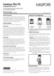
hemi<strong>chordates</strong> (Figure 3). Those in <strong>the</strong> midbrain <strong>of</strong><br />
<strong>chordates</strong> are expressed in <strong>the</strong> mesosome <strong>and</strong> anterior<br />
metasome <strong>of</strong> hemi<strong>chordates</strong>, stopping at <strong>the</strong> first gill slit.<br />
Those in <strong>the</strong> chordate hindbrain <strong>and</strong> spinal cord are<br />
expressed in <strong>the</strong> hemichordate metasome, entirely posterior<br />
to <strong>the</strong> first gill slit [25 ]. Finally, those in <strong>the</strong><br />
chordate tail are expressed in <strong>the</strong> hemichordate post-anal<br />
extension, mentioned above. For <strong>the</strong> first time, <strong>the</strong> body<br />
plans <strong>of</strong> <strong>chordates</strong> <strong>and</strong> hemi<strong>chordates</strong> can be aligned on<br />
this shared map. The first gill slit <strong>of</strong> hemi<strong>chordates</strong> <strong>and</strong><br />
first branchial arch <strong>of</strong> <strong>chordates</strong> — ventral to <strong>the</strong> midbrain–hindbrain<br />
boundary — fall at <strong>the</strong> same domain<br />
intersections. Presumably, <strong>the</strong> deuterostome ancestor<br />
had this detailed map.<br />
About 60% <strong>of</strong> <strong>the</strong> domains are shared with protostomes,<br />
indicating <strong>the</strong>ir likely presence in <strong>the</strong> bilateral ancestor.<br />
The o<strong>the</strong>rs are unique to deuterostomes, particularly<br />
those encoding signaling proteins. For example, fgf8<br />
<strong>and</strong> wnt1 are expressed in neurectoderm at <strong>the</strong> midbrain–hindbrain<br />
boundary <strong>of</strong> <strong>chordates</strong>, which is just<br />
dorsal to <strong>the</strong> first branchial arch (Figure 3), <strong>and</strong> genes<br />
for similar signals are expressed in <strong>the</strong> ectoderm <strong>of</strong><br />
hemi<strong>chordates</strong> at <strong>the</strong> level <strong>of</strong> <strong>the</strong> first gill slit (C Lowe,<br />
unpublished) [33]. In <strong>the</strong> posterior forebrain <strong>of</strong> <strong>chordates</strong>,<br />
wnt2b <strong>and</strong> wnt8 are expressed, <strong>and</strong> similar genes<br />
areexpressedat<strong>the</strong>base<strong>of</strong><strong>the</strong>hemichordateprosome.<br />
In <strong>the</strong> anterior forebrain <strong>of</strong> <strong>chordates</strong>, sfrp <strong>and</strong> various fgfs<br />
are expressed, <strong>and</strong> similar genes are expressed at <strong>the</strong><br />
anterior tip <strong>of</strong> <strong>the</strong> hemichordate prosome (C Lowe,<br />
unpublished) [33]. Protostomes have no counterparts<br />
<strong>of</strong> <strong>the</strong>se centers.<br />
Figure 3<br />
vax<br />
chx10<br />
tbr<br />
rx<br />
six3<br />
ptf<br />
bf-1<br />
dlx<br />
nkx2.1<br />
pax6<br />
emx<br />
otx<br />
irx<br />
en<br />
pax2<br />
gbx<br />
hox1<br />
hox3<br />
hox4<br />
hox5<br />
hox7/8<br />
hox9/10<br />
cdx<br />
dmbx<br />
tll<br />
otp<br />
dbx<br />
barH<br />
lim1/5<br />
Prosome<br />
Mesosome<br />
Metasome<br />
1st gill slit<br />
fgf3,4,8<br />
sfrp<br />
Mouth<br />
Forebrain<br />
wnt2b,8<br />
Midbrain<br />
Hemichordate<br />
fgf8, wnt1<br />
Hindbrain<br />
Spinal cord<br />
Anus<br />
hox11/13a<br />
hox11/13b<br />
hox11/13c<br />
fgf4.8<br />
wnt1,3a<br />
Chordate<br />
Current Opinion in Genetics & Development<br />
The anteroposterior map <strong>of</strong> expression domains for genes important in chordate neural patterning, encoding transcription factors (in black) <strong>and</strong><br />
signaling proteins (in red). Note <strong>the</strong> alignment <strong>of</strong> <strong>the</strong> bodies: prosome with ventral forebrain; mesosome <strong>and</strong> anterior metasome with dorsal<br />
forebrain <strong>and</strong> midbrain; posterior metasome with hindbrain <strong>and</strong> spinal cord; <strong>and</strong> post-anal tails toge<strong>the</strong>r. The gill slits <strong>of</strong> both <strong>chordates</strong> <strong>and</strong><br />
hemi<strong>chordates</strong> develop at <strong>the</strong> same domain intersections. Signaling centers (red bars), which are important in patterning <strong>the</strong> chordate nervous<br />
system, are similar in hemi<strong>chordates</strong>.<br />
Current Opinion in Genetics & Development 2005, 15:461–467<br />
www.sciencedirect.com
<strong>Hemi<strong>chordates</strong></strong> <strong>and</strong> <strong>the</strong> <strong>origin</strong> <strong>of</strong> <strong>chordates</strong> Gerhart, Lowe <strong>and</strong> Kirschner 465<br />
Although hemi<strong>chordates</strong> <strong>and</strong> <strong>chordates</strong> share this map,<br />
<strong>the</strong>y develop different morphologies: a diffuse versus a<br />
centralized nervous system. The map is clearly more<br />
ancient <strong>and</strong> conserved than are <strong>the</strong> particular anatomies<br />
<strong>and</strong> physiologies that develop from particular domains in<br />
different lineages <strong>of</strong> animals.<br />
Two differences st<strong>and</strong> out. First, domains encircle <strong>the</strong><br />
hemichordate body, each as a b<strong>and</strong>, whereas in <strong>chordates</strong><br />
<strong>the</strong>y are patches within <strong>the</strong> neural ectoderm. This correlates<br />
with <strong>the</strong> nervous systems; in hemi<strong>chordates</strong> it<br />
encircles <strong>the</strong> body, whereas in <strong>chordates</strong> it is centralized<br />
within a subregion <strong>of</strong> ectoderm. Second, most <strong>of</strong> <strong>the</strong>se<br />
genes are expressed only in <strong>the</strong> hemichordate ectoderm,<br />
whereas in <strong>chordates</strong> <strong>the</strong>y are also expressed in <strong>the</strong><br />
mesoderm, such as Hox genes in somites.<br />
Our data, combined with those on larvae, by o<strong>the</strong>rs,<br />
disfavor Garstang’s hypo<strong>the</strong>sis. S. kowalevskii has no larval<br />
stage, but ano<strong>the</strong>r hemichordate, Ptychodera flava, does.<br />
Of <strong>the</strong> few domains investigated, <strong>the</strong> larva lacks some <strong>and</strong><br />
places o<strong>the</strong>rs (otx, nkx2.1) at locations unlike those in <strong>the</strong><br />
map <strong>of</strong> S. kowalevskii or <strong>chordates</strong> [34,35]. Assuming that<br />
<strong>the</strong> P. flava adult map resembles that <strong>of</strong> S. kowalevskii, <strong>and</strong><br />
assuming that <strong>the</strong> deuterostome ancestor already had<br />
modern larval <strong>and</strong> adult expression patterns, <strong>the</strong> ancestral<br />
larva would seem to be a poor c<strong>and</strong>idate for <strong>the</strong> evolution<br />
<strong>of</strong> <strong>the</strong> chordate nervous system, compared with <strong>the</strong><br />
ancestral adult. Fur<strong>the</strong>rmore, echinoderm larvae lack<br />
Hox domains, whereas metamorphosing adults have <strong>the</strong>m<br />
[36] although not in neurogenic ectoderm. Garstang’s<br />
hypo<strong>the</strong>sis differs from o<strong>the</strong>rs in starting with an ancestral<br />
diffuse nervous system <strong>and</strong> proposing centralization in<br />
<strong>the</strong> chordate line. Unknown to him, <strong>the</strong> hemichordate<br />
adult, not just <strong>the</strong> larva, has such a system.<br />
Dorsoventral organization in hemi<strong>chordates</strong><br />
Dorsal <strong>and</strong> ventral positions are hard to define because<br />
many hemi<strong>chordates</strong> live vertically in burrows, in uniform<br />
surroundings. If <strong>the</strong> mouth is defined as ventral, a differentiated<br />
dorsoventral dimension can be designated<br />
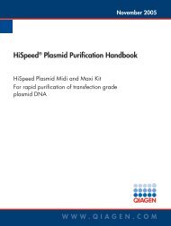
(Figures 1,4), for example, with dorsolateral gill slits.<br />
In this dimension, hemi<strong>chordates</strong> <strong>and</strong> <strong>chordates</strong> differ<br />
considerably in <strong>the</strong>ir domain maps. <strong>Hemi<strong>chordates</strong></strong><br />
express bmp2/4 <strong>and</strong> bmp7 in an ectodermal stripe at <strong>the</strong><br />
dorsal midline (Figure 4), <strong>and</strong> also genes (xolloid, twg,<br />
crossveinless <strong>and</strong> bambi) encoding agents for transmitting<br />
<strong>and</strong> regulating Bmp signals. In a ventral midline ectodermal<br />
stripe, chordin is expressed, encoding a Bmp<br />
antagonist (C Lowe et al., unpublished). admp is also<br />
<strong>the</strong>re, encoding a Bmp complement. <strong>Hemi<strong>chordates</strong></strong><br />
clearly have a Bmp–Chordin axis, as do Drosophila <strong>and</strong><br />
<strong>chordates</strong>, but, when referred to <strong>the</strong> location <strong>of</strong> <strong>the</strong><br />
mouth, it is oriented like that <strong>of</strong> Drosophila <strong>and</strong> <strong>the</strong><br />
inverse <strong>of</strong> <strong>chordates</strong>. In hemi<strong>chordates</strong>, several dorsoventral<br />
domains are placed relative to Bmp (Figure 4). tbx2/3,<br />
dlx, slit, unc5 <strong>and</strong> dcc/neogenin span <strong>the</strong> dorsal midline, <strong>and</strong><br />
mnh, sim <strong>and</strong> netrin span <strong>the</strong> ventral midline. Fur<strong>the</strong>r<br />
resemblance to protostomes is revealed by sim <strong>and</strong> netrin,<br />
which are expressed at <strong>the</strong> Drosophila ventral midline, <strong>and</strong><br />
by slit, which is expressed at <strong>the</strong> dorsal midline in Caenorhabditis<br />
elegans. Netrin <strong>and</strong> Slit might repel <strong>and</strong> attract<br />
axons to <strong>the</strong> dorsal <strong>and</strong> ventral tracts in hemi<strong>chordates</strong>.<br />
Table 1 summarizes <strong>the</strong> organs <strong>and</strong> domains that are<br />
inversely located in hemi<strong>chordates</strong> <strong>and</strong> <strong>chordates</strong>. The<br />
Figure 4<br />
Dorsal axon tract<br />
Dorsal vessel/heart (nkx2.5)<br />
Gill slit (pax1/9; six1) (mt)<br />
bmp2/4, bmp7,<br />
twg, crv, tolloid,<br />
bambi, slit<br />
dlx, tbx2/3,<br />
unc5, dcc/neo;<br />
pitx(pro)<br />
Diffuse nerve net<br />
<strong>and</strong> epidermal<br />
ectoderm<br />
Axon matPharynx/gut<br />
endoderm (ms; mt)<br />
Mesoderm<br />
Ventral muscle (mt)<br />
Ventral vessel<br />
Ventral axon tract (mt)<br />
chordin, admp,<br />
netrin<br />
mnx, sim (mt);<br />
nkx2.1, brn2/4 (pro);<br />
gsc (ms)<br />
Current Opinion in Genetics & Development<br />
Anatomy <strong>of</strong> <strong>the</strong> dorsoventral axis <strong>of</strong> hemi<strong>chordates</strong>, <strong>and</strong>, superimposed, <strong>the</strong> map <strong>of</strong> expression domains <strong>of</strong> genes encoding transcription<br />
factors (black) <strong>and</strong> signaling proteins (red). Ventral is defined by <strong>the</strong> location <strong>of</strong> <strong>the</strong> mouth. The section crosses <strong>the</strong> pharynx in <strong>the</strong> metasome<br />
(mt), but dorsoventral domains have been included from <strong>the</strong> prosome (pro) <strong>and</strong> mesosome (ms).<br />
www.sciencedirect.com Current Opinion in Genetics & Development 2005, 15:461–467
466 Pattern formation <strong>and</strong> developmental mechanisms<br />
Table 1<br />
Dorsoventral order <strong>of</strong> organs <strong>and</strong> tissues in hemi<strong>chordates</strong> <strong>and</strong><br />
<strong>chordates</strong>.<br />
Bmp expression domain<br />
Heart/contractile vessel (nkx2.5), forward blood flow<br />
ttf2 expression domain in <strong>the</strong> anterior endoderm<br />
Gill slits ( pax1/9)<br />
Body wall muscle<br />
Vessel with backward blood flow<br />
Post-anal tail (posterior Hox genes)<br />
chordin/admp expression domains<br />
For hemi<strong>chordates</strong>, <strong>the</strong> top <strong>of</strong> <strong>the</strong> list corresponds to <strong>the</strong> most<br />
dorsal position <strong>and</strong> <strong>the</strong> bottom to <strong>the</strong> most ventral. For <strong>chordates</strong>,<br />
<strong>the</strong> order is in reverse. The nervous system is not included, because<br />
it is both dorsal <strong>and</strong> ventral in hemi<strong>chordates</strong>.<br />
nervous system must be omitted because it is diffuse in<br />
hemi<strong>chordates</strong>, although sensory <strong>and</strong> motoneurons might<br />
differ dorsoventrally.<br />
Body inversion can explain <strong>the</strong> inverse relationship, as<br />
long as centralization <strong>of</strong> <strong>the</strong> nervous system is kept a<br />
separate question. Holl<strong>and</strong> [37 ] recently summarized<br />
arguments that <strong>the</strong> bilateral <strong>and</strong> deuterostome ancestors<br />
were diffuse, <strong>and</strong> that centralization occurred independently<br />
in <strong>chordates</strong>, arthropods <strong>and</strong> several o<strong>the</strong>r protostome<br />
lines. Chordate ancestors had to segregate<br />
neurogenic ectoderm from epidermis for centralization.<br />
This could have happened after body inversion. Centralization<br />
might have been an easy morphological modification<br />
to achieve once a diffuse ancestor had a rich domain<br />
map <strong>and</strong> <strong>the</strong> means to segregate axon tracts, as <strong>the</strong><br />
deuterostome ancestor probably had.<br />
Alternatively, perhaps <strong>the</strong> Bmp–Chordin axis, <strong>and</strong> not <strong>the</strong><br />
‘body’, inverted, <strong>and</strong> <strong>the</strong> mouth stayed put. In <strong>chordates</strong>,<br />
Chordin is produced, not by ectoderm but by <strong>the</strong> notochord<br />
<strong>and</strong> Spemann’s organizer, which derive from endomesoderm<br />
<strong>of</strong> <strong>the</strong> archenteron ro<strong>of</strong>. Given that <strong>the</strong> <strong>origin</strong><br />
<strong>of</strong> <strong>the</strong> chordate notochord is still unknown, who can say<br />
whe<strong>the</strong>r it arose dorsally or ventrally If it arose on <strong>the</strong> old<br />
dorsal side, it would reverse <strong>the</strong> Bmp–Chordin axis <strong>and</strong>,<br />
with it, <strong>the</strong> development <strong>of</strong> <strong>the</strong> anatomy <strong>of</strong> all three germ<br />
layers. Ano<strong>the</strong>r alternative, not yet made explicit, is <strong>the</strong><br />
Bateson–Goodrich [8] hypo<strong>the</strong>sis that various parts <strong>of</strong> an<br />
uninverted ancestor moved around <strong>the</strong> anterior <strong>and</strong> posterior<br />
tips to give chordate organization.<br />
Conclusions<br />
Chordate evolution, we suggest, entailed little or no<br />
change <strong>of</strong> domain organization from that already present<br />
in <strong>the</strong> anteroposterior axis <strong>of</strong> <strong>the</strong> deuterostome ancestor.<br />
Gill slits <strong>and</strong> <strong>the</strong> post-anal tail might be ancestral deuterostome<br />
traits <strong>of</strong> this conserved dimension. Considerable<br />
change from <strong>the</strong> ancestor has occurred in <strong>the</strong> chordate<br />
line in <strong>the</strong> dorsoventral dimension, particularly in <strong>the</strong><br />
centralization <strong>of</strong> <strong>the</strong> nervous system <strong>and</strong> <strong>the</strong> <strong>origin</strong>ation <strong>of</strong><br />
Current Opinion in Genetics & Development 2005, 15:461–467<br />
<strong>the</strong> notochord; an inversion <strong>of</strong> <strong>the</strong> Bmp–Chordin axis<br />
might also have occurred. Although <strong>the</strong> hemichordate<br />
nervous system is diffuse, it is extensively patterned.<br />
None <strong>of</strong> <strong>the</strong> old hypo<strong>the</strong>ses <strong>of</strong> chordate <strong>origin</strong>s seems<br />
wholly apt, but all have elements worth pursuing. Fur<strong>the</strong>r<br />
studies <strong>of</strong> hemichordate development will help to assess<br />
<strong>the</strong>se suggestions <strong>and</strong> to devise new ones. Of particular<br />
interest are <strong>the</strong> means by which <strong>the</strong> embryo establishes<br />
six signaling centers important in its patterning: dorsal<br />
<strong>and</strong> ventral midlines; anterior <strong>and</strong> posterior termini; <strong>the</strong><br />
prosome base; <strong>and</strong> <strong>the</strong> first gill slit.<br />
Acknowledgements<br />
The authors thank <strong>the</strong> United States Public Health Service (USPHS<br />
grant HD42724 to JG <strong>and</strong> HD37277 to MK) <strong>and</strong> NASA (grant<br />
FDNAG2-1605 to JG <strong>and</strong> MK) for research support, <strong>and</strong> thank Dr Eric<br />
L<strong>and</strong>er (MIT/Whitehead/Broad Institute) for valuable ESTs <strong>and</strong><br />
Dr Chris Gruber (Express Genomics) for excellent libraries.<br />
References <strong>and</strong> recommended reading<br />
Papers <strong>of</strong> particular interest, published within <strong>the</strong> annual period <strong>of</strong><br />
review, have been highlighted as:<br />
<strong>of</strong> special interest<br />
<strong>of</strong> outst<strong>and</strong>ing interest<br />
1. Hyman LH: The enterocoelous coelomates — <strong>the</strong><br />
Hemichordata. InThe Invertebrates, Volume 5: McGraw-Hill;<br />
1959:72-207.<br />
2. Van der Horst CJ: Hemichordata. InKlassen und Ordnungen des<br />
Tierreichs, Volume 4, Abt 4, Buch 2, Teil 2. Edited by HG Bronn;<br />
1939:1-739.<br />
3. Hadfield MG: Hemichordata. InReproduction <strong>of</strong> Marine<br />
Invertebrates, II. Edited by Giese AC, Pearse JS. Academic Press;<br />
1975:185-240.<br />
4. Bateson W: The early stages in <strong>the</strong> development <strong>of</strong><br />
Balanoglossus (sp. Incert.). Quart J Microscop Sci 1884,<br />
24:208-236.<br />
5. Bateson W: The later stages in <strong>the</strong> development <strong>of</strong><br />
Balanoglossus kowalevskii, with a suggestion as to <strong>the</strong><br />
affinities <strong>of</strong> <strong>the</strong> enteropneusta. Quart J Microscop Sci 1885,<br />
25:81-128.<br />
6. Bateson W: Continued account <strong>of</strong> <strong>the</strong> later stages in <strong>the</strong><br />
development <strong>of</strong> Balanoglossus kowalevskii, <strong>and</strong> <strong>of</strong> <strong>the</strong><br />
morphology <strong>of</strong> <strong>the</strong> enteropneusta. Quart J Microscop Sci 1886,<br />
26:511-534.<br />
7. Burdon-Jones C: Development <strong>and</strong> biology <strong>of</strong> <strong>the</strong> larva <strong>of</strong><br />
Saccoglossus horsti (Enteropneusta). Phil Trans Roy Soc<br />
London 1952, 236B:553-589.<br />
8. Goodrich ES: ‘Proboscis pores’ in craniate vertebrates, a<br />
suggestion concerning <strong>the</strong> prem<strong>and</strong>ibular somites <strong>and</strong><br />
hypophysis. Quart J Microscop Sci 1917, 62:539-553.<br />
9. Colwin AL, Colwin LH: Relationships between <strong>the</strong> egg <strong>and</strong> larva<br />
<strong>of</strong> Saccoglossus kowalevskii: axes <strong>and</strong> planes; general<br />
prospective significance <strong>of</strong> <strong>the</strong> early blastomeres.<br />
J Exp Zool 1951, 117:111-137.<br />
10. Colwin AL, Colwin LH: The normal embryology <strong>of</strong> Saccoglossus<br />
kowalevskii. J Morphol 1953, 92:401-453.<br />
11. Lowe CJ, Tagawa K, Humphreys T, Kirschner M, Gerhart J:<br />
Hemichordate embryos: procurement, culture, <strong>and</strong> basic<br />
methods. Methods Cell Biol 2004, 74:171-194.<br />
12. Halanych KM: The phylogenetic position <strong>of</strong> <strong>the</strong> pterobranch<br />
hemi<strong>chordates</strong> based on 18S rDNA sequence data.<br />
Mol Phylogenet Evol 1995, 4:72-76.<br />
13. Cameron CB, Garey JR, Swalla BJ: Evolution <strong>of</strong> <strong>the</strong> chordate<br />
body plan: new insights from phylogenetic analyses <strong>of</strong><br />
www.sciencedirect.com
<strong>Hemi<strong>chordates</strong></strong> <strong>and</strong> <strong>the</strong> <strong>origin</strong> <strong>of</strong> <strong>chordates</strong> Gerhart, Lowe <strong>and</strong> Kirschner 467<br />
deuterostome phyla. Proc Natl Acad Sci USA 2000,<br />
97:4469-4474.<br />
14. Adoutte A, Balavoine G, Lartillot N, Lespinet O, Prud’homme B, de<br />
Rosa R: The new animal phylogeny: reliability <strong>and</strong> implications.<br />
Proc Natl Acad Sci USA 2000, 97:4453-4456.<br />
15. Bourlat SJ, Nielsen C, Lockyer AE, Littlewood DT, Telford MJ:<br />
Xenoturbella is a deuterostome that eats molluscs. Nature<br />
2003, 424:925-928.<br />
16. Shu D-G, Conway Morris S, Han J, Chen L, Zhang X-L, Zhang Z-F,<br />
Liu H-Q, Li Y, Liu J-N: Primitive deuterostomes from <strong>the</strong><br />
Chengjiang Lagerstätte (Lower Cambrian, China). Nature 2001,<br />
414:419-424.<br />
17. Bateson W: The ancestry <strong>of</strong> <strong>the</strong> chordata. Quart J Microscop Sci<br />
1886, 26:535-571.<br />
18. Garstang W: The <strong>origin</strong> <strong>and</strong> evolution <strong>of</strong> larval forms. Rep Brit<br />
Ass Adv Sci 1928:77-98.<br />
19. McGinnis W, Krumlauf R: Homeobox genes <strong>and</strong> axial<br />
patterning. Cell 1992, 68:283-302.<br />
20. Scott MP: Intimations <strong>of</strong> a creature. Cell 1994, 79:1121-1124.<br />
21. Arendt D, Nubler-Jung K: Inversion <strong>of</strong> dorsoventral axis<br />
Nature 1994, 371:26.<br />
22. De Robertis EM, Sasai Y: A common plan for dorsoventral<br />
patterning in bilateria. Nature 1996, 380:37-40.<br />
23. Nübler-Jung K, Arendt D: Enteropneusts <strong>and</strong> chordate<br />
evolution. Curr Biol 1996, 6:352-353.<br />
24. Ogasawara M, Wada H, Peters H, Satoh N: Developmental<br />
expression <strong>of</strong> Pax1/9 genes in urochordate <strong>and</strong> hemichordate<br />
gills: insight into function <strong>and</strong> evolution <strong>of</strong> <strong>the</strong> pharyngeal<br />
epi<strong>the</strong>lium. Development 1999, 126:2539-2550.<br />
25.<br />
<br />
Lowe CJ, Wu M, Salic A, Evans L, L<strong>and</strong>er E, Stange-Thomann N,<br />
Gruber CE, Gerhart J, Kirschner M: Anteroposterior patterning in<br />
hemi<strong>chordates</strong> <strong>and</strong> <strong>the</strong> <strong>origin</strong>s <strong>of</strong> <strong>the</strong> chordate nervous<br />
system. Cell 2003, 113:853-865.<br />
The domains <strong>of</strong> 22 neural patterning genes, as revealed by in situ<br />
hybridization staining, are described in addition to those <strong>of</strong> pan-neural<br />
gene expression throughout <strong>the</strong> ectoderm.<br />
26. Peterson KJ: Isolation <strong>of</strong> Hox <strong>and</strong> Parahox genes in <strong>the</strong><br />
hemichordate Ptychodera flava <strong>and</strong> <strong>the</strong> evolution <strong>of</strong><br />
deuterostome Hox genes. Mol Phylogenet Evol 2004,<br />
31:1208-1215.<br />
27. Balser EJ, Ruppert EE: Structure, ultrastructure, <strong>and</strong> function <strong>of</strong><br />
<strong>the</strong> preoral heart-kidney on Saccoglossus kowalevskii<br />
(Hemichordata, Enteropneusta) including new data on <strong>the</strong><br />
stomochord. Acta Zool 1990, 71:235-249.<br />
28. Peterson KJ, Cameron RA, Tagawa K, Satoh N, Davidson EH:<br />
A comparative molecular approach to mesodermal patterning<br />
in basal deuterostomes: <strong>the</strong> expression pattern <strong>of</strong> Brachyury<br />
in <strong>the</strong> enteropneust hemichordate Ptychodera flava.<br />
Development 1999, 126:85-95.<br />
29. Yu JK, Holl<strong>and</strong> LZ, Jamrich M, Blitz IL, Holl<strong>and</strong> ND: AmphiFoxE4,<br />
an amphioxus winged helix/forkhead gene encoding a<br />
protein closely related to vertebrate thyroid transcription<br />
factor-2: expression during pharyngeal development.<br />
Evol Dev 2002, 4:9-15.<br />
30. Bullock TH: The nervous system <strong>of</strong> hemi<strong>chordates</strong>.<br />
In Structure <strong>and</strong> Function in <strong>the</strong> Nervous Systems <strong>of</strong> Invertebrates.<br />
WH Freeman <strong>and</strong> Co.; 1965:1567-1577.<br />
31. Knight-Jones E: The nervous system <strong>of</strong> Saccoglossus<br />
cambriensis (Enteropneusta). Phil Trans Roy Soc London 1952,<br />
236B:315-354.<br />
32. Cameron CB, Mackie GO: Conduction pathways in <strong>the</strong><br />
nervous system <strong>of</strong> Saccoglossus sp. (Enteropneusta).<br />
Can J Zool 1996, 74:15-19.<br />
33. Wilson SW, Houart C: Early steps in <strong>the</strong> development <strong>of</strong> <strong>the</strong><br />
forebrain. Dev Cell 2004, 6:167-181.<br />
34. Harada Y, Okai N, Taguchi S, Tagawa K, Humphreys T, Satoh N:<br />
Developmental expression <strong>of</strong> <strong>the</strong> hemichordate otx<br />
ortholog. Mech Dev 2000, 91:337-339.<br />
35. Takacs CM, Moy VN, Peterson KJ: Testing putative<br />
hemichordate homologues <strong>of</strong> <strong>the</strong> chordate dorsal nervous<br />
system <strong>and</strong> endostyle: expression <strong>of</strong> NK2.1 (TTF-1) in<strong>the</strong><br />
acorn worm Ptychodera flava (Hemichordata, Ptychoderidae).<br />
Evol Dev 2002, 4:405-417.<br />
36. Arenas-Mena C, Cameron AR, Davidson EH: Spatial expression<br />
<strong>of</strong> Hox cluster genes in <strong>the</strong> ontogeny <strong>of</strong> a sea urchin.<br />
Development 2000, 127:4631-4643.<br />
37. Holl<strong>and</strong> ND: Early central nervous system evolution: an era <strong>of</strong><br />
skin brains Nat Rev Neurosci 2003, 4:617-627.<br />
The author summarizes arguments for <strong>the</strong> possibility that <strong>the</strong> bilateral<br />
ancestor had a diffuse nervous system, <strong>and</strong> that centralization occurred<br />
independently in several lines.<br />
www.sciencedirect.com Current Opinion in Genetics & Development 2005, 15:461–467