Breast cancer risk factors - American Nurse Today
Breast cancer risk factors - American Nurse Today
Breast cancer risk factors - American Nurse Today

Create successful ePaper yourself
Turn your PDF publications into a flip-book with our unique Google optimized e-Paper software.
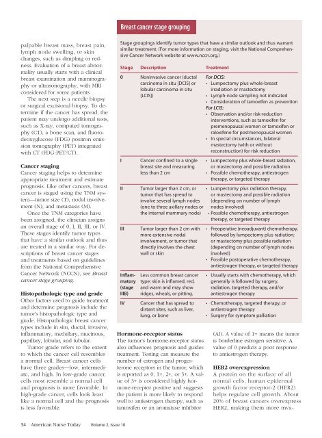
<strong>Breast</strong> <strong>cancer</strong> stage grouping<br />
palpable breast mass, breast pain,<br />
lymph node swelling, or skin<br />
changes, such as dimpling or redness.<br />
Evaluation of a breast abnormality<br />
usually starts with a clinical<br />
breast examination and mammography<br />
or ultrasonography, with MRI<br />
considered for some patients.<br />
The next step is a needle biopsy<br />
or surgical excisional biopsy. To determine<br />
if the <strong>cancer</strong> has spread, the<br />
patient may undergo additional tests,<br />
such as X-ray, computed tomography<br />
(CT), a bone scan, and fluorodeoxyglucose<br />
(FDG) positron emission<br />
tomography (PET) integrated<br />
with CT (FDG-PET/CT).<br />
Cancer staging<br />
Cancer staging helps to determine<br />
appropriate treatment and estimate<br />
prognosis. Like other <strong>cancer</strong>s, breast<br />
<strong>cancer</strong> is staged using the TNM system—tumor<br />
size (T), nodal involvement<br />
(N), and metastasis (M).<br />
Once the TNM categories have<br />
been assigned, the clinician assigns<br />
an overall stage of 0, I, II, III, or IV.<br />
These stages identify tumor types<br />
that have a similar outlook and thus<br />
are treated in a similar way. For descriptions<br />
of breast <strong>cancer</strong> stages<br />
and treatments based on guidelines<br />
from the National Comprehensive<br />
Cancer Network (NCCN), see <strong>Breast</strong><br />
<strong>cancer</strong> stage grouping.<br />
Histopathologic type and grade<br />
Other <strong>factors</strong> used to guide treatment<br />
and determine prognosis include the<br />
tumor’s histopathologic type and<br />
grade. Histopathologic breast <strong>cancer</strong><br />
types include in situ, ductal, invasive,<br />
inflammatory, medullary, mucinous,<br />
papillary, lobular, and tubular.<br />
Tumor grade refers to the extent<br />
to which the <strong>cancer</strong> cell resembles<br />
a normal cell. <strong>Breast</strong> <strong>cancer</strong> cells<br />
have three grades—low, intermediate,<br />
and high. In low-grade <strong>cancer</strong>,<br />
cells most resemble a normal cell<br />
and prognosis is more favorable. In<br />
high-grade <strong>cancer</strong>, cells look least<br />
like a normal cell and the prognosis<br />
is less favorable.<br />
Stage groupings identify tumor types that have a similar outlook and thus warrant<br />
similar treatment. (For more information on staging, visit the National Comprehensive<br />
Cancer Network website at www.nccn.org.)<br />
Stage Description Treatment<br />
0 Noninvasive <strong>cancer</strong> (ductal For DCIS:<br />
carcinoma in situ [DCIS] or • Lumpectomy plus whole-breast<br />
lobular carcinoma in situ irradiation or mastectomy<br />
[LCIS]) • Lymph-node sampling not indicated<br />
• Consideration of tamoxifen as prevention<br />
For LCIS:<br />
• Observation and/or <strong>risk</strong>-reduction<br />
interventions, such as tamoxifen for<br />
premenopausal women or tamoxifen or<br />
raloxifene for postmenopausal women<br />
• In special circumstances, bilateral<br />
mastectomy (with or without<br />
reconstruction) for <strong>risk</strong> reduction<br />
I Cancer confined to a single • Lumpectomy plus whole-breast radiation,<br />
breast site and measuring or mastectomy and possible radiation<br />
less than 2 cm • Possible chemotherapy, antiestrogen<br />
therapy, or targeted therapy<br />
II Tumor larger than 2 cm, or • Lumpectomy plus radiation therapy,<br />
tumor that has spread to or mastectomy and possible radiation<br />
involve several lymph nodes (depending on number of lymph<br />
(one to three axillary nodes or nodes involved)<br />
the internal mammary node) • Possible chemotherapy, antiestrogen<br />
therapy, or targeted therapy<br />
III Tumor larger than 2 cm with • Preoperative (neoadjuvant) chemotherapy,<br />
more extensive nodal<br />
followed by lumpectomy plus radiation;<br />
involvement, or tumor that or mastectomy plus possible radiation<br />
directly involves the chest (depending on number of lymph nodes<br />
wall or skin<br />
involved)<br />
• Possible postoperative chemotherapy,<br />
antiestrogen therapy, or targeted therapy<br />
Inflam- Less common breast <strong>cancer</strong> • Usually starts with chemotherapy, which<br />
matory type; skin is inflamed, red, generally is followed by surgery,<br />
(stage and warm and may show radiation, targeted therapy, and/or<br />
IIIB) ridges, wheals, or pitting. antiestrogen therapy<br />
IV Cancer that has spread to • Chemotherapy, targeted therapy, or<br />
distant sites, such as liver, antiestrogen therapy<br />
lung, or bone • Surgery for symptom palliation<br />
Hormone-receptor status<br />
The tumor’s hormone-receptor status<br />
also influences prognosis and guides<br />
treatment. Testing can measure the<br />
number of estrogen and progesterone<br />
receptors in the tumor, which<br />
is reported as 0, 1+, 2+, or 3+. A value<br />
of 3+ is considered highly hormone-receptor<br />
positive and suggests<br />
the patient is more likely to respond<br />
well to antiestrogen therapy, such as<br />
tamoxifen or an aromatase inhibitor<br />
(AI). A value of 1+ means the tumor<br />
is borderline estrogen sensitive. A<br />
value of 0 predicts a poor response<br />
to antiestrogen therapy.<br />
HER2 overexpression<br />
A protein on the surface of all<br />
normal cells, human epidermal<br />
growth factor receptor-2 (HER2)<br />
helps regulate cell growth. About<br />
20% of breast <strong>cancer</strong>s overexpress<br />
HER2, making them more inva-<br />
34 <strong>American</strong> <strong>Nurse</strong> <strong>Today</strong> Volume 2, Issue 10