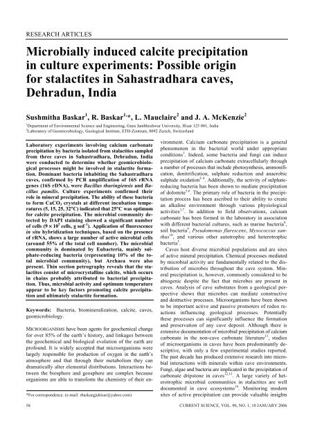
Microbially induced calcite precipitation in culture experiments ... - IISc
Microbially induced calcite precipitation in culture experiments ... - IISc
Microbially induced calcite precipitation in culture experiments ... - IISc

You also want an ePaper? Increase the reach of your titles
YUMPU automatically turns print PDFs into web optimized ePapers that Google loves.
RESEARCH ARTICLES<br />
<strong>Microbially</strong> <strong><strong>in</strong>duced</strong> <strong>calcite</strong> <strong>precipitation</strong><br />
<strong>in</strong> <strong>culture</strong> <strong>experiments</strong>: Possible orig<strong>in</strong><br />
for stalactites <strong>in</strong> Sahastradhara caves,<br />
Dehradun, India<br />
Sushmitha Baskar 1 , R. Baskar 1, *, L. Mauclaire 2 and J. A. McKenzie 2<br />
1 Department of Environmental Science and Eng<strong>in</strong>eer<strong>in</strong>g, Guru Jambheshwar University, Hisar 125 001, India<br />
2 Laboratory of Geomicrobiology, Geological Institute, ETH-Zentrum, 8092 Zurich, Switzerland<br />
Laboratory <strong>experiments</strong> <strong>in</strong>volv<strong>in</strong>g calcium carbonate<br />
<strong>precipitation</strong> by bacteria isolated from stalactites sampled<br />
from three caves <strong>in</strong> Sahastradhara, Dehradun, India<br />
were conducted to determ<strong>in</strong>e whether geomicrobiological<br />
processes might be <strong>in</strong>volved <strong>in</strong> stalactite formation.<br />
Dom<strong>in</strong>ant bacteria <strong>in</strong>habit<strong>in</strong>g the Sahastradhara<br />
caves, confirmed by PCR amplification of 16S rRNA<br />
genes (16S rDNA), were Bacillus thur<strong>in</strong>giensis and Bacillus<br />
pumilis. Culture <strong>experiments</strong> confirmed their<br />
role <strong>in</strong> m<strong>in</strong>eral <strong>precipitation</strong>. The ability of these bacteria<br />
to form CaCO 3 crystals at different <strong>in</strong>cubation temperatures<br />
(5, 15, 25, 32°C) <strong>in</strong>dicated that 25°C was optimum<br />
for <strong>calcite</strong> <strong>precipitation</strong>. The microbial community detected<br />
by DAPI sta<strong>in</strong><strong>in</strong>g showed a significant number<br />
of cells (9 × 10 5 cells, g sed –1 ). Application of fluorescence<br />
<strong>in</strong> situ hybridization techniques, based on the presence<br />
of rRNA, shows a large number of active microbial cells<br />
(around 55% of the total cell number). The microbial<br />
community is dom<strong>in</strong>ated by Eubacteria, ma<strong>in</strong>ly sulphate-reduc<strong>in</strong>g<br />
bacteria (represent<strong>in</strong>g 10% of the total<br />
microbial community), but Archaea were also<br />
present. Th<strong>in</strong> section petrography reveals that the stalactites<br />
consist of microcrystall<strong>in</strong>e <strong>calcite</strong>, which occurs<br />
<strong>in</strong> cha<strong>in</strong>s probably attributed to bacterial <strong>precipitation</strong>.<br />
Thus, microbial activity and optimum temperature<br />
appear to be key factors promot<strong>in</strong>g <strong>calcite</strong> <strong>precipitation</strong><br />
and ultimately stalactite formation.<br />
Keywords: Bacteria, biom<strong>in</strong>eralization, <strong>calcite</strong>, caves,<br />
geomicrobiology.<br />
*For correspondence. (e-mail: rbaskargjuhisar@yahoo.com)<br />
58<br />
MICROORGANISMS have been agents for geochemical change<br />
for over 85% of the earth’s history, and l<strong>in</strong>kages between<br />
the geochemical and biological evolution of the earth are<br />
profound. It is widely accepted that microorganisms were<br />
largely responsible for production of oxygen <strong>in</strong> the earth’s<br />
atmosphere and that through their metabolism they can<br />
dramatically alter elemental distributions. Interactions between<br />
the biosphere and geosphere are complex because<br />
organisms are able to transform the chemistry of their environment.<br />
Calcium carbonate <strong>precipitation</strong> is a general<br />
phenomenon <strong>in</strong> the bacterial world under appropriate<br />
conditions 1 . Indeed, some bacteria and fungi can <strong>in</strong>duce<br />
<strong>precipitation</strong> of calcium carbonate extracellularly through<br />
a number of processes that <strong>in</strong>clude photosynthesis, ammonification,<br />
denitrification, sulphate reduction and anaerobic<br />
sulphide oxidation 2–4 . Additionally, the activity of sulphatereduc<strong>in</strong>g<br />
bacteria has been shown to mediate <strong>precipitation</strong><br />
of dolomite 5,6 . The primary role of bacteria <strong>in</strong> the <strong>precipitation</strong><br />
process has been ascribed to their ability to create<br />
an alkal<strong>in</strong>e environment through various physiological<br />
activities 3,7 . In addition to field observations, calcium<br />
carbonate has been formed <strong>in</strong> the laboratory <strong>in</strong> association<br />
with different bacterial <strong>culture</strong>s, such as mar<strong>in</strong>e bacteria 8 ,<br />
soil bacteria 9 , Pseudomonas flurescens, Myxococcus xanthus<br />
10 , and various other autotrophic and heterotrophic<br />
bacteria 3 .<br />
Caves host diverse microbial populations and are sites<br />
of active m<strong>in</strong>eral <strong>precipitation</strong>. Chemical processes mediated<br />
by microbial activity are fundamentally related to the distribution<br />
of microbes throughout the cave system. M<strong>in</strong>eral<br />
<strong>precipitation</strong> is, however, commonly considered to be<br />
abiogenic despite the fact that microbes are present <strong>in</strong><br />
caves. Analysis of cave substrates from a geological perspective<br />
shows that microbes can mediate constructive<br />
and destructive processes. Microorganisms have been shown<br />
to be important active and passive promoters of redox reactions<br />
<strong>in</strong>fluenc<strong>in</strong>g geological processes. Potentially<br />
these processes can significantly <strong>in</strong>fluence the formation<br />
and preservation of any cave deposit. Although there is<br />
extensive documentation of microbial <strong>precipitation</strong> of calcium<br />
carbonate <strong>in</strong> the non-cave carbonate literature 11 , studies<br />
of microorganisms <strong>in</strong> caves have been predom<strong>in</strong>antly descriptive,<br />
with only a few experimental studies reported.<br />
The past decade has produced extensive research <strong>in</strong>to microbial<br />
<strong>in</strong>teractions with m<strong>in</strong>erals with<strong>in</strong> cave environments.<br />
Fungi, algae and bacteria are implicated <strong>in</strong> the <strong>precipitation</strong> of<br />
carbonate dripstone <strong>in</strong> caves 12,13 . A large variety of heterotrophic<br />
microbial communities <strong>in</strong> stalactites are well<br />
documented <strong>in</strong> cave ecosystems 14 . Monitor<strong>in</strong>g modern<br />
sites of active <strong>precipitation</strong> can provide valuable <strong>in</strong>sights<br />
CURRENT SCIENCE, VOL. 90, NO. 1, 10 JANUARY 2006
RESEARCH ARTICLES<br />
<strong>in</strong>to the factors that control <strong>precipitation</strong> and evolution of<br />
various cave deposits 15 . Various microbiological techniques<br />
have also been used to show that microbes are present <strong>in</strong><br />
most cave environments and modify the composition of<br />
fluids and/or <strong>in</strong>fluence <strong>precipitation</strong> of various m<strong>in</strong>erals,<br />
<strong>in</strong>clud<strong>in</strong>g <strong>calcite</strong> 16 .<br />
Microbial diversity <strong>in</strong> selected habitats cannot be comprehensively<br />
studied by traditional approaches. Because<br />
DNA analysis of bacteria provides no <strong>in</strong>formation on metabolism,<br />
physiology, ecology, biochemistry or geomicrobiology<br />
of a stra<strong>in</strong> 16 , only by grow<strong>in</strong>g microorganisms <strong>in</strong><br />
the laboratory under controlled conditions is it possible to<br />
del<strong>in</strong>eate their ability to alter the chemistry of their microenvironment<br />
and produce biom<strong>in</strong>erals. Hence laboratory-based<br />
<strong>culture</strong> <strong>experiments</strong>, as well as geochemical and molecular<br />
biological techniques were used <strong>in</strong> this study to understand<br />
the possible extent of microbial <strong>in</strong>volvement <strong>in</strong> stalactite<br />
formations <strong>in</strong> the Sahastradhara caves.<br />
Geological sett<strong>in</strong>g<br />
The study area is situated <strong>in</strong> the Dehradun Valley, a crescentshaped<br />
<strong>in</strong>termontane valley formed with<strong>in</strong> the Siwalik<br />
Formations <strong>in</strong> Garhwal Himalaya (Figure 1). The valley<br />
is separated from the Lesser Himalayan Formations <strong>in</strong> the<br />
north by a major <strong>in</strong>traplate thrust, the Ma<strong>in</strong> Boundary<br />
Thrust (MBT). The Siwalik group underly<strong>in</strong>g the valley<br />
and the Southern Siwalik Range are thrust over the Indo-<br />
Gangetic Pla<strong>in</strong> sediments along the Himalayan Frontal<br />
Fault, form<strong>in</strong>g the Dehradun Valley as a piggyback bas<strong>in</strong> 17 .<br />
The study area (lat. 30°17′ to 30°24′N; long. 78°00′ to<br />
78°06′S) is between the Lesser Himalayan hills <strong>in</strong> the north<br />
and the topographic relief of the Siwalik ranges <strong>in</strong> the<br />
south, enclosed by the rivers Ganga <strong>in</strong> the east and Yamuna<br />
<strong>in</strong> the west. The thick alluvium deposit that fills the syncl<strong>in</strong>al<br />
depression consists of eastern, central and western<br />
fans descend<strong>in</strong>g from the Lesser Himalayan hills, occupy<strong>in</strong>g a<br />
major portion of the area as surficial deposits. Coalescence<br />
of these fans has resulted <strong>in</strong> a piedmont pla<strong>in</strong> 18 . Caves<br />
and circulat<strong>in</strong>g groundwater are an essential part of this<br />
terra<strong>in</strong>.<br />
Sahastradhara, mean<strong>in</strong>g place of the ‘thousand-fold<br />
spr<strong>in</strong>g’, is situated at a distance of 14 km from Dehradun.<br />
Of the 1545 caves throughout India 19 , the Sahastradhara<br />
spr<strong>in</strong>gs (Figure 2) are pH neutral as well as be<strong>in</strong>g exposed<br />
to light, allow<strong>in</strong>g photosynthetic activity to take place.<br />
These caves are rather small <strong>in</strong> size (10 m long, 2 m<br />
wide), but they are important because of their exceptional<br />
beauty and the associated spr<strong>in</strong>gs, which are of high medic<strong>in</strong>al<br />
value. The spr<strong>in</strong>g hosts a complex microbial community.<br />
The cold spr<strong>in</strong>g water flows downward about 9 m, leav<strong>in</strong>g<br />
an <strong>in</strong>crustation of carbonate on the surface. Accumulat<strong>in</strong>g<br />
over the centuries, the <strong>in</strong>crustations form project<strong>in</strong>g<br />
ledges with caves, from the roofs of which falls a perpetual<br />
shower.<br />
Methodology<br />
Sample collection and preservation<br />
Samples were taken from small stalactites, which were<br />
approximately 15–20 cm <strong>in</strong> length and 3–4 cm <strong>in</strong> diameter.<br />
For the geomicrobiological study, samples were brought<br />
to the laboratory (Geomicrobiology Laboratory, ETH,<br />
Zurich, Switzerland) and preserved at 4°C before aseptic<br />
cutt<strong>in</strong>g to collect uncontam<strong>in</strong>ated sub-samples. Classical<br />
cultivation methods select specific organisms and are <strong>in</strong>adequate<br />
to quantify active cells. To overcome this ‘plate<br />
count anomaly’, the molecular approach with rRNA<br />
analysis was employed. Samples collected from different<br />
caves were fixed for 6 h <strong>in</strong> 4% para formaldehyde <strong>in</strong><br />
phosphate buffer sal<strong>in</strong>e (PBS; 0.13 M NaCl, 7 mM Na 2 HPO 4 ,<br />
Figure 1. Regional geological map of the study area. YTF, Yamuna<br />
Tear Fault; GTF, Ganga Tear Fault; MBT, Ma<strong>in</strong> Boundary Thrust;<br />
HFF, Himalayan Frontal Fault. Dra<strong>in</strong>age areas of the Donga fan (Do),<br />
Dehradun fan (De), Song River (So) and Bhogpur fan (Bh) (after S<strong>in</strong>gh<br />
et al. 18 ).<br />
Figure 2.<br />
Stalactites from Sahastradhara caves, Dehradun.<br />
CURRENT SCIENCE, VOL. 90, NO. 1, 10 JANUARY 2006 59
RESEARCH ARTICLES<br />
3 mM NaH 2 PO 4 , pH 7.2 <strong>in</strong> water). Samples were washed<br />
<strong>in</strong> PBS and stored at –20°C <strong>in</strong> ethanol : PBS (1 : 1) before<br />
subsequent analysis.<br />
Isolation of calcify<strong>in</strong>g bacteria<br />
Stalactite samples (1 g) were powdered <strong>in</strong> a mortar and<br />
pestle, suspended <strong>in</strong> 9 ml sterile sal<strong>in</strong>e solution and vortexed.<br />
Triplicate B-4 medium (2.5 g calcium acetate, 4 g yeast<br />
extract, 10 g glucose and 18 g agar per litre of distilled<br />
water 1 ) spread plates were <strong>in</strong>oculated with sample dilutions<br />
rang<strong>in</strong>g from 10 1 to 10 6 and <strong>in</strong>cubated at 32°C for two<br />
weeks. Individual colonies were selected and purified by<br />
repeated streak<strong>in</strong>g on B-4 agar. For short-term preservation,<br />
the isolates were streaked on B-4 agar slants and<br />
stored at 4°C. Bacteria were identified by DNA sequenc<strong>in</strong>g<br />
us<strong>in</strong>g identification methods – PCR amplification of 16S<br />
rRNA genes (16S rDNA) and Gapped BLAST and PSI–<br />
BLAST prote<strong>in</strong> database search programs 20 at IMD (Institut<br />
für mediz<strong>in</strong>ische Diagnostik), Zurich.<br />
Calcite <strong>precipitation</strong> by bacteria<br />
Bacterial isolates <strong>culture</strong>d on B-4 agar were <strong>in</strong>cubated<br />
aerobically at different temperatures (5, 15, 25, 32°C) for<br />
two weeks. Petri plates were exam<strong>in</strong>ed for the presence<br />
of crystals by optical microscopy periodically for up to 25<br />
days. Controls consisted of un<strong>in</strong>oculated <strong>culture</strong> medium<br />
along with experimental samples. The morphology and<br />
characteristics of the bacteria and crystals were studied<br />
by SEM–EDAX (Geophysics Laboratory, ETH) gold-shadowed<br />
and analysed.<br />
Analysis of microbial communities<br />
Total number of microorganisms was <strong>in</strong>vestigated us<strong>in</strong>g<br />
DAPI sta<strong>in</strong><strong>in</strong>g (4′,6-diamid<strong>in</strong>o-2-phenyl<strong>in</strong>dole) 21 . Microbial<br />
community structure was studied by fluorescence <strong>in</strong> situ<br />
hybridization (FISH) us<strong>in</strong>g three general oligonucleotide<br />
probes target<strong>in</strong>g the doma<strong>in</strong> level of Eubacteria (Eub338)<br />
and Archaea (Arch915), and most of the members of the<br />
δ-subclass of Proteobacteria, which are sulphate reducers<br />
(SRB385) 22 . Next 0.5 g of fixed samples was unfrozen<br />
and diluted with 20–40 ml of 0.1% pyrophosphate. The<br />
<strong>calcite</strong> present was dissolved us<strong>in</strong>g 0.1 M EDTA (pH 4.5)<br />
followed by centrifugation. Bacteria were detached us<strong>in</strong>g<br />
an ultrasonic homogenizer (2 m<strong>in</strong>, power 7, Sonifer B-12<br />
Branson sonic power). Then 20 µl aliquots were spotted onto<br />
gelat<strong>in</strong>-coated slides and air-dried. Next 10 µl of hybridization<br />
buffer (0.9 M NaCl, 20 mM Tris-HCl, 5 mM<br />
EDTA, 0.01% SDS, 30% formamide, pH 7.2), DAPI solution<br />
(f<strong>in</strong>al concentration of 20 ng ml –1 ) and one of the<br />
oligonucleotide probes labelled with Cy3 fluorochrome<br />
(25 ng µl –1 ) were placed onto the slides and <strong>in</strong>cubated for<br />
2 h at 40°C <strong>in</strong> the dark. Excess sta<strong>in</strong> was washed <strong>in</strong><br />
60<br />
buffer (10 mM Tris, 5 mM EDTA, 0.01% SDS and 0.9 M<br />
NaCl) for 20 m<strong>in</strong> at 48°C. Slides were air-dried and mounted<br />
with citifluor. The preparations were exam<strong>in</strong>ed at 1000 x<br />
magnification (Zeiss Plan-Neofluar) with a Zeiss Axiophot<br />
microscope fitted for epifluorescence with a 50 W<br />
high-pressure mercury bulb and filter set for Cy3 (G 546,<br />
FT 560, BP 575-640) and filter set for DAPI (G365,<br />
FT395, LP420). Four hundred bacteria (10 × 40 fields)<br />
were counted per slide triplicate.<br />
M<strong>in</strong>eralogy and geochemistry<br />
Us<strong>in</strong>g a Sc<strong>in</strong>tag X-ray diffractometer (XRD) (M<strong>in</strong>eralogy/<br />
Petrology Laboratory, ETH), powdered stalactite samples<br />
were scanned at a rate of 1° per m<strong>in</strong>ute <strong>in</strong> the scann<strong>in</strong>g<br />
range of 2θ values between 4° and 50° and the spectra of<br />
m<strong>in</strong>erals produced were identified us<strong>in</strong>g the ICDD database<br />
(JCPDS) 23 . Electron microscope studies were conducted<br />
us<strong>in</strong>g a SEM CamScan CS44LB equipped with an<br />
energy dispersive X-ray system (SEM–EDAX). Representative<br />
chip samples were mounted on stubs and coated<br />
with gold of thickness 100–150 Å and then transferred to<br />
the sample chamber of the <strong>in</strong>strument prior to imag<strong>in</strong>g.<br />
Major element geochemistry was analysed us<strong>in</strong>g XRF<br />
(Geochemistry Laboratory, ETH).<br />
Results and discussion<br />
Direct microscopy revealed the presence of a variety of<br />
<strong>in</strong>digenous heterotrophic bacteria, act<strong>in</strong>omycetes, cyanobacteria<br />
and mosses. Bacillus thur<strong>in</strong>giensis and Bacillus<br />
pumilis stra<strong>in</strong>s were the dom<strong>in</strong>ant bacteria identified.<br />
Colonies that formed m<strong>in</strong>erals were isolated by repeated<br />
streak<strong>in</strong>g. These pure <strong>culture</strong>s precipitated <strong>calcite</strong> <strong>in</strong> vitro<br />
(Figure 3 a–f ). Act<strong>in</strong>omycetes <strong>culture</strong>s were also observed<br />
to precipitate <strong>calcite</strong> (Figure 4). Predom<strong>in</strong>ance of bacilli<br />
is <strong>in</strong> agreement with the established hypothesis that Bacillus<br />
species play a major role <strong>in</strong> carbonate deposition <strong>in</strong> natural<br />
habitats 1,24,25 .<br />
The temperature, number of stra<strong>in</strong>s and time required<br />
for <strong>calcite</strong> <strong>precipitation</strong> were studied by cultivat<strong>in</strong>g the stra<strong>in</strong>s<br />
at different <strong>in</strong>cubation temperatures (5, 15, 25, 32°C).<br />
The number of bacterial stra<strong>in</strong>s <strong>in</strong>creased from 5 to 25°C<br />
and the maximum number of stra<strong>in</strong>s was observed at 25°C.<br />
There was a decrease <strong>in</strong> the number of stra<strong>in</strong>s at 32°C<br />
(Figure 5). The optimum temperature for <strong>calcite</strong> <strong>precipitation</strong><br />
was 25°C. Crystal <strong>precipitation</strong> at 25°C started after a<br />
week of <strong>in</strong>oculation (Figure 6). Petri plates were exam<strong>in</strong>ed<br />
periodically up to 25 days by optical microscopy for<br />
the presence of crystals. At lower temperatures (5–15°C),<br />
crystal <strong>precipitation</strong> started after 15 days of <strong>in</strong>oculation<br />
(Table 1). In contrast, control <strong>experiments</strong> without bacteria<br />
precipitated no carbonate m<strong>in</strong>erals.<br />
Microscopic observations showed that this <strong>precipitation</strong><br />
takes place <strong>in</strong> a microenvironment provided by bacterial<br />
CURRENT SCIENCE, VOL. 90, NO. 1, 10 JANUARY 2006
RESEARCH ARTICLES<br />
a<br />
Label A:<br />
CaKa<br />
b<br />
Untitled: 1<br />
CaKb<br />
2.00 4.00 6.00 8.00 10.00 12.00 14.00 16.00 18.00<br />
c<br />
Label A:<br />
d<br />
Untitled: 1<br />
CaKa<br />
CaKb<br />
2.00 4.00 6.00 8.00 10.00 12.00 14.00 16.00 18.00<br />
e<br />
f<br />
Figure 3. a, c, Calcite precipitated by Bacilli sp. b, d, SEM–EDAX of <strong>calcite</strong> crystals <strong>in</strong> (a) and (c) respectively. e, Bacterial <strong>calcite</strong> seen as oval<br />
structures. f, Bacteria across the crystal.<br />
growth and metabolism. The size and quantity of crystals<br />
<strong>in</strong>creased with time (Figure 7 a, b). These changes show<br />
the <strong>in</strong>fluence of bacteria <strong>in</strong> the geochemical alteration of<br />
their closed environment and undoubtedly, <strong>in</strong> the mediation<br />
of <strong>calcite</strong> <strong>precipitation</strong>. The nucleation and growth of<br />
<strong>calcite</strong> crystals occurred <strong>in</strong> an organic matrix and bacteria<br />
were found to be associated with m<strong>in</strong>eral surfaces (Figure<br />
3f ), <strong>in</strong>dicat<strong>in</strong>g that microbial mediation could be an active<br />
process. The organic mucus encapsulat<strong>in</strong>g the bacteria<br />
might be promot<strong>in</strong>g diffusion gradients, whereby ions can<br />
diffuse through it and enable the development of physicochemical<br />
conditions enhanc<strong>in</strong>g m<strong>in</strong>eral <strong>precipitation</strong>. Culture<br />
CURRENT SCIENCE, VOL. 90, NO. 1, 10 JANUARY 2006 61
RESEARCH ARTICLES<br />
Table 1.<br />
Calcite <strong>precipitation</strong> at different temperatures<br />
Incubation temperatures (°C)<br />
Calcite precipitated at various temperatures (°C)<br />
Microorganism 5 15 25 32 5 15 25 32<br />
Bacillus sp. +/– ++ +++ ++ – – +++ ++<br />
Act<strong>in</strong>omycetes – + ++ ++ – – + +<br />
–, No growth; +/–, Poor growth; +, Moderate growth; ++, Good growth; +++, Optimal growth.<br />
a<br />
b<br />
Figure 4. Calcite precipitated by act<strong>in</strong>omycetes.<br />
35<br />
30<br />
25<br />
No. of Stra<strong>in</strong>s<br />
20<br />
15<br />
Figure 7.<br />
Bacterial <strong>calcite</strong>: a, after a week and b, after 20 days.<br />
10<br />
5<br />
0<br />
0 5 10 15 20 25 30 35<br />
Temperature (°C)<br />
Figure 5.<br />
Temperature vs bacterial stra<strong>in</strong>s.<br />
Figure 8.<br />
Microbes detected by DAPI sta<strong>in</strong><strong>in</strong>g.<br />
Figure 6.<br />
M<strong>in</strong>eral <strong>precipitation</strong> by bacilli.<br />
<strong>experiments</strong> us<strong>in</strong>g sulphate-reduc<strong>in</strong>g bacteria <strong>in</strong> the mediation<br />
of dolomite formation <strong>in</strong> a synthetic anoxic hyper<br />
sal<strong>in</strong>e medium demonstrated the same 6,26 .<br />
An abundant microbial community (9 × 10 5 cells, g sed –1 )<br />
was detected by direct microscopic observation after<br />
DAPI sta<strong>in</strong><strong>in</strong>g (Figure 8). FISH demonstrates the presence<br />
of a large number of active microbial cells represent<strong>in</strong>g<br />
55% of the total cell number. The percentage of hybridization<br />
obta<strong>in</strong>ed with the two doma<strong>in</strong> probes (Eub and<br />
62<br />
CURRENT SCIENCE, VOL. 90, NO. 1, 10 JANUARY 2006
RESEARCH ARTICLES<br />
Arch) depends on the content of ribosomes; FISH detects<br />
reliably only the physiologically active cells. The microbial<br />
community is dom<strong>in</strong>ated by Eubacteria, ma<strong>in</strong>ly sulphatereduc<strong>in</strong>g<br />
bacteria (Figure 9; represent<strong>in</strong>g 10% of the total microbial<br />
community), but Archaea were also present (Figure<br />
10). A significant fraction of these cells are active,<br />
<strong>in</strong>dicat<strong>in</strong>g the high probability of their participation <strong>in</strong><br />
biom<strong>in</strong>eralization processes <strong>in</strong>volved <strong>in</strong> stalactite formation<br />
27 . A large number (about 26%) of detected Eubacteria<br />
belong to the δ-subgroup of Proteobacteria, which is<br />
the largest group of organisms capable of dissimilatory sulphate<br />
reduction 28 .<br />
XRD <strong>in</strong>dicated that <strong>calcite</strong> is the dom<strong>in</strong>ant m<strong>in</strong>eral,<br />
with aragonite and dolomite be<strong>in</strong>g present <strong>in</strong> trace amounts.<br />
EDAX analysis <strong>in</strong>dicated that the Ca peak is strong, and<br />
rhombic morphology of <strong>calcite</strong> is observed. Major element<br />
geochemistry showed the presence of 42.86% CaO. Th<strong>in</strong>section<br />
petrography reveals the presence of microcrystall<strong>in</strong>e<br />
<strong>calcite</strong>. Microcrystall<strong>in</strong>e <strong>calcite</strong> deposition observed<br />
<strong>in</strong> cha<strong>in</strong>s may be related to bacterial <strong>in</strong>volvement <strong>in</strong> the<br />
<strong>precipitation</strong> process (Figure 11).<br />
Calcium carbonate <strong>precipitation</strong> is governed by the calcium<br />
concentration, concentration of dissolved <strong>in</strong>organic<br />
carbon, pH and availability of nucleation sites 3,29–32 . Microbial<br />
m<strong>in</strong>eral <strong>precipitation</strong> result<strong>in</strong>g from metabolic activities<br />
of microorganisms can occur <strong>in</strong>side or outside the<br />
cells. Often, bacterial activity simply triggers a change <strong>in</strong><br />
Figure 9. SRB detected after hybridization with Cy3-labelled sulphate-reduc<strong>in</strong>g<br />
bacteria probes.<br />
Figure 11.<br />
Micrite deposition observed <strong>in</strong> cha<strong>in</strong>s.<br />
solution chemistry that leads to over-saturation and m<strong>in</strong>eral<br />
<strong>precipitation</strong>.<br />
The role of heterotrophic bacteria (for example Bacillus<br />
sp.) <strong>in</strong> carbonate formation may be twofold. First, actively<br />
grow<strong>in</strong>g cells (or cell aggregates) alter their local<br />
environment by establish<strong>in</strong>g a concentration gradient of<br />
calcium, magnesium, carbonate ions and pH, all of which<br />
are necessary to promote ideal conditions for carbonate<br />
formation. This is comparable to the role of the metabolic<br />
pathway of heterotrophic bacteria proposed for calcium<br />
carbonate formation, which consists of a passive process<br />
<strong>in</strong>volv<strong>in</strong>g change <strong>in</strong> pH and <strong>in</strong>crease <strong>in</strong> metabolic end products,<br />
such as carbonate ions 3,33 , Secondly, the cell surfaces<br />
of metabolically active bacteria (SRB) are <strong>in</strong>volved <strong>in</strong> the<br />
concentration of Ca 2+ and Mg 2+ ions around the cell 26 .<br />
In the present study, the metabolic activity of bacterial<br />
stra<strong>in</strong>s and temperature are key factors <strong>in</strong> carbonate deposition.<br />
The optimum temperatures have a positive effect<br />
on bacterial <strong>precipitation</strong> of <strong>calcite</strong>, <strong>in</strong>creas<strong>in</strong>g the ability<br />
of the stra<strong>in</strong> to form crystals. Calcium carbonate <strong>precipitation</strong><br />
chemically favours bacterial survival and proliferation<br />
33 . Seepage of water saturated with carbonates has a<br />
partial pressure of CO 2 greater than that of the cave atmosphere.<br />
Degass<strong>in</strong>g of CO 2 takes place from water to air<br />
lead<strong>in</strong>g to a decrease <strong>in</strong> the solubility of calcium carbonate,<br />
and hence saturation with respect to calcium carbonate<br />
and ultimately its deposition. The classical model is based<br />
on <strong>in</strong>organic processes associated with carbonate solubility,<br />
shown by the follow<strong>in</strong>g equation:<br />
Ca 2+ + 2HCO – 3 ⇔ CaCO 3 ↓ + CO 2 ↑ + H 2 O.<br />
Figure 10. DAPI-sta<strong>in</strong>ed cells (%) detectable after hybridization with<br />
Cy3-labelled sulphate-reduc<strong>in</strong>g bacteria (SRB), Eubacteria (Eub) and<br />
Archaea (Arch) probes.<br />
The equilibrium constant expression for CaCO 3 dissolution/<strong>precipitation</strong><br />
is: K = [Ca ++ ] [CO – 3 – ]. When K = Ksp*<br />
(solubility product constant <strong>in</strong> water at any given temperature,<br />
pressure and sal<strong>in</strong>ity), the solution is considered<br />
to be exactly saturated. Carbonate <strong>precipitation</strong> occurs when<br />
the product of the concentration of calcium and carbonate<br />
exceeds Ksp*. The metabolic activity of <strong>in</strong> situ microbes<br />
facilitates <strong>in</strong> exceed<strong>in</strong>g the solubility product of carbonate<br />
m<strong>in</strong>erals and also by provid<strong>in</strong>g nucleation sites that promote<br />
stability of m<strong>in</strong>eral nuclei.<br />
CURRENT SCIENCE, VOL. 90, NO. 1, 10 JANUARY 2006 63
RESEARCH ARTICLES<br />
Conclusions<br />
The <strong>culture</strong> <strong>experiments</strong> demonstrate that B. thur<strong>in</strong>giensis<br />
and B. pumilis mediate the <strong>precipitation</strong> of <strong>calcite</strong> under<br />
well-def<strong>in</strong>ed conditions. The optimum temperature for <strong>calcite</strong><br />
<strong>precipitation</strong> was 25°C. The presence of an active microbial<br />
community with high biodiversity coloniz<strong>in</strong>g the Sahastradhara<br />
caves, suggests that carbonate <strong>precipitation</strong><br />
could be mediated or catalysed by microbes, s<strong>in</strong>ce microbial<br />
metabolism can <strong>in</strong>fluence the bicarbonate–carbonate equilibrium.<br />
Thus we propose that microbial activity and optimum<br />
temperature appear to be key factors promot<strong>in</strong>g <strong>calcite</strong><br />
<strong>precipitation</strong> and ultimately stalactite formation.<br />
1. Boquet, E., Boronat, A. and Ramos-Cormenza, A., Production of<br />
<strong>calcite</strong> (calcium carbonate) crystals by soil bacteria is a general<br />
phenomenon. Nature, 1973, 246, 527–529.<br />
2. Castanier, S., Le M’etayer-Levrel, G. and Perthuisot, J. P., Bacterial<br />
roles <strong>in</strong> the <strong>precipitation</strong> of carbonate m<strong>in</strong>erals. In Microbial<br />
Sediments (eds Rid<strong>in</strong>g, R. E. and Awramik, S. M.), Heidelberg,<br />
Spr<strong>in</strong>ger-Verlag, 2000, pp. 32–39.<br />
3. Castanier, S., Le M’etayer-Levrel, G. and Perthuisot, J. P., Cacarbonates<br />
<strong>precipitation</strong> and limestone genesis – the microbiologist<br />
po<strong>in</strong>t of view. Sediment. Geol., 1999, 126, 9–23.<br />
4. Rid<strong>in</strong>g, R., Microbial carbonates: the geological record of calcified<br />
bacterial–algal mats and biofilms. Sedimentology, 2000, 47,<br />
179–214.<br />
5. Vasconcelos, C., McKenzie, J. A., Bernasconi, S., Grujic, D. and<br />
Tien, A. J., Microbial mediation as a possible mechanism for natural<br />
dolomite formation at low temperature. Nature, 1995, 377, 220–<br />
222.<br />
6. Warthmann, R., Lith, Y. V., Vasconcelos, C., McKenzie, J. A. and<br />
Karpoff, A. M., Bacterially <strong><strong>in</strong>duced</strong> dolomite <strong>precipitation</strong> <strong>in</strong> anoxic<br />
<strong>culture</strong> <strong>experiments</strong>. Geology, 2000, 28, 1091–1094.<br />
7. Douglas, S. and Beveridge, T. J., M<strong>in</strong>eral formation by bacteria <strong>in</strong><br />
natural communities. FEMS Microbiol. Ecol., 1998, 26, 79–88.<br />
8. Buczynski, C. and Chafetz, H. S., Habit of bacterially <strong><strong>in</strong>duced</strong><br />
precipitates of calcium carbonate: examples from laboratory <strong>experiments</strong><br />
and recent sediments. In Carbonate Microfabrics (eds<br />
Rezak, R. and Lavoie, D. L.), Spr<strong>in</strong>ger-Verlag, New York, 1993,<br />
pp. 105–116.<br />
9. Rivadeneyra, M. A., Delgado, G., Ramos-Cormenza, A. and<br />
Delgado, R., Biom<strong>in</strong>eralization of carbonates by Halomonas eurihal<strong>in</strong>a<br />
<strong>in</strong> solid and liquid media with different sal<strong>in</strong>ities: crystal<br />
formation sequences. Res. Microbiol., 1985, 149, 277–287.<br />
10. Gonzalaz-Munoz, M. T., Chekroun, K. B., Aboud, A. B., Arias, J.<br />
M. and Rodriguez-Gallego, M., Bacterially <strong><strong>in</strong>duced</strong> Mg-<strong>calcite</strong><br />
formation: Role of Mg 2 <strong>in</strong> development of crystal morphology. J.<br />
Sediment. Res., 2000, 70, 559–564.<br />
11. Ehrlich, H. L., In Geomicrobiology, Marcel Dekker, New York,<br />
1996, 3rd edn, p. 719.<br />
12. Danielli, H. M. C. and Ed<strong>in</strong>gton, M. A., Bacterial calcification <strong>in</strong><br />
limestone caves. Geomicrobiol. J., 1983, 3, 1–16.<br />
13. Cox, G., James, J. M., Leggett, K. E. A. and Osborne, R. A. L.,<br />
Cyanobacterially deposited speleothems: subaerial stromatolites.<br />
Geomicrobiol. J., 1989, 7, 245–252.<br />
14. Laiz, L., Groth, I., Schumann, P., Zezza, F., Felske, A., Hermos<strong>in</strong>,<br />
B. and Saiz-Jimene, C., Microbiology of the stalactites from Grotta<br />
dei Cervi, Porto Badisco, Italy. Int. Microbiol., 2000, 3, 25–30.<br />
15. Borsata, A., Frisia, S., Jones, B. and van der Borg, K., Calcite<br />
moonmilk: crystal morphology and environment of formation <strong>in</strong><br />
caves <strong>in</strong> the Italian Alps. J. Sediment. Res., 2000, 70, 1179–1190.<br />
16. Northup, D. E., Reysenbach, A. L. and Pace, N. R., Microorganisms<br />
and speleothems. In Cave M<strong>in</strong>erals of the World (eds Hill, C.<br />
and Forti, P.), National Speleogical Society, Huntsville, A. L.,<br />
1997, 2nd edn, pp. 261–266.<br />
64<br />
17. Ori, G. G. and Friend, F. P., Sedimentary bas<strong>in</strong>s formed and carried<br />
piggy-back on active thrust sheets. Geology, 1984, 12, 18–50.<br />
18. S<strong>in</strong>gh, A. K., Parkash, B., Moh<strong>in</strong>dra, R., Thomas, J. V. and S<strong>in</strong>ghvi,<br />
A. K., Quaternary alluvial fan sedimentation <strong>in</strong> the Dehradun Valley<br />
piggyback bas<strong>in</strong>, NW Himalaya: tectonic and paleoclimatic<br />
implications. Bas<strong>in</strong> Res., 2001, 13, 449–471.<br />
19. Deshmukh, M., Influence of geology on the localization of ancient<br />
caves. J. Geol. Soc. India, 1994, 44, 213–217.<br />
20. Altschul, S. F., Madden, T. L., Schäffer, A. A., J<strong>in</strong>ghui Zhang,<br />
Zheng Zhang, Miller, W. and Lipman, D. A., Gapped BLAST and<br />
PSI–BLAST: a new generation of prote<strong>in</strong> database search programs.<br />
Nucleic Acids Res., 1997, 25, 3389–3402.<br />
21. Porter, K. and Feig, Y., The use of DAPI for identify<strong>in</strong>g and<br />
count<strong>in</strong>g aquatic microflora. Limnol. Oceanogr., 1980, 25, 943–948.<br />
22. Amann, R. I., Ludwig, W. and Schleifer, K. H., Phylogenetic identification<br />
and <strong>in</strong> situ detection of <strong>in</strong>dividual microbial cells without<br />
cultivation. Microbiol. Rev., 1995, 559, 13–19.<br />
23. JCPDS–ICDD, XRPD database. International Centre for Diffraction<br />
Data, PA, USA. Powder diffraction file cards no. 5, p. 586.<br />
24. Rivadeneyra, M. A., Delgado, R., Delgado, G., Del Moral, A.,<br />
Ferrer, M. R. and Ramos-Cormenza, A., Precipitation of carbonate<br />
by Bacillus sp. isolated from sal<strong>in</strong>e soils. Geomicrobiol. J., 1993,<br />
11, 175–184.<br />
25. Ferrer, M. R., Quevedo-Sarmiento, J., Bejar, V., Delgado, R.,<br />
Ramos-Cormenza, A. and Rivadeneyra, M. A., Calcium carbonate<br />
formation by Deleya halophila: effect of salt concentration and <strong>in</strong>cubation<br />
temperature. Geomicrobiol. J., 1988, 6, 49–57.<br />
26. van Lith, Y., Warthmann, R., Vasconcelos, C. and McKenzie, J.<br />
A., Sulfate-reduc<strong>in</strong>g bacteria <strong>in</strong>duce low-temperature Ca-dolomite<br />
and high Mg-<strong>calcite</strong> formation. Geobiology, 2003, 1, 71–79.<br />
27. Sushmitha, B., Baskar, R., Mauclaire, L. and McKenzie, J. A.,<br />
Role of microbial community <strong>in</strong> stalactite formation, Sahastradhara<br />
caves, Dehradun, India. Curr. Sci., 2005, 88, 1305–1308.<br />
28. Northup, D. E. and Lavoie, K. H., Geomicrobiology of caves: A<br />
review. Geomicrobiol. J., 2001, 18, 199–222.<br />
29. Sanchez-Moral, S., Soler, V., Canaveras, J. C., Sanz-Rubio, E.,<br />
Van Grieken, R. and Gysels, K., Inorganic deterioration affect<strong>in</strong>g<br />
the Altamira Cave, N Spa<strong>in</strong>: quantitative approach to wall-corrosion<br />
(solutional etch<strong>in</strong>g) processes <strong><strong>in</strong>duced</strong> by visitors. Sci. Total<br />
Environ., 1999, 243, 67–84.<br />
30. Kile, D. E., Eberl, D. D., Hoch, A. R. and Reddy, M. M., An assessment<br />
of <strong>calcite</strong> crystal growth mechanisms based on crystal<br />
size distributions. Geochim. Cosmchim. Acta, 2000, 64, 2937–2950.<br />
31. Mastromei, G., Biagiotti, L., Daly, S., Perito, B. and Tiano, P.,<br />
Stone re<strong>in</strong>forcement by biomediated <strong>calcite</strong> crystal <strong>precipitation</strong>.<br />
In International Conference on Microbiology and Conservation,<br />
Florene, 1999, pp. 253–256.<br />
32. Banfield, J. F. and Nealson, K. H., Geomicrobiology. In Reviews<br />
<strong>in</strong> M<strong>in</strong>eralogy, Geological Society of America, Boulder, CO,<br />
USA, 1997, vol. 35, p. 448.<br />
33. Hammes, F. and Verstraete, W., Key roles of pH and calcium metabolism<br />
<strong>in</strong> microbial carbonate <strong>precipitation</strong>. Rev. Environ. Sci.<br />
Biotechnol., 2002, 1, 3–7.<br />
ACKNOWLEDGEMENTS. S.B. and R.B. thank the Department of<br />
Earth Sciences, Swiss Federal Institute of Technology (ETH Zentrum<br />
and ETH Hönggerberg, Zurich, Switzerland) for generous access to<br />
laboratories (XRF, SEM–EDAX, XRD, Geomicrobiology) and library<br />
facilities. Institut für mediz<strong>in</strong>ische Diagnostik, Zurich, Switzerland is<br />
acknowledged for bacterial identification. S.B. received f<strong>in</strong>ancial assistance<br />
from the Federal Commission of Scholarships (FCS), Switzerland.<br />
R.B. thanks Prof. Judith McKenzie for <strong>in</strong>vitation as an Academic<br />
Guest at ETH, Zurich enabl<strong>in</strong>g successful completion of this work. Drs<br />
Crisogono Vasconcelos and Rolf Warthmann (Geomicrobiology group,<br />
ETH) are thanked for shar<strong>in</strong>g their expertise.<br />
Received 27 April 2005; revised accepted 17 September 2005<br />
CURRENT SCIENCE, VOL. 90, NO. 1, 10 JANUARY 2006