Download Full Paper - Daniel Burkhoff MD PhD
Download Full Paper - Daniel Burkhoff MD PhD
Download Full Paper - Daniel Burkhoff MD PhD

You also want an ePaper? Increase the reach of your titles
YUMPU automatically turns print PDFs into web optimized ePapers that Google loves.
Ann Thorac Surg<br />
KOHMOTO ET AL<br />
1998;65:1360–7 MYOCARDIAL VASCULAR GROWTH AFTER TMR<br />
1365<br />
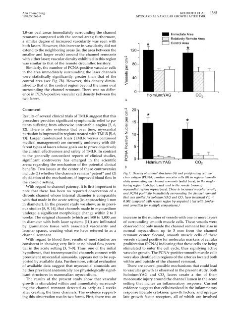
1.0-cm oval areas immediately surrounding the channel<br />
remnants compared with the control areas; furthermore,<br />
a similar degree of increased vascularity was seen with<br />
both lasers. However, this increase in vascularity did not<br />
extend to the neighboring areas (ie, the area between the<br />
smaller and larger ovals) around the channel remnants<br />
with either laser; vascular density exhibited in this region<br />
was similar to that of the remote circumflex territory.<br />
Similarly, the number of PCNA-positive vascular cells<br />
in the area immediately surrounding the laser channels<br />
were statistically significantly greater than that of the<br />
control area (see Fig 7B). However, this density diminished<br />
to that of the control region beyond the inner oval<br />
surrounding the channel remnant. There was no difference<br />
in PCNA-positive vascular cell density between the<br />
two lasers.<br />
Comment<br />
Results of several clinical trials of TMLR suggest that this<br />
procedure provides significant symptomatic relief to patients<br />
suffering from otherwise untreatable angina [5, 6,<br />
12]. There is also evidence that over time, myocardial<br />
perfusion is improved in regions treated with TMLR [5, 6,<br />
13]. Larger randomized trials (TMLR versus continued<br />
medical management) are currently underway with different<br />
types of lasers whose goals are to prove objectively<br />
the clinical effectiveness and safety of TMLR. In contrast<br />
to the generally concordant reports of clinical studies,<br />
significant controversy has emerged in the scientific<br />
arena regarding the mechanism of the potential clinical<br />
benefits. Two issues at the center of these controversies<br />
include (1) whether the channels remain “patent” and (2)<br />
elucidation of the mechanisms of improved blood flow in<br />
the chronic setting.<br />
With regard to channel patency, it is first important to<br />
note that there has been no reported observation of a<br />
chronic channel whose internal diameter is comparable<br />
with that made in the acute setting (ie, approaching 1 mm<br />
in diameter). In the present study we show, as in previous<br />
studies [8, 9, 14], that channels made in myocardium<br />
undergo a significant morphologic change within 2 to 3<br />
weeks. The original channels (which are 800 to 1,000 m<br />
in diameter with both laser systems [11]) are infiltrated<br />
by granulation tissue with associated vascularity and<br />
lacunar spaces, creating what we have referred to as a<br />
channel remnant.<br />
With regard to blood flow, results of most studies are<br />
consistent in showing very little or no blood flow potential<br />
in the acute setting [3, 7–9]. Thus, one of the initial<br />
hypotheses, that transmyocardial channels connect with<br />
preexistent myocardial sinusoids, appears not to be supported<br />
by available data. Furthermore, critical evaluation<br />
of available data suggest that myocardial sinusoids are<br />
neither prevalent anatomically nor physiologically significant<br />
structures in mammalian myocardium.<br />
The results of the present study show that vascular<br />
growth is stimulated within and immediately surrounding<br />
the channel remnant detected as early as 2 weeks<br />
after creating the laser channels. The evidence supporting<br />
this observation was in two forms. First, there was an<br />
Fig 7. Density of arterial structures (A) and proliferating cell nuclear<br />
antigen (PCNA) positive vascular cells (B) in regions immediately<br />
surrounding the channel remnants (solid bars), in the neighboring<br />
region (hatched bars), and in the remote (normal)<br />
myocardial regions (open bars). There is increased vascular density<br />
and PCNA positivity immediately surrounding the channel remnant<br />
that was similar for holmium:YAG and CO 2 laser treatment (*p <br />
0.001 compared with remote region by unpaired t test with Bonferroni<br />
correction for multiple comparisons.)<br />
increase in the number of vessels with one or more layers<br />
of surrounding smooth muscle cells. These vessels were<br />
observed not only inside the channel remnant but also in<br />
normal myocardium up to 3 mm from the channel<br />
remnant center. Second, smooth muscle cells of these<br />
vessels stained positive for molecular markers of cellular<br />
proliferation (PCNA) indicating that these cells are being<br />
stimulated to enter the cell cycle, thus signifying active<br />
vascular growth. The PCNA-positive smooth muscle cells<br />
were also identified in regions of the arteries located both<br />
within and outside of the channel remnant.<br />
There are several possible mechanisms that could lead<br />
to vascular growth as observed in the present study. Both<br />
holmium:YAG and CO 2 lasers create a rim of thermoacoustic<br />
injury around the channel lumen in the acute<br />
setting that incites an inflammatory response. Current<br />
evidence suggests that cells involved in the inflammatory<br />
response liberate cytokines, growth factors, and upregulate<br />
growth factor receptors, all of which are involved