Tricky Optic Nerves and OCT Analysis
Tricky Optic Nerves and OCT Analysis
Tricky Optic Nerves and OCT Analysis
Create successful ePaper yourself
Turn your PDF publications into a flip-book with our unique Google optimized e-Paper software.
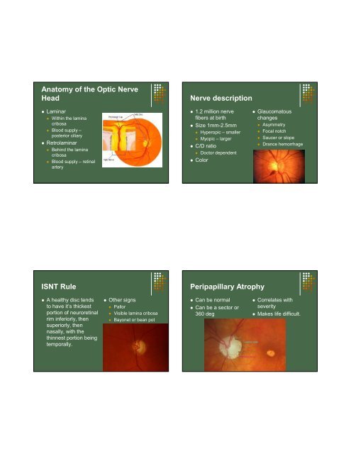
Anatomy of the <strong>Optic</strong> Nerve<br />
Head<br />
Laminar<br />
Within the lamina<br />
cribosa<br />
Blood supply –<br />
posterior ciliary<br />
Retrolaminar<br />
Behind the lamina<br />
cribosa<br />
Blood supply – retinal<br />
artery<br />
Nerve description<br />
1.2 million nerve<br />
fibers at birth<br />
Size 1mm-2.5mm<br />
Hyperopic – smaller<br />
Myopic – larger<br />
C/D ratio<br />
Doctor dependent<br />
Color<br />
Glaucomatous<br />
changes<br />
Asymmetry<br />
Focal notch<br />
Saucer or slope<br />
Drance hemorrhage<br />
ISNT Rule<br />
Peripapillary Atrophy<br />
A healthy disc tends<br />
to have it’s thickest<br />
portion of neuroretinal<br />
rim inferiorly, then<br />
superiorly, then<br />
nasally, with the<br />
thinnest portion being<br />
temporally.<br />
Other signs<br />
Pallor<br />
Visible lamina cribosa<br />
Bayonet or bean pot<br />
Can be normal<br />
Can be a sector or<br />
360 deg<br />
Correlates with<br />
severity<br />
Makes life difficult.


