

Management of Subluxated Lens
Management of Subluxated Lens
Management of Subluxated Lens

Create successful ePaper yourself
Turn your PDF publications into a flip-book with our unique Google optimized e-Paper software.
REVIEW/UPDATE: MANAGEMENT OF SUBLUXATED CRYSTALLINE LENS<br />
1907<br />
Figure 3. Crossed-swords capsule-pinch technique for initiating<br />
capsulotomy in the presence <strong>of</strong> a weak zonule.<br />
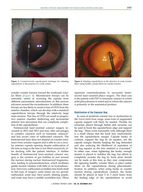
Figure 4. Initiating capsulorhexis in the direction <strong>of</strong> weak zonular<br />
fibers using healthy zonular fibers for counter traction.<br />
zonular counter traction toward the weakened zonular<br />
fibers (Figure 4). Microincision forceps can be<br />
extremely useful in accessing the capsule from<br />
different paracentesis microincisions as this process<br />
advances around the circumference. In addition, these<br />
forceps are less likely to result in loss <strong>of</strong> OVD from the<br />
anterior chamber, which can develop with a standard<br />
capsulorhexis forceps through the relatively large<br />
main incision. This loss <strong>of</strong> OVD can result in progressive<br />
anterior chamber shallowing and incremental<br />
anterior lens movement that can complicate completion<br />
<strong>of</strong> the capsulorhexis.<br />
Femtosecond laser–assisted cataract surgery increased<br />
in 2012 and 2013 and may <strong>of</strong>fer advantages<br />
in complex cataracts such as traumatic cataracts 8<br />
and less severe cases <strong>of</strong> subluxated cataracts. The<br />
femtosecond laser does not depend on counter resistance<br />
from zonular support and is able to cut a circular<br />
anterior capsule opening despite subluxation <strong>of</strong><br />
the lens as long as the lens is not tilted excessively after<br />
docking with the patient interface. A further<br />
benefit <strong>of</strong> femtosecond laser–assisted cataract surgery<br />
is the creation <strong>of</strong> gas bubbles in and around<br />
the nucleus during nuclear femtosecond fragmentation,<br />
leading to pneumodissection, which facilitates<br />
gentle nuclear rotation with little or no hydrodissection.<br />
Not every subluxated crystalline lens lends itself<br />
to this type <strong>of</strong> surgery; some lenses are too grossly<br />
subluxated, some may have poorly dilating pupils,<br />
and some may have a mobile crystalline lens. These<br />
represent contraindications to successful femtosecond<br />
laser–assisted phaco surgery. The ideal case<br />
is the patient with PXF or traumatic cataract in which<br />
mild phacodonesis is noted and in whom the cataract<br />
is primarily in the anatomical position.<br />
Stabilization <strong>of</strong> the Capsular Bag<br />
In cases <strong>of</strong> moderate zonular loss or dysfunction in<br />
the 3-to-6 clock hour range, some form <strong>of</strong> augmented<br />
capsule support will likely be needed. Flexible iris<br />
retractors placed through limbal stab incisions can<br />
be used to hook the capsulorhexis edge and support<br />
the bag. 9 These work reasonably well, although there<br />
is a small chance that the hook may inadvertently<br />
tear the capsulorhexis margin. Capsule hooks, in<br />
contrast, support the bag by its equator, not the<br />
capsule margin, thereby keeping the bag distended<br />
and also reducing the likelihood <strong>of</strong> aspiration <strong>of</strong><br />
the bag equator as the lens material is evacuated. B<br />
In either case, when tightening the hooks, enough<br />
tension is placed to stabilize the bag, but efforts to<br />
completely recenter the bag by hook alone should<br />
not be made at this time as they may compromise<br />
the opposing healthy zonular fibers or place undue<br />
stress on the capsulorhexis during phacoemulsification.<br />
In addition, when using hooks as counter<br />
traction during capsulorhexis creation, the hooks<br />
should be placed at least 2 to 3 clock hours from<br />
the leading edge <strong>of</strong> the capsulorhexis to avoid<br />
J CATARACT REFRACT SURG - VOL 39, DECEMBER 2013