- Page 2 and 3:
1 Topics in Medicinal Chemistry Edi
- Page 4 and 5:
Drug research requires interdiscipl
- Page 6 and 7:
Topics in Medicinal Chemistry Also
- Page 8 and 9:
Preface to Volume 1 With supreme ir
- Page 10 and 11:
Contents Overview R.H.Bradbury ....
- Page 12 and 13:
Top Med Chem (2007) 1: 1-17 DOI 10.
- Page 14 and 15:
Overview 3 led to animal experiment
- Page 16 and 17:
Overview 5 electrodes, an effect la
- Page 18 and 19:
Overview 7 Fig. 3 Acquired capabili
- Page 20 and 21:
Overview 9 Table 3 Marketed targete
- Page 22 and 23:
Overview 11 pathways mediating one
- Page 24 and 25:
Overview 13 Table 4 Targeted drugs
- Page 26 and 27:
Overview 15 10. Brock N (1989) Canc
- Page 28 and 29:
Overview 17 83. Daniel KG, Kuhn DJ,
- Page 30 and 31:
20 J. Hoffmann · A. Sommer with in
- Page 32 and 33:
22 J. Hoffmann · A. Sommer Most ta
- Page 34 and 35:
24 J. Hoffmann · A. Sommer classic
- Page 36 and 37:
26 J. Hoffmann · A. Sommer Fig. 3
- Page 38 and 39:
28 J. Hoffmann · A. Sommer PR is h
- Page 40 and 41:
30 J. Hoffmann · A. Sommer activat
- Page 42 and 43:
32 J. Hoffmann · A. Sommer of ERα
- Page 44 and 45:
34 J. Hoffmann · A. Sommer in brea
- Page 46 and 47:
36 J. Hoffmann · A. Sommer Fig. 7
- Page 48 and 49:
38 J. Hoffmann · A. Sommer cantly
- Page 50 and 51:
40 J. Hoffmann · A. Sommer pared w
- Page 52 and 53:
42 J. Hoffmann · A. Sommer in the
- Page 54 and 55:
44 J. Hoffmann · A. Sommer Fig. 9
- Page 56 and 57:
46 J. Hoffmann · A. Sommer the syn
- Page 58 and 59:
48 J. Hoffmann · A. Sommer ture [1
- Page 60 and 61:
50 J. Hoffmann · A. Sommer isation
- Page 62 and 63:
52 J. Hoffmann · A. Sommer droloxi
- Page 64 and 65:
54 J. Hoffmann · A. Sommer Non-ste
- Page 66 and 67:
56 J. Hoffmann · A. Sommer genic,
- Page 68 and 69:
58 J. Hoffmann · A. Sommer The pur
- Page 70 and 71:
60 J. Hoffmann · A. Sommer cacy of
- Page 72 and 73:
62 J. Hoffmann · A. Sommer Fig. 15
- Page 74 and 75:
64 J. Hoffmann · A. Sommer Neverth
- Page 76 and 77:
66 J. Hoffmann · A. Sommer in the
- Page 78 and 79:
68 J. Hoffmann · A. Sommer been de
- Page 80 and 81:
70 J. Hoffmann · A. Sommer High-do
- Page 82 and 83:
72 J. Hoffmann · A. Sommer Table 1
- Page 84 and 85:
74 J. Hoffmann · A. Sommer Blockin
- Page 86 and 87:
76 J. Hoffmann · A. Sommer 55. Rod
- Page 88 and 89:
78 J. Hoffmann · A. Sommer 122. Hi
- Page 90 and 91:
80 J. Hoffmann · A. Sommer 174. Is
- Page 92 and 93:
82 J. Hoffmann · A. Sommer 234. Go
- Page 94 and 95:
84 E.M. Wallace et al. Abstract App
- Page 96 and 97:
86 E.M. Wallace et al. Fig. 1 Targe
- Page 98 and 99:
88 E.M. Wallace et al. There are th
- Page 100 and 101:
90 E.M. Wallace et al. participates
- Page 102 and 103:
92 E.M. Wallace et al. ture, most o
- Page 104 and 105:
94 E.M. Wallace et al. Fig. 5 The b
- Page 106 and 107:
96 E.M. Wallace et al. 4.1 Selectiv
- Page 108 and 109:
98 E.M. Wallace et al. care or supp
- Page 110 and 111:
100 E.M. Wallace et al. trials. The
- Page 112 and 113:
102 E.M. Wallace et al. in three hu
- Page 114 and 115:
104 E.M. Wallace et al. Table 1 Cel
- Page 116 and 117:
106 E.M. Wallace et al. 4.2.3 ARRY-
- Page 118 and 119:
108 E.M. Wallace et al. HKI-272 inh
- Page 120 and 121:
110 E.M. Wallace et al. crease hepa
- Page 122 and 123:
112 E.M. Wallace et al. respectivel
- Page 124 and 125:
114 E.M. Wallace et al. Fig. 9 Stru
- Page 126 and 127:
116 E.M. Wallace et al. end of this
- Page 128 and 129:
118 E.M. Wallace et al. compound do
- Page 130 and 131:
120 E.M. Wallace et al. 6.2 PD03259
- Page 132 and 133:
122 E.M. Wallace et al. sies, but t
- Page 134 and 135:
124 E.M. Wallace et al. Despite the
- Page 136 and 137:
126 E.M. Wallace et al. are designe
- Page 138 and 139:
128 E.M. Wallace et al. 42. Fabian
- Page 140 and 141:
130 E.M. Wallace et al. Perrier M,
- Page 142 and 143:
132 E.M. Wallace et al. nowski S, O
- Page 144 and 145:
134 D.W. End et al. logical maligna
- Page 146 and 147:
136 D.W. End et al. growth. In part
- Page 148 and 149:
138 D.W. End et al. C-A-A-X recogni
- Page 150 and 151:
140 D.W. End et al. Fig. 3 Structur
- Page 152 and 153:
142 D.W. End et al. Fig. 5 CAAX pep
- Page 154 and 155:
144 D.W. End et al. mediates all of
- Page 156 and 157:
146 D.W. End et al. 3.6 FTase Knock
- Page 158 and 159:
148 D.W. End et al. acid giving a c
- Page 160 and 161:
150 D.W. End et al. Fig. 7 Target d
- Page 162 and 163:
152 D.W. End et al. GTase II. Altho
- Page 164 and 165:
154 D.W. End et al. Fig. 11 Aryl cy
- Page 166 and 167:
156 D.W. End et al. Fig. 14 Structu
- Page 168 and 169:
158 D.W. End et al. ness, Chagas di
- Page 170 and 171:
160 D.W. End et al. References 1. Q
- Page 172 and 173:
162 D.W. End et al. 68. Chen Z, Sun
- Page 174 and 175:
164 D.W. End et al. 122. Shaikenov
- Page 176 and 177:
166 D.W. End et al. 156. Dinsmore C
- Page 178 and 179:
168 D.W. End et al. 201. Gelb MH, V
- Page 180 and 181:
170 C. Garcia-Echeverria Keywords H
- Page 182 and 183:
172 C. Garcia-Echeverria 2 Insulin-
- Page 184 and 185:
174 C. Garcia-Echeverria 2.2 Modula
- Page 186 and 187:
176 C. Garcia-Echeverria lesions ar
- Page 188 and 189:
178 C. Garcia-Echeverria the divers
- Page 190 and 191:
180 C. Garcia-Echeverria Fig. 3 Rep
- Page 192 and 193:
182 C. Garcia-Echeverria cal assays
- Page 194 and 195:
184 C. Garcia-Echeverria one within
- Page 196 and 197:
186 C. Garcia-Echeverria Epidemiolo
- Page 198 and 199:
188 C. Garcia-Echeverria Fig. 5 Rep
- Page 200 and 201:
190 C. Garcia-Echeverria allosteric
- Page 202 and 203:
192 C. Garcia-Echeverria Currently,
- Page 204 and 205:
194 C. Garcia-Echeverria 24 h. No i
- Page 206 and 207:
196 C. Garcia-Echeverria Fig. 8 Rep
- Page 208 and 209:
198 C. Garcia-Echeverria Fig. 9 PI3
- Page 210 and 211:
200 C. Garcia-Echeverria References
- Page 212 and 213: 202 C. Garcia-Echeverria 64. Ihle N
- Page 214 and 215: 204 C. Garcia-Echeverria 119. Jones
- Page 216 and 217: 206 C. Garcia-Echeverria 175. Posad
- Page 218 and 219: 208 K.J. Moriarty et al. 3.3.3 Thie
- Page 220 and 221: 210 K.J. Moriarty et al. erodimeric
- Page 222 and 223: 212 K.J. Moriarty et al. inhibitors
- Page 224 and 225: 214 K.J. Moriarty et al. Structure
- Page 226 and 227: 216 K.J. Moriarty et al. K i = 0.00
- Page 228 and 229: 218 K.J. Moriarty et al. Structure
- Page 230 and 231: 220 K.J. Moriarty et al. lectivity
- Page 232 and 233: 222 K.J. Moriarty et al. 2.3.5 3,5-
- Page 234 and 235: 224 K.J. Moriarty et al. A variety
- Page 236 and 237: 226 K.J. Moriarty et al. Fig. 3 Com
- Page 238 and 239: 228 K.J. Moriarty et al. a benefici
- Page 240 and 241: 230 K.J. Moriarty et al. Structure
- Page 242 and 243: 232 K.J. Moriarty et al. Structure
- Page 244 and 245: 234 K.J. Moriarty et al. This compo
- Page 246 and 247: 236 K.J. Moriarty et al. IC 50 = 0.
- Page 248 and 249: 238 K.J. Moriarty et al. Structure
- Page 250 and 251: 240 K.J. Moriarty et al. over CDK1.
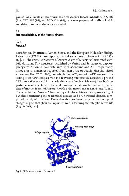
- Page 252 and 253: 242 K.J. Moriarty et al. Structure
- Page 254 and 255: 244 K.J. Moriarty et al. with an al
- Page 256 and 257: 246 K.J. Moriarty et al. Fig. 7 Com
- Page 258 and 259: 248 K.J. Moriarty et al. studies, 6
- Page 260 and 261: 250 K.J. Moriarty et al. Structure
- Page 264 and 265: 254 K.J. Moriarty et al. Fig. 10 Di
- Page 266 and 267: 256 K.J. Moriarty et al. Fig. 13 Co
- Page 268 and 269: 258 K.J. Moriarty et al. Structure
- Page 270 and 271: 260 K.J. Moriarty et al. Structure
- Page 272 and 273: 262 K.J. Moriarty et al. (70% inhib
- Page 274 and 275: 264 K.J. Moriarty et al. Structure
- Page 276 and 277: 266 K.J. Moriarty et al. Structure
- Page 278 and 279: 268 K.J. Moriarty et al. Structure
- Page 280 and 281: 270 K.J. Moriarty et al. of DNA syn
- Page 282 and 283: 272 K.J. Moriarty et al. Structure
- Page 284 and 285: 274 K.J. Moriarty et al. 4.3.2 Pyri
- Page 286 and 287: 276 K.J. Moriarty et al. Structure
- Page 288 and 289: 278 K.J. Moriarty et al. Structure
- Page 290 and 291: 280 K.J. Moriarty et al. 4.3.8 3-Et
- Page 292 and 293: 282 K.J. Moriarty et al. 29. Brothe
- Page 294 and 295: 284 K.J. Moriarty et al. 81. Yue EW
- Page 296 and 297: 286 K.J. Moriarty et al. 122. Brams
- Page 298 and 299: 288 K.J. Moriarty et al. Hercend T,
- Page 300 and 301: 290 K.J. Moriarty et al. 229. Esced
- Page 302 and 303: Top Med Chem (2007) 1: 293-331 DOI
- Page 304 and 305: HDAC Inhibition in Cancer Therapy 2
- Page 306 and 307: HDAC Inhibition in Cancer Therapy 2
- Page 308 and 309: HDAC Inhibition in Cancer Therapy 2
- Page 310 and 311: HDAC Inhibition in Cancer Therapy 3
- Page 312 and 313:
HDAC Inhibition in Cancer Therapy 3
- Page 314 and 315:
HDAC Inhibition in Cancer Therapy 3
- Page 316 and 317:
HDAC Inhibition in Cancer Therapy 3
- Page 318 and 319:
HDAC Inhibition in Cancer Therapy 3
- Page 320 and 321:
HDAC Inhibition in Cancer Therapy 3
- Page 322 and 323:
HDAC Inhibition in Cancer Therapy 3
- Page 324 and 325:
HDAC Inhibition in Cancer Therapy 3
- Page 326 and 327:
HDAC Inhibition in Cancer Therapy 3
- Page 328 and 329:
HDAC Inhibition in Cancer Therapy 3
- Page 330 and 331:
HDAC Inhibition in Cancer Therapy 3
- Page 332 and 333:
HDAC Inhibition in Cancer Therapy 3
- Page 334 and 335:
HDAC Inhibition in Cancer Therapy 3
- Page 336 and 337:
HDAC Inhibition in Cancer Therapy 3
- Page 338 and 339:
HDAC Inhibition in Cancer Therapy 3
- Page 340 and 341:
HDAC Inhibition in Cancer Therapy 3
- Page 342 and 343:
334 K. Paz · Z. Zhu application of
- Page 344 and 345:
336 K. Paz · Z. Zhu mor cells [33-
- Page 346 and 347:
338 K. Paz · Z. Zhu of vascular in
- Page 348 and 349:
340 K. Paz · Z. Zhu DC101 has been
- Page 350 and 351:
342 K. Paz · Z. Zhu cemia. In Marc
- Page 352 and 353:
344 K. Paz · Z. Zhu but there were
- Page 354 and 355:
346 K. Paz · Z. Zhu effects after
- Page 356 and 357:
348 K. Paz · Z. Zhu adverse effect
- Page 358 and 359:
350 K. Paz · Z. Zhu the first comp
- Page 360 and 361:
352 K. Paz · Z. Zhu detanib doses
- Page 362 and 363:
354 K. Paz · Z. Zhu 3.4.4 Neovasta
- Page 364 and 365:
356 K. Paz · Z. Zhu monotherapy tr
- Page 366 and 367:
358 K. Paz · Z. Zhu 13736), a pote
- Page 368 and 369:
360 K. Paz · Z. Zhu Phase I clinic
- Page 370 and 371:
362 K. Paz · Z. Zhu 3.4.10 BAY57-9
- Page 372 and 373:
364 K. Paz · Z. Zhu stabilization
- Page 374 and 375:
366 K. Paz · Z. Zhu cles. Common a
- Page 376 and 377:
368 K. Paz · Z. Zhu ovary, and fal
- Page 378 and 379:
370 K. Paz · Z. Zhu 6. Creamer D,
- Page 380 and 381:
372 K. Paz · Z. Zhu 73. Sato Y, Ka
- Page 382 and 383:
374 K. Paz · Z. Zhu 132. Takahashi
- Page 384 and 385:
376 K. Paz · Z. Zhu 189. Graeven U
- Page 386 and 387:
378 K. Paz · Z. Zhu Hawtin R, Tang
- Page 388 and 389:
380 K. Paz · Z. Zhu 287. Carlomagn
- Page 390 and 391:
382 K. Paz · Z. Zhu 345. Yu JL, Ra
- Page 392 and 393:
384 T.K. Sawyer Keywords Src · Src
- Page 394 and 395:
386 T.K. Sawyer Table 2 Some functi
- Page 396 and 397:
388 T.K. Sawyer pathways has been l
- Page 398 and 399:
390 T.K. Sawyer nate proteins, and
- Page 400 and 401:
392 T.K. Sawyer Fig. 6 Some known S
- Page 402 and 403:
394 T.K. Sawyer interactions betwee
- Page 404 and 405:
396 T.K. Sawyer collagen type-I-ind
- Page 406 and 407:
398 T.K. Sawyer AP23451 administrat
- Page 408 and 409:
400 T.K. Sawyer ery. This chapter h
- Page 410 and 411:
402 T.K. Sawyer 48. Lutz MP, Esser
- Page 412 and 413:
404 T.K. Sawyer 96. Nimmanapalli R,
- Page 414 and 415:
Top Med Chem (2007) 1: 407-444 DOI
- Page 416 and 417:
Bcr-Abl Kinase Inhibitors 409 2 Ima
- Page 418 and 419:
Bcr-Abl Kinase Inhibitors 411 3 New
- Page 420 and 421:
Bcr-Abl Kinase Inhibitors 413 sista
- Page 422 and 423:
Bcr-Abl Kinase Inhibitors 415 3.1.2
- Page 424 and 425:
Bcr-Abl Kinase Inhibitors 417 Schem
- Page 426 and 427:
Bcr-Abl Kinase Inhibitors 419 Schem
- Page 428 and 429:
Bcr-Abl Kinase Inhibitors 421 group
- Page 430 and 431:
Bcr-Abl Kinase Inhibitors 423 Altho
- Page 432 and 433:
Bcr-Abl Kinase Inhibitors 425 4.4 S
- Page 434 and 435:
Bcr-Abl Kinase Inhibitors 427 of CM
- Page 436 and 437:
Bcr-Abl Kinase Inhibitors 429 both
- Page 438 and 439:
Bcr-Abl Kinase Inhibitors 431 five
- Page 440 and 441:
Bcr-Abl Kinase Inhibitors 433 ity o
- Page 442 and 443:
Bcr-Abl Kinase Inhibitors 435 cal t
- Page 444 and 445:
Bcr-Abl Kinase Inhibitors 437 The l
- Page 446 and 447:
Bcr-Abl Kinase Inhibitors 439 44. M
- Page 448 and 449:
Bcr-Abl Kinase Inhibitors 441 92. h
- Page 450 and 451:
Bcr-Abl Kinase Inhibitors 443 134.
- Page 452 and 453:
Author Index Volume 1 Thevolumenumb
- Page 454 and 455:
Subject Index Abiraterone 50 Ableso
- Page 456 and 457:
Subject Index 449 Hormone response
- Page 458:
Subject Index 451 Testosterone 47,