

History-Evolution-of-Foot-and-Lower-Extremity-Biomechanics-and-Foot-Orthoses_Kevin-Kirby
History-Evolution-of-Foot-and-Lower-Extremity-Biomechanics-and-Foot-Orthoses_Kevin-Kirby
History-Evolution-of-Foot-and-Lower-Extremity-Biomechanics-and-Foot-Orthoses_Kevin-Kirby

Create successful ePaper yourself
Turn your PDF publications into a flip-book with our unique Google optimized e-Paper software.
3/26/2015<br />
<strong>History</strong> <strong>and</strong> <strong>Evolution</strong> <strong>of</strong> <strong>Foot</strong> <strong>and</strong><br />
<strong>Lower</strong> <strong>Extremity</strong> <strong>Biomechanics</strong><br />
<strong>and</strong> <strong>Foot</strong> <strong>Orthoses</strong><br />
<strong>Kevin</strong> A. <strong>Kirby</strong>, DPM<br />
Adjunct Associate Pr<strong>of</strong>essor<br />
Department <strong>of</strong> Applied <strong>Biomechanics</strong><br />
California School <strong>of</strong> Podiatric Medicine<br />
Oakl<strong>and</strong>, California<br />
Private Practice, Sacramento, California<br />
2015 PAC Symposium: “<strong>Biomechanics</strong> <strong>and</strong> the Future <strong>of</strong> <strong>Foot</strong>care”<br />
Vancouver, BC<br />
April 17-18, 2015<br />
What is the <strong>History</strong> <strong>of</strong> <strong>Foot</strong><br />
<strong>Biomechanics</strong> <strong>and</strong> <strong>Foot</strong> <strong>Orthoses</strong>?<br />
<strong>Biomechanics</strong> <strong>and</strong> foot orthoses have<br />
evolved over the centuries<br />
Many talented individuals have increased<br />
our knowledge in foot <strong>and</strong> lower extremity<br />
biomechanics <strong>and</strong> about foot orthoses<br />
What people <strong>and</strong> events have shaped<br />
history <strong>and</strong> evolution <strong>of</strong> podiatric<br />
biomechanics <strong>and</strong> orthoses?<br />
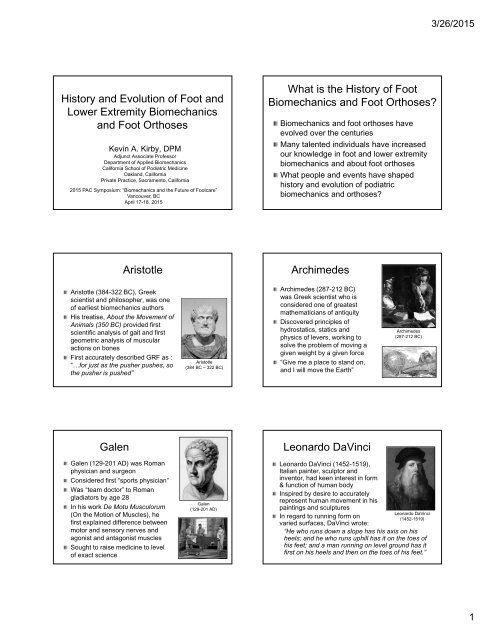
Aristotle<br />
Archimedes<br />
Aristotle (384-322 BC), Greek<br />
scientist <strong>and</strong> philosopher, was one<br />
<strong>of</strong> earliest biomechanics authors<br />
His treatise, About the Movement <strong>of</strong><br />
Animals (350 BC) provided first<br />
scientific analysis <strong>of</strong> gait <strong>and</strong> first<br />
geometric analysis <strong>of</strong> muscular<br />
actions on bones<br />
First accurately described GRF as :<br />
“…for just as the pusher pushes, so<br />
the pusher is pushed”<br />
Aristotle<br />
(384 BC – 322 BC)<br />
Archimedes (287-212 BC)<br />
was Greek scientist who is<br />
considered one <strong>of</strong> greatest<br />
mathematicians <strong>of</strong> antiquity<br />
Discovered principles <strong>of</strong><br />
hydrostatics, statics <strong>and</strong><br />
physics <strong>of</strong> levers, working to<br />
solve the problem <strong>of</strong> moving a<br />
given weight by a given force<br />
“Give me a place to st<strong>and</strong> on,<br />
<strong>and</strong> I will move the Earth”<br />
Archimedes<br />
(287-212 BC)<br />
Galen<br />
Leonardo DaVinci<br />
Galen (129-201 AD) was Roman<br />
physician <strong>and</strong> surgeon<br />
Considered first “sports physician”<br />
Was “team doctor” to Roman<br />
gladiators by age 28<br />
In his work De Motu Musculorum<br />
(On the Motion <strong>of</strong> Muscles), he<br />
first explained difference between<br />
motor <strong>and</strong> sensory nerves <strong>and</strong><br />
agonist <strong>and</strong> antagonist muscles<br />
Sought to raise medicine to level<br />
<strong>of</strong> exact science<br />
Galen<br />
(129-201 AD)<br />
Leonardo DaVinci (1452-1519),<br />
Italian painter, sculptor <strong>and</strong><br />
inventor, had keen interest in form<br />
& function <strong>of</strong> human body<br />
Inspired by desire to accurately<br />
represent human movement in his<br />
paintings <strong>and</strong> sculptures<br />
In regard to running form on<br />
varied surfaces, DaVinci wrote:<br />
“He who runs down a slope has his axis on his<br />
Leonardo DaVinci<br />
(1452-1519)<br />
heels; <strong>and</strong> he who runs uphill has it on the toes <strong>of</strong><br />
his feet; <strong>and</strong> a man running on level ground has it<br />
first on his heels <strong>and</strong> then on the toes <strong>of</strong> his feet.”<br />
1
3/26/2015<br />
Giovanni Borelli<br />
Isaac Newton<br />
Giovanni Borelli (1608-1679),<br />
Italian physicist, physiologist <strong>and</strong><br />
mathematician, wrote first book on<br />
biomechanics, De Motu<br />
Animalium, in 1685<br />
First to fully describe geometrical<br />
principles <strong>of</strong> levers <strong>of</strong> musculoskeletal<br />
system <strong>and</strong> that muscles<br />
produce much larger forces than<br />
resisting external forces<br />
Borelli found forces required for<br />
equilibrium in various joints <strong>of</strong><br />
human body before Newton<br />
Borelli is <strong>of</strong>ten called:<br />
“Father <strong>of</strong> <strong>Biomechanics</strong>”<br />
Giovanni Borelli<br />
(1608-1679)<br />
Isaac Newton (1642-1727),<br />
British mathematician <strong>and</strong><br />
physicist, is considered by many<br />
to be greatest scientist <strong>of</strong> all time<br />
Invented calculus at age 24<br />
Published Philosophiae Naturalis<br />
Principia Mathematica in 1686<br />
which contained his now famous<br />
Three Laws <strong>of</strong> Motion<br />
Credited with creating the laws <strong>of</strong><br />
inertia, acceleration, action <strong>and</strong><br />
reaction forces <strong>and</strong> <strong>of</strong> gravity<br />
Isaac Newton<br />
(1642-1727)<br />
Nicolas Andry<br />
Petrus Camper<br />
Nicolas Andry (1658-1742), a French<br />
physician, first coined the term<br />
“orthopedics” meaning “straight<br />
child”<br />
Published book, Orthopaedia: The<br />
Art <strong>of</strong> Correcting <strong>and</strong> Preventing<br />
Deformities in Children in1740<br />
“If the feet incline too much to one<br />
side, you must give the child shoes<br />
that are higher on that side, both in<br />
the sole <strong>and</strong> heel, which will make<br />
him incline to the opposite side.“<br />
Nicolas Andry<br />
(1658-1742)<br />
Petrus Camper (1722-1789) was<br />
a Dutch physician <strong>and</strong> pioneer in<br />
pediatrics<br />
Published one <strong>of</strong> first books on<br />
foot deformities, “On the Best<br />
Form <strong>of</strong> Shoe” in 1781, which<br />
was reprinted into 14 editions<br />
Camper’s book stimulated<br />
interest in placing archsupporting<br />
orthoses into shoes<br />
for children’s flatfoot<br />
Petrus Camper<br />
(1722-1789)<br />
“It is surprising that while mankind in all<br />
ages have bestowed the greatest attention<br />
upon the feet <strong>of</strong> horses, mules, oxen, <strong>and</strong><br />
other animals <strong>of</strong> burthen <strong>of</strong> draught, they<br />
have entirely neglected those <strong>of</strong> their own<br />
species, ab<strong>and</strong>oning them to the ignorance<br />
<strong>of</strong> workmen, who, in general, can only<br />
make a shoe upon routine principles, <strong>and</strong><br />
according to the absurdities <strong>of</strong> fashion, or<br />
the depraved taste <strong>of</strong> the day. Thus, from<br />
our earliest infancy, shoes, as at present<br />
worn, serve but to deform the toes <strong>and</strong><br />
cover the feet with corns, which not only<br />
render walking painful, but, in some cases,<br />
absolutely impossible.” P. Camper, 1781<br />
Petrus Camper<br />
Shoe Fashions<br />
from 1760<br />
Lewis Durlacher<br />
Lewis Durlacher (1792-1864), a<br />
British chiropodist, was royal<br />
chiropodist appointed to Queen<br />
Victoria<br />
In 1845, he developed leather<br />
foot orthosis to correct for<br />
“plantar pressure lesions” <strong>and</strong><br />
“foot imbalances”<br />
Durlacher first described<br />
intermetatarsal neuroma, over<br />
30 years before T.G. Morton<br />
2
3/26/2015<br />
Hugh Owen Thomas<br />
Newton Melman Shaffer<br />
Hugh Owen Thomas (1834-1891)<br />
was a British orthopedic surgeon<br />
with interest in treating feet<br />
In 1874, Thomas suggested using<br />
a “few pennies worth <strong>of</strong> leather”<br />
for lifts, bars, <strong>and</strong> wedges on<br />
shoes to treat foot problems<br />
Invented, in 1876, “crooking” <strong>of</strong><br />
shoe heel, to extend the heel<br />
under the antero-medial aspect <strong>of</strong><br />
shoe sole for treating pronated<br />
feet (now called “Thomas heel”)<br />
Hugh Owen Thomas<br />
(1834-1891)<br />
Newton M. Shaffer (1846-<br />
1928), a New York City<br />
orthopedist, first described<br />
high arched foot with<br />
multiple clawtoes<br />
Became widely known as<br />
“Shaffer’s <strong>Foot</strong>”<br />
Also designed a highmedial<br />
arched orthosis<br />
with a heel cup which<br />
became known as a<br />
“Shaffer Plate”<br />
Newton M. Shaffer<br />
(1846-1928)<br />
Royal Whitman<br />
Whitman’s “Weak <strong>Foot</strong>”<br />
Royal Whitman (1857-<br />
1946) was a 1882<br />
Harvard Medical School<br />
graduate <strong>and</strong> New York<br />
City orthopedic surgeon<br />
that had special interest<br />
in foot function<br />
Wrote numerous<br />
textbooks on orthopedic<br />
surgery <strong>and</strong> taught<br />
orthopedics for 40 years<br />
Royal Whitman<br />
(1857-1946)<br />
Whitman’s description <strong>of</strong><br />
“weak foot” very closely<br />
matches our current<br />
description <strong>of</strong> the pronated,<br />
flat-arched foot with an<br />
internally rotated tibia<br />
Whitman’s Three Grades <strong>of</strong> “Weak <strong>Foot</strong>”<br />
1 st Degree: The normal foot improperly used, as<br />
shown by the method <strong>of</strong> st<strong>and</strong>ing <strong>and</strong> walking<br />
2 nd Degree: The foot in which the range <strong>of</strong> voluntary<br />
motion is restricted, showing disuse <strong>of</strong> function, <strong>and</strong><br />
in which the elements <strong>of</strong> deformity are apparent<br />
when weight is borne<br />
3 rd Degree: That in which the passive range <strong>of</strong><br />
motion is restricted, or in which there are evident<br />
weakness <strong>and</strong> deformity. This limitation <strong>of</strong> motion<br />
depends, as a rule, on accommodative changes in<br />
structure to the habitual postures or to the deformity<br />
Whitman R: A study <strong>of</strong> the weak foot, with reference to its causes, its diagnosis, <strong>and</strong> its cure; with an<br />
analysis <strong>of</strong> a thous<strong>and</strong> cases <strong>of</strong> so-called flat-foot. JBJS, 8:42-77, 1896.<br />
Whitman’s <strong>Foot</strong> Brace<br />
Developed in 1885<br />
Made from plaster cast<br />
taken with foot in<br />
supinated position<br />
18-20 gauge sheet<br />
steel was formed into a<br />
high medial arch brace<br />
Goal was to raise<br />
medial arch so foot<br />
would be less pronated<br />
3
3/26/2015<br />
Percy Willard Roberts<br />
1895 - Beginning <strong>of</strong> Chiropody<br />
Percy W. Roberts (1867-1937), American orthopedic<br />
surgeon, developed metal foot orthosis in 1912<br />
Roberts foot orthosis had deep inverted heel cup that<br />
attempted to tilt rearfoot into inverted position<br />
Had medial <strong>and</strong> lateral clips <strong>and</strong> narrow heel cup<br />
Roberts brace applied too much force over too little<br />
area <strong>and</strong> tended to cause irritation to plantar foot<br />
During most <strong>of</strong> 19 th century, medical doctors<br />
showed little interest in treating foot problems<br />
Barbers, families <strong>and</strong> practically anyone that<br />
showed an interest in treating feet adopted the<br />
“craft” <strong>of</strong> foot care to fill the void in healthcare <strong>of</strong><br />
the foot that was left by medical doctors<br />
In 1895, group <strong>of</strong> dedicated practitioners in New<br />
York successfully appealed to NY State<br />
Legislature to first establish chiropody (now<br />
podiatry) as a licensed pr<strong>of</strong>ession<br />
First Podiatry Society<br />
& School Formed<br />
In 1895, Pedic Society <strong>of</strong> New<br />
York became first <strong>of</strong>ficial society<br />
for chiropody/podiatry<br />
First school <strong>of</strong> chiropody<br />
established in New York in 1911<br />
In 1912, first national podiatry<br />
association, <strong>and</strong> precursor to<br />
current APMA, American National<br />
Chiropody Association, was<br />
formed <strong>and</strong> is now 100 years old<br />
First Issue <strong>of</strong> Pedic Society<br />
Items – First “Podiatry Journal”<br />
Alfred Joseph, First<br />
President <strong>of</strong> National<br />
Association <strong>of</strong><br />
Chiropodists”<br />
Podiatric Approach to <strong>Foot</strong><br />
<strong>Biomechanics</strong> in Early 20 th Century<br />
Approach to foot mechanics in<br />
early 20 th century was based<br />
largely on shape <strong>of</strong> medial<br />
longitudinal arch with “normal foot”<br />
being viewed as having “normally<br />
arched architecture” with<br />
treatments geared toward<br />
restoring a “normal arch”<br />
Many podiatrists relied on<br />
orthopedic shoe makers to make<br />
custom leather foot or steel<br />
appliances to treat “weak feet”<br />
Early 20 th Century<br />
Treatment <strong>of</strong> “Weak<br />
Feet” Used Whitman’s<br />
Concepts<br />
Otto Frederick Schuster<br />
Otto F. Schuster (1881-<br />
1936) arrived in US in 1906<br />
from Hamburg, Germany<br />
where he had trained as a<br />
brace maker<br />
Schuster started making<br />
Whitman braces for Royal<br />
Whitman <strong>and</strong> other<br />
orthopedists in NY in 1909<br />
Became a podiatrist in 1911<br />
Otto F. Schuster<br />
(1881-1936)<br />
First Podiatric<br />
Orthopedics Text<br />
Otto F. Schuster became<br />
pr<strong>of</strong>essor <strong>of</strong> orthopedics at<br />
NY Podiatry School<br />
In 1927, published first<br />
podiatric orthopedics text,<br />
<strong>Foot</strong> Orthopedics, later<br />
rewritten by his podiatristson,<br />
Otto N. Schuster<br />
<strong>Foot</strong> Orthopedics remained<br />
in use as a valuable<br />
textbook until 1950s<br />
4
3/26/2015<br />
Roberts-Whitman Brace<br />
Dudley J. Morton<br />
In the 1920s, Otto F. Schuster<br />
combined ideas <strong>of</strong> Roberts<br />
brace with Whitman brace to<br />
make Roberts-Whitman brace<br />
Roberts-Whitman brace had<br />
deep inverted heel cup, high<br />
medial arch <strong>and</strong> wider pr<strong>of</strong>ile<br />
that provided better pronation<br />
control <strong>and</strong> allowed for more<br />
comfortable medial arch<br />
Otto Schuster created<br />
Roberts-Whitman Brace<br />
Dudley Joy Morton (1884-1960)<br />
was physician, anatomist <strong>and</strong><br />
anthropologist<br />
Work focused on shortened 1st<br />
metatarsal, “hypermobility” <strong>of</strong> 1st<br />
metatarsal segment <strong>and</strong><br />
correlation <strong>of</strong> 1 st ray mechanics to<br />
excessive foot pronation<br />
Published book, The Human<br />
<strong>Foot</strong>, It’s <strong>Evolution</strong>, Physiology<br />
<strong>and</strong> Functional Disorders, in 1935<br />
Dudley J. Morton<br />
(1884-1960)<br />
Morton Was First to Describe Load-<br />
Deformation <strong>of</strong> 1 st Ray in 1935<br />
“If the plantar ligaments <strong>of</strong><br />
any segment are slack<br />
when the head <strong>of</strong> its<br />
metatarsal bone lies on<br />
the same plane as the<br />
others whose ligaments<br />
are taut, that segment will<br />
fail to share in the carriage<br />
<strong>of</strong> body weight….”<br />
Morton DJ: The Human <strong>Foot</strong>: Its <strong>Evolution</strong>, Physiology<br />
<strong>and</strong> Functional Disorders. Columbia University Press.<br />
Morningside Heights, New York, 1935.<br />
“…the plantar ligaments <strong>of</strong> the first metatarsal<br />
segment in these feet were lax when the other<br />
ligaments had become tense under body weight;<br />
hence the first metatarsal still retained a margin <strong>of</strong><br />
dorsal extension <strong>and</strong> therefore was ineffective as a<br />
weightbearing structure.”<br />
Morton DJ: The Human <strong>Foot</strong>: Its <strong>Evolution</strong>, Physiology <strong>and</strong> Functional Disorders. Columbia<br />
University Press. Morningside Heights, New York, 1935.<br />
Morton DJ: Physiological<br />
considerations in the<br />
treatment <strong>of</strong> foot<br />
deformities. JBJS,<br />
19:1052-1056, 1937.<br />
Morton believed “hypermobility <strong>of</strong> first<br />
metatarsal” affects foot in three ways:<br />
1. Second metatarsal has “increased burden”<br />
since first metatarsal “fails to assume” its<br />
normal share <strong>of</strong> weight<br />
2. <strong>Foot</strong> pronates because “medial buttress” is<br />
ineffective until “slack in its ligaments is taken<br />
up” as pronation increases<br />
3. As pronation advances, “functional stresses are<br />
thrown increasingly on muscles on inner side <strong>of</strong><br />
ankle, imposing them undue strain”<br />
Morton’s Compensating Insole<br />
In 1932, Morton designed a<br />
“compensating insole” that<br />
focused on elevating first<br />
metatarsal head <strong>and</strong><br />
preventing pronation<br />
compensation for short,<br />
“hypermobile” first ray<br />
Morton also designed inshoe<br />
support with high<br />
medial arch-flange to resist<br />
pronation<br />
5
3/26/2015<br />
First Plaster Splint Impression<br />
Casting <strong>of</strong> <strong>Foot</strong><br />
Edward Reed, MD, an<br />
orthopedic surgeon from<br />
Santa Monica, California,<br />
was first to describe<br />
plaster splint impression<br />
casting for foot orthoses<br />
in 1933<br />
Reed EN: A simple method for making plaster casts <strong>of</strong><br />
feet. JBJS, 17:1007, 1933.<br />
Alan Murray & Benjamin Levy<br />
In 1930s, Alan Murray, ice<br />
skater, developed shoe molded<br />
directly onto cast <strong>of</strong> foot called<br />
the Murray Space Shoe<br />
NY podiatrist, Benjamin Levy,<br />
developed idea <strong>of</strong> using inner<br />
sole from Murray Space Shoe as<br />
a removable cork <strong>and</strong> leather<br />
insole with a toe crest, became<br />
known as Levy mold.<br />
Levy B: An appliance to induce toe flexion on weight bearing. J<br />
Natl Assoc <strong>of</strong> Chiropodists, 40(6):24-33, 1950.<br />
John Tinkham Manter<br />
John T. Manter (1910-1968), was<br />
pr<strong>of</strong>essor <strong>of</strong> zoology at Columbia<br />
University <strong>and</strong> pr<strong>of</strong>essor <strong>of</strong> anatomy<br />
at University <strong>of</strong> South Dakota<br />
Wrote numerous articles on zoology,<br />
animal locomotion <strong>and</strong><br />
biomechanics<br />
In 1941, Manter wrote first English<br />
language scientific paper on subtalar<br />
joint <strong>and</strong> midtarsal joint axes<br />
Manter JT: Movements <strong>of</strong> the subtalar <strong>and</strong> transverse tarsal joints. Anat<br />
Rec, 80:397-410, 1941.<br />
John T. Manter<br />
(1910-1968)<br />
Manter’s Mean STJ Axis Location<br />
in 16 Cadaver Feet<br />
Manter JT: Movements <strong>of</strong> the subtalar <strong>and</strong> transverse tarsal joints. Anat Rec, 80:397-<br />
410, 1941.<br />
Manter’s Study Determined STJ Axis<br />
to Transverse <strong>and</strong> Sagittal Planes<br />
Manter’s 1941 study found that mean position<br />
<strong>of</strong> STJ axis in 16 cadaver feet was angulated<br />
16 0 from sagittal plane <strong>and</strong> 42 0 from<br />
transverse plane<br />
Manter’s study only determined angular<br />
deviation <strong>of</strong> STJ axis from cardinal planes,<br />
but did not determine spatial location <strong>of</strong> STJ<br />
axis in relation to plantar foot (i.e. whether<br />
STJ axis was medially or laterally positioned)<br />
Manter JT: Movements <strong>of</strong> the subtalar <strong>and</strong> transverse tarsal joints. Anat Rec, 80:397-410,<br />
1941.<br />
Manter was also one <strong>of</strong> first researchers to study<br />
axes <strong>of</strong> rotation <strong>of</strong> MTJ<br />
Calcaneus clamped in apparatus to stabilize<br />
rearfoot <strong>and</strong> rods <strong>and</strong> pointers driven into unknown<br />
bones in forefoot<br />
Pointers aligned against glass plates to determine<br />
axes <strong>of</strong> rotation<br />
Forefoot moved with unknown magnitude <strong>and</strong><br />
direction <strong>of</strong> input force to determine axes <strong>of</strong> rotation<br />
Manter JT: Movements <strong>of</strong> the subtalar <strong>and</strong> transverse tarsal joints. Anat Rec, 80:397-410, 1941.<br />
6
3/26/2015<br />
Oblique axis<br />
John H. Hicks<br />
Longitudinal<br />
axis<br />
Manter’s Midtarsal Joint Axes<br />
Longitudinal transverse tarsal joint axis<br />
–9 0 from transverse plane<br />
–15 0 from sagittal plane<br />
Oblique transverse tarsal joint axis<br />
–52 0 from transverse plane<br />
–57 0 from sagittal plane<br />
John H. Hicks<br />
(1915-1992)<br />
John H. Hicks (1915-1992), orthopedic surgeon from<br />
Birmingham, UK, had great interest in foot<br />
biomechanics <strong>and</strong> performed pioneering research<br />
Wrote series <strong>of</strong> classic scientific papers from 1953-<br />
1961 on biomechanics <strong>of</strong> foot, plantar fascial function<br />
<strong>and</strong> biomechanics <strong>of</strong> balance relative to GRF<br />
Also determined axes <strong>of</strong> motion <strong>of</strong> ankle joint, STJ,<br />
MTJ, first ray <strong>and</strong> fifth ray<br />
Hicks’ Midtarsal Joint Axes<br />
In 1953, Hicks found two distinct MTJ axes<br />
– Oblique midtarsal joint (OMTJ) axis<br />
– Antero-posterior midtarsal joint (APMTJ) axis<br />
However, Hicks’ MTJ axes were in different<br />
locations to Manter’s MTJ axes<br />
Hicks JH: The mechanics <strong>of</strong> the foot. I. The joints. Journal <strong>of</strong> Anatomy. 87:25-31, 1953.<br />
Hicks was first to describe the “Windlass Effect”<br />
<strong>of</strong> hallux dorsiflexion in 1954<br />
Hicks, JH: The mechanics <strong>of</strong> the foot. II. The plantar aponeurosis <strong>and</strong> the arch. Journal <strong>of</strong> Anatomy.<br />
88:24-31, 1954.<br />
Hallux dorsiflexion in cadavers <strong>and</strong> live subjects<br />
produced following simultaneous responses:<br />
Increase in medial longitudinal arch height<br />
Inversion <strong>of</strong> rearfoot<br />
External rotation <strong>of</strong> leg<br />
Appearance <strong>of</strong> tight b<strong>and</strong> in region <strong>of</strong> plantar<br />
aponeurosis<br />
Windlass <strong>and</strong> Reverse Windlass Effect<br />
Herbert Oliver Elftman<br />
Windlass Effect<br />
Reverse Windlass Effect<br />
Hicks also noted that body weight flattened arch <strong>and</strong><br />
plantarflexed hallux more forcefully into ground<br />
Effect <strong>of</strong> hallux forcefully pressing into ground was<br />
noted to nearly disappear with transection <strong>of</strong> plantar<br />
fascia in cadaver foot<br />
Hicks JH: The mechanics <strong>of</strong> the foot. II. The plantar aponeurosis <strong>and</strong> the arch. Journal <strong>of</strong> Anatomy.<br />
88:24-31, 1954.<br />
Herbert O. Elftman, PhD,<br />
pr<strong>of</strong>essor <strong>of</strong> anatomy at<br />
Columbia University, wrote<br />
many papers on foot <strong>and</strong><br />
lower extremity biomechanics<br />
from 1934 to 1970<br />
Elftman proposed that MTJ<br />
has joint axes <strong>and</strong> motion<br />
that may be affected by<br />
subtalar joint pronation <strong>and</strong><br />
supination<br />
Elftman H: The transverse tarsal joint <strong>and</strong> its control. Clin.<br />
Orthop., 16:41-44, 1960.<br />
Herbert O. Elftman<br />
(1902-1988)<br />
7
3/26/2015<br />
Elftman thought that in the pronated foot major<br />
axis <strong>of</strong> TNJ (TN-1) was directly in line with CC-1:<br />
“allows the forepart <strong>of</strong> the foot to move freely<br />
with respect to hindpart without movement <strong>of</strong> the<br />
talus with respect to the calcaneus”<br />
Thought that MTJ axis changes in location as<br />
STJ rotates from pronated to supinated position:<br />
“In passing from pronated to supinated position<br />
there is a continuous change in positions <strong>of</strong><br />
significant joint elements <strong>and</strong>, consequently, <strong>of</strong><br />
instantaneous transverse tarsal axis.”<br />
Verne Thompson Inman<br />
Verne T. Inman, MD, PhD<br />
(1905-1980) began to<br />
research lower extremity<br />
biomechanics in 1957 at UC<br />
Berkeley due to need for<br />
better prostheses in post-<br />
WW II amputees<br />
His two books: The Joints <strong>of</strong><br />
the Ankle <strong>and</strong> Human<br />
Walking are classics in foot<br />
<strong>and</strong> lower extremity<br />
biomechanics<br />
Verne T. Inman<br />
(1905-1980)<br />
Howard Davis Eberhart<br />
UC <strong>Biomechanics</strong> Lab<br />
Howard D. Eberhart was pr<strong>of</strong>essor in<br />
civil engineering at UC Berkeley, when<br />
accident required BK amputation<br />
Verne Inman, pr<strong>of</strong>essor <strong>of</strong> orthopedic<br />
surgery at UC Berkeley, was surgeon<br />
that amputated Eberhart’s leg in 1944<br />
In order to research lower limb<br />
biomechanics <strong>and</strong> develop better<br />
prosthesis designs, Eberhart teamed<br />
with Inman <strong>and</strong> became director <strong>of</strong><br />
Prosthetics Devices Research Project<br />
at UC Berkeley Dept. <strong>of</strong> Engineering<br />
Howard D. Eberhart<br />
(1906-1993)<br />
UCBL was center <strong>of</strong><br />
research in foot <strong>and</strong> lower<br />
extremity biomechanics<br />
<strong>and</strong> prosthetics research<br />
from 1957 to 1974 at<br />
UCSF <strong>and</strong> UC Berkeley<br />
During its time, UCBL<br />
produced more research<br />
<strong>and</strong> notable researchers<br />
in foot <strong>and</strong> lower<br />
extremity biomechanics<br />
than any other in world<br />
Dr. Verne Inman (center), Pr<strong>of</strong>essor<br />
Howard Eberhart (right), <strong>and</strong> W.H.<br />
Henderson (in wheelchair) in<br />
<strong>Biomechanics</strong> Laboratory at U.C.<br />
Berkeley.<br />
D. Gilbert Wright<br />
John B. Saunders<br />
Researcher at UC <strong>Biomechanics</strong><br />
Lab who, in 1964, performed<br />
classic studies <strong>of</strong> range <strong>of</strong> motion<br />
<strong>of</strong> STJ during walking <strong>and</strong> on<br />
elastic properties <strong>of</strong> plantar fascia<br />
Wright DG, Desai SM, Henderson WH: Action <strong>of</strong> the subtalar <strong>and</strong><br />
ankle-joint complex during the stance phase <strong>of</strong> walking.<br />
JBJS, 46 (A): 361, 1964.<br />
Wright DG, Rennels DC:<br />
A study <strong>of</strong> the elastic<br />
properties <strong>of</strong> plantar<br />
fascia. JBJS, 46<br />
(A):482-492, 1964.<br />
Chairman <strong>of</strong> Dept <strong>of</strong> Anatomy at UC<br />
Berkeley from 1938-1956<br />
Coauthored classic article on<br />
determinants <strong>of</strong> gait, described how<br />
locomotion mechanisms minimize<br />
excursion <strong>of</strong> CoM during gait<br />
John B. Saunders<br />
(1903-1991)<br />
Saunders JB, Inman<br />
VT, Eberhart HD: The<br />
major determinants in<br />
normal <strong>and</strong><br />
pathological gait. JBJS,<br />
35A:543-558, 1953.<br />
8
3/26/2015<br />
Henderson & Campbell: UCBL<br />
In 1969, dental student, R.E. Isman, did study<br />
with Verne Inman at UC <strong>Biomechanics</strong> Lab that<br />
measured STJ axis in 46 cadaver feet<br />
STJ was 42 0 inclinated from transverse plane<br />
<strong>and</strong> 23 0 adducted from sagittal plane<br />
Isman RE, Inman VT: Anthropometric studies <strong>of</strong> the human foot <strong>and</strong> ankle. Bull Pros Research,<br />
10:97-129, 1969.<br />
Henderson <strong>and</strong> Campbell<br />
developed high heel-cupped<br />
polypropylene orthosis with<br />
high medial <strong>and</strong> lateral flanges<br />
at UC <strong>Biomechanics</strong> Lab in<br />
1967<br />
Henderson WH, Campbell JW: U.C.B.L. shoe insert casting <strong>and</strong><br />
fabrication. Technical Report 53. <strong>Biomechanics</strong> Laboratory,<br />
University <strong>of</strong> California at San Francisco <strong>and</strong> Berkeley, 1967.<br />
UCBL orthosis primarily used<br />
by orthopedic community for<br />
treatment <strong>of</strong> pediatric flexible<br />
flatfoot deformity<br />
Merton Louis Root<br />
Merton L. Root (1922-2002)<br />
became interested in research as a<br />
WW II army paratrooper<br />
After war decided to pursue career<br />
in podiatry in 1948 after seeing<br />
need for better research in podiatry<br />
Graduated from California College<br />
<strong>of</strong> Chiropody in 1952<br />
Started world’s first Department <strong>of</strong><br />
Podiatric <strong>Biomechanics</strong> in 1966 at<br />
CCPM in San Francisco<br />
Merton L. Root<br />
1922-2002<br />
San Francisco Bay Area became hub for foot<br />
<strong>and</strong> lower extremity biomechanics research <strong>and</strong><br />
development in late 1950s <strong>and</strong> 1960s<br />
Root’s Eight Biophysical<br />
Criteria for “Normalcy”<br />
Eight biophysical criteria for<br />
“normalcy” proposed by Root, et<br />
al in 1971<br />
Root ML, Orien WP, Weed JH, Hughes RJ: Biomechanical<br />
Examination <strong>of</strong> the <strong>Foot</strong>, Volume 1. Clinical <strong>Biomechanics</strong><br />
Corporation, Los Angeles, 1971.<br />
Root, et al proposed that all feet<br />
<strong>and</strong> lower extremities which did<br />
not meet criteria for “normal”<br />
had structural defects <strong>and</strong> were,<br />
therefore, “abnormal”<br />
Root’s “Deformities”<br />
Based on STJ Neutral<br />
Root developed biomechanical<br />
classification system based on<br />
concept that STJ neutral was<br />
ideal foot position during gait<br />
Root classified “foot types” with<br />
frontal plane positions <strong>of</strong><br />
rearfoot to tibia, forefoot to<br />
rearfoot, <strong>and</strong> first ray position<br />
relative to 1 st -5 th metatarsal<br />
9
3/26/2015<br />
Root Developed “Neutral<br />
Suspension Casting Method”<br />
In 1971, Root, Weed <strong>and</strong> Orien<br />
described “neutral suspension casting<br />
technique” with plaster splints to<br />
accomplish the following objectives:<br />
Capture STJ in neutral position<br />
Dorsiflex the 4 th <strong>and</strong> 5 th metatarsals<br />
Pronate midtarsal joint to maximum to<br />
“lock” forefoot against rearfoot<br />
Preserve NWB plantar contour <strong>of</strong> foot<br />
Root ML, Weed JH, Orien WP: Neutral Position Casting Techniques,<br />
Clinical <strong>Biomechanics</strong> Corp., Los Angeles, 1971.<br />
Root Functional<br />
Orthosis<br />
Root was one <strong>of</strong> first to experiment with new<br />
materials called thermoplastics <strong>and</strong> began to<br />
develop his Root Functional Orthosis in 1958<br />
RFO had lower MLA than previous orthoses since<br />
Root didn’t believe that high MLA or arch pain was<br />
necessary to control abnormal pronation<br />
Root designed his RFO to allow the STJ to function<br />
in neutral position by preventing “compensation” for<br />
rearfoot <strong>and</strong> forefoot “deformities”<br />
Root’s Lectures Notes<br />
Published by Sgarlato<br />
Thomas E. Sgarlato, DPM,<br />
took over as chairman <strong>of</strong><br />
world’s first Department <strong>of</strong><br />
Podiatric <strong>Biomechanics</strong> from<br />
Root (due to illness) in 1969<br />
Sgarlato worked with podiatry<br />
students <strong>and</strong> biomechanics<br />
faculty to publish Root’s<br />
lecture notes as “A<br />
Compendium <strong>of</strong> Podiatric<br />
<strong>Biomechanics</strong>” in March 1971<br />
Thomas E. Sgarlato<br />
Normal <strong>and</strong> Abnormal Function <strong>of</strong><br />
the <strong>Foot</strong>, by Root, Orien <strong>and</strong> Weed<br />
in 1977, was foot <strong>and</strong> lower<br />
extremity biomechanics textbook<br />
Text described normal <strong>and</strong><br />
abnormal mechanics <strong>of</strong> foot <strong>and</strong><br />
lower extremity, biomechanics <strong>of</strong><br />
foot pathologies <strong>and</strong> how foot<br />
structure may predict foot function<br />
John Herbert Weed<br />
(1938-1992)<br />
William Phillip Orien<br />
E.J. Van Langelaan<br />
E.J. Van Langelaan, orthopedic<br />
surgeon in Netherl<strong>and</strong>s, did first<br />
modern study on motions <strong>of</strong> STJ<br />
<strong>and</strong> MTJ in 1983 in his l<strong>and</strong>mark<br />
PhD thesis using 3D xray<br />
photogrammetry<br />
Performed under guidance <strong>of</strong><br />
Antony Huson, PhD at University<br />
<strong>of</strong> Leiden, Netherl<strong>and</strong>s<br />
Van Langelaan EJ: A kinematical analysis <strong>of</strong> the tarsal joints. Acta<br />
Orthop Sc<strong>and</strong>, 54:Suppl. 204, 135-229, 1983.<br />
E.J. Van Langelaan<br />
Apparatus Used in Van<br />
Langelaan Study<br />
(Videos courtesy <strong>of</strong> A. Huson, MD, PhD)<br />
10
3/26/2015<br />
Thoughts from Van Langelaan<br />
“ ...intertarsal joints are not ‘complicatedly structured but<br />
functionally simple hinge joints’. Movements are found to<br />
take place around an axis which moves continuously,<br />
<strong>and</strong> the position <strong>of</strong> which could be approximated with the<br />
aid <strong>of</strong> a bundle <strong>of</strong> discrete helical axes.”<br />
“The relative axes within a bundle proved to approach<br />
each other more closely at characteristic sites in the<br />
tarsus.” TNJ: central portion <strong>of</strong> talar head, CCJ: dorsalmedial-superior<br />
corner <strong>of</strong> CCJ<br />
Benno M. Nigg<br />
Benno Nigg, trained as nuclear<br />
physicist, became interested in<br />
biomechanics in 1971<br />
Founded <strong>and</strong> developed<br />
world’s largest biomechanics<br />
research facility at University <strong>of</strong><br />
Calgary in 1981<br />
Has authored/edited 10 books<br />
<strong>and</strong> has authored over 290<br />
scientific papers on sports<br />
shoes <strong>and</strong> foot <strong>and</strong> lower<br />
extremity biomechanics<br />
Benno M. Nigg<br />
Howard J. Dananberg<br />
Howard Dananberg<br />
popularized concept that<br />
functional hallux limitus was<br />
key to abnormal foot function<br />
FnHL thought to produce<br />
“sagittal plane blockade“<br />
during walking that caused<br />
pronation <strong>of</strong> foot as result<br />
Patented “kinetic wedge” to<br />
address his theory <strong>of</strong> “sagittal<br />
plane blockade”<br />
Dananberg, HJ: Gait style as an etiology to chronic postural pain.<br />
Part I. Functional hallux limitus. JAPMA, 83:433-441, 1993.<br />
Howard J. Dananberg<br />
Richard L. Blake<br />
Richard Blake, a 1981 CCPM<br />
<strong>Biomechanics</strong> Fellowship<br />
graduate, developed the<br />
Blake Inverted Orthosis from<br />
1981-1982<br />
Blake RL, Denton J: Functional foot orthoses for athletic injuries.<br />
JAPMA, 75:359-362,1985.<br />
Blake RL, Ferguson H: <strong>Foot</strong> orthosis for the severe flatfoot in sports.<br />
JAPMA, 81:549, 1991.<br />
Blake RL: Inverted functional orthoses. JAPMA, 76:275-276, 1986.<br />
Blake RL, Ferguson H: "The inverted orthotic technique:”, in Valmassy,<br />
R.L.(editor), Clinical <strong>Biomechanics</strong> <strong>of</strong> the <strong>Lower</strong> Extremities, Mosby-<br />
Year Book, St. Louis, 1996.<br />
Richard Blake<br />
Blake Inverted Orthosis<br />
As Dr. Blake developed his BIO, he discovered<br />
that increasing levels <strong>of</strong> cast inversion increased<br />
pronation control from his inverted orthosis due<br />
to increased MLA height <strong>and</strong> inverted heel cup<br />
However, as cast inversion approached 10 0 , Dr.<br />
Blake noted new orthosis problems:<br />
– Plantar fascial irritation<br />
– <strong>Foot</strong> slid laterally <strong>of</strong>f <strong>of</strong> orthosis plate<br />
– Excessive orthosis arch height caused late<br />
midstance pronation, instead <strong>of</strong> late midstance<br />
supination, during walking<br />
Blake Inverted<br />
Orthosis<br />
Positive cast <strong>of</strong> BIO inverted<br />
15, 25 or 35 0 which causes<br />
varus heel cup <strong>and</strong> higher<br />
medial arch to orthosis<br />
Modified medial arch fill <strong>and</strong><br />
plantar fascial accommodation<br />
are added to prevent plantar<br />
fascial irritation<br />
Heel cups <strong>of</strong> 20 mm <strong>and</strong> flat<br />
rearfoot posts are st<strong>and</strong>ard<br />
11
3/26/2015<br />
New Research on STJ Axis<br />
Location Emphasized STJ Kinetics<br />
In 1987, concept that STJ axis location may be<br />
determined clinically <strong>and</strong> that GRF medial to STJ axis<br />
causes supination moments <strong>and</strong> GRF lateral to axis<br />
causes STJ pronation moments was first introduced<br />
<strong>Kirby</strong> KA: Methods for determination <strong>of</strong> positional variations in the subtalar joint axis. JAPMA, 77: 228-<br />
234, 1987.<br />
In 1989, concept <strong>of</strong> rotational equilibrium used to<br />
explain how balance <strong>of</strong> STJ moments may<br />
explain abnormal internal forces within foot <strong>and</strong><br />
different types <strong>of</strong> foot pathology<br />
<strong>Kirby</strong> KA: Rotational equilibrium across the subtalar joint axis. JAPMA, 79: 1-14, 1989.<br />
In 1992, medial heel skive technique, was first<br />
described as method to increase varus shape <strong>of</strong><br />
heel cup in functional foot orthosis<br />
Designed to increase STJ supination moment from<br />
orthosis <strong>and</strong> more effectively treat symptoms<br />
caused by excessive pronation moments such as<br />
PTTD <strong>and</strong> children’s flatfoot deformity<br />
<strong>Kirby</strong> KA: The medial heel skive technique: improving pronation control in foot orthoses. JAPMA, 82:<br />
177-188, 1992.<br />
In 2001, new theory <strong>of</strong> foot<br />
function, Subtalar Joint Axis<br />
Location <strong>and</strong> Rotational<br />
Equilibrium (SALRE) Theory was<br />
first introduced<br />
Described how concepts <strong>of</strong> STJ<br />
axis spatial location <strong>and</strong> rotational<br />
equilibrium may be combined to<br />
explain many observations<br />
relating to biomechanical function<br />
Also <strong>of</strong>fered explanation for<br />
biomechanical effect <strong>of</strong> certain<br />
foot surgeries<br />
<strong>Kirby</strong> KA: Subtalar joint axis location <strong>and</strong> rotational equilibrium<br />
theory <strong>of</strong> foot function. JAPMA, 91:465-488, 2001.<br />
SALRE Theory Explains How<br />
<strong>Orthoses</strong> Alter STJ Moments<br />
If orthosis is designed to<br />
“control pronation”, ORF<br />
will be shifted medially so<br />
STJ supination moment<br />
is increased<br />
If orthosis is designed to<br />
“control STJ supination”,<br />
then ORF will be shifted<br />
laterally so STJ pronation<br />
moment is increased<br />
<strong>Kirby</strong> KA: Subtalar joint axis location <strong>and</strong> rotational<br />
equilibrium theory <strong>of</strong> foot function. JAPMA,<br />
91:465-488, 2001.<br />
ORF<br />
ORF<br />
ORF<br />
ORF<br />
Supination Area<br />
<strong>of</strong> <strong>Foot</strong> Orthosis<br />
Normal STJ Axis Location<br />
As STJ axis becomes<br />
medially deviated, orthosis<br />
has decreased area medial<br />
to STJ axis to cause STJ<br />
supination moments<br />
Medial deviation <strong>of</strong> STJ<br />
decreases orthosis ability<br />
to supinate STJ<br />
Supination Area<br />
<strong>of</strong> <strong>Foot</strong> Orthosis<br />
Child with Flatfoot Deformity<br />
12
3/26/2015<br />
Tissue Stress Model<br />
Tissue stress model first proposed as<br />
a model for mechanical foot therapy<br />
in 1995 by McPoil <strong>and</strong> Hunt<br />
McPoil TG, Hunt GC: Evaluation <strong>and</strong> management <strong>of</strong> foot <strong>and</strong> ankle disorders:<br />
Present problems <strong>and</strong> future directions. JOSPT, 21:381-388, 1995.<br />
Tissue stress model is not a novel<br />
idea since it is based on same ideas<br />
are already in current use in<br />
treatment <strong>of</strong> parts <strong>of</strong> body other than<br />
foot <strong>and</strong> lower extremity<br />
Tissue stress model doesn’t rely on<br />
“unreliable measurement techniques”<br />
Thomas G. McPoil<br />
Gary C. Hunt<br />
Tissue Stress Model<br />
Tissue stress model <strong>and</strong> its clinical<br />
applications have been discussed by<br />
other authors as a clinical method by<br />
which to more effectively prescribe<br />
custom foot orthoses<br />
Fuller EA: Center <strong>of</strong> pressure <strong>and</strong> its theoretical relationship to foot pathology. JAPMA, 89<br />
(6):278-291, 1999.<br />
Fuller EA: Reinventing biomechanics. Podiatry Today, 13:(3), December 2000.<br />
<strong>Kirby</strong> KA: Tissue stress approach to mechanical foot therapy. Precision Intricast Newsletter,<br />
February 2002.<br />
<strong>Kirby</strong> KA: The biomechanics <strong>of</strong> tissue stress. Precision Intricast Newsletter, Payson, Arizona,<br />
March 2002.<br />
<strong>Kirby</strong> KA: Using the tissue stress approach in clinical practice. Precision Intricast Newsletter,<br />
April 2002.<br />
Tissue Stress Allows Efficient<br />
Determination <strong>of</strong> Orthosis Goals<br />
Orthosis goals are best achieved by<br />
specifically designing CFO to :<br />
– reduce stress on injured structural components<br />
within foot <strong>and</strong> lower extremity<br />
– optimize function <strong>of</strong> foot <strong>and</strong> lower extremity for<br />
specific weightbearing activities<br />
– prevent other injuries or pathologies from<br />
occurring due to foot orthosis therapy<br />
Steps to Using Tissue Stress<br />
1. Specifically identify anatomical<br />
structures which are source <strong>of</strong><br />
patient’s complaints<br />
2. Determine structural <strong>and</strong>/or<br />
functional variables which may be<br />
source <strong>of</strong> pathological forces on<br />
injured structures<br />
3. Design orthosis/shoe treatment plan<br />
which will most effectively reduce<br />
pathological forces on injured<br />
structural components, will optimize<br />
gait function <strong>and</strong> will not cause other<br />
pathology or symptoms<br />
Paul Robert Scherer<br />
Paul Scherer, DPM, is former pr<strong>of</strong>essor<br />
<strong>of</strong> biomechanics at CCPM <strong>and</strong> current<br />
pr<strong>of</strong>essor <strong>of</strong> biomechanics at CSPM<br />
Scientific chair for first ten PFOLA<br />
Conferences which were world-class<br />
international events that presented<br />
latest knowledge in biomechanics <strong>and</strong><br />
foot orthosis therapy<br />
His book “Recent Advances in Orthotic<br />
Therapy” is premier educational<br />
resource detailing scientific research<br />
evidence for orthosis therapy<br />
Benno Nigg’s<br />
Preferred Movement<br />
Pathway Model<br />
In 2001, Benno Nigg proposed “preferred<br />
movement pathway model” <strong>of</strong> orthosis function<br />
Nigg proposes that orthoses do not function by<br />
realigning skeleton but rather alter input signals into<br />
plantar foot that change “muscle tuning<br />
Nigg proposed that if foot orthosis counteracts<br />
preferred movement path, then muscle activity will<br />
increase <strong>and</strong> that optimal shoe or foot orthosis<br />
design will reduce or minimize muscle activity<br />
Nigg BM: The role <strong>of</strong> impact forces <strong>and</strong> foot pronation: a new paradigm. Clin J Sport Med, 11:2-9,<br />
2001.<br />
13
3/26/2015<br />
Chris Nester<br />
Chris Nester, first a podiatrist<br />
then a PhD in biomechanics,<br />
became foot biomechanics<br />
researcher at University <strong>of</strong><br />
Salford, UK<br />
Along with coworkers, Nester<br />
has performed important<br />
pioneering bone pin research<br />
in kinematics <strong>of</strong> foot joints in<br />
both cadaver <strong>and</strong> live feet<br />
Chris Nester<br />
Nester <strong>and</strong> coworkers were first to suggest that<br />
previous model <strong>of</strong> simultaneously occurring oblique<br />
<strong>and</strong> longitudinal MTJ axes can not occur <strong>and</strong> should<br />
be replaced by a single moving MTJ axis<br />
Simple concept described by Nester et al is very<br />
important for underst<strong>and</strong>ing MTJ biomechanics:<br />
“axes <strong>of</strong> rotation do not determine the motion at a<br />
joint; rather, the motion determines the axis”<br />
Nester CJ, Findlow A, Bowker P: Scientific approach to the axis <strong>of</strong> rotation <strong>of</strong> the midtarsal joint.<br />
JAPMA, 91(2):68-73, 2001.<br />
Important Coworkers Have<br />
Further Developed STJ Theory<br />
Also proposed three “reference axes” for MTJ:<br />
– Medial-lateral MTJ axis (z-axis)<br />
– Anterior-posterior MTJ axis (x-axis)<br />
– Vertical MTJ axis (y-axis)<br />
Nester CJ, Findlow AH: Clinical <strong>and</strong> experimental models <strong>of</strong> the midtarsal joint. Proposed terms <strong>of</strong><br />
reference <strong>and</strong> associated terminology. JAPMA, 96:24-31, 2006.<br />
Eric Fuller, introduced concept that<br />
CoP position relative to STJ axis<br />
can be used to predict how<br />
different tissues will be stressed<br />
Fuller EA: Center <strong>of</strong> pressure <strong>and</strong> its theoretical relationship to foot<br />
pathology. JAPMA, 89 (6):278-291, 1999.<br />
Co-developed concept <strong>of</strong> STJ<br />
axis/tissue stress approach to<br />
biomechanical therapy<br />
Fuller EA, <strong>Kirby</strong> KA: Subtalar joint equilibrium <strong>and</strong> tissue stress<br />
approach to biomechanical therapy <strong>of</strong> the foot <strong>and</strong> lower<br />
extremity. In Albert SF, Curran SA (eds): <strong>Biomechanics</strong> <strong>of</strong> the<br />
<strong>Lower</strong> <strong>Extremity</strong>: Theory <strong>and</strong> Practice, Volume 1. Bipedmed,<br />
LLC, Denver, 2013, pp. 205-264.<br />
Eric Fuller<br />
Simon Spooner, PhD, developed STJ<br />
axis locator <strong>and</strong> promoted analyzing<br />
orthosis function with FEA<br />
Spooner SK, <strong>Kirby</strong> KA: The subtalar joint axis locator: A preliminary<br />
report. JAPMA, 96:212-219, 2006.<br />
Craig Payne <strong>and</strong> coworkers showed<br />
correlation <strong>of</strong> STJ axis location to<br />
supination resistance<br />
Payne C, Munteaunu S, Miller K: Position <strong>of</strong> the subtalar joint axis <strong>and</strong><br />
resistance <strong>of</strong> the rearfoot to supination. JAPMA, 93(2):131-135, 2003.<br />
Javier Pascual Huerta, PhD <strong>and</strong><br />
coworkers demonstrated how max<br />
pronated STJ position affects foot<br />
resistance to supination with orthoses<br />
Pascual Huerta J, Ropa Moreno JM, <strong>Kirby</strong> KA: Static response <strong>of</strong><br />
maximally pronated <strong>and</strong> nonmaximally pronated feet to frontal plane<br />
wedging <strong>of</strong> foot orthoses. JAPMA, 99:13-19, 2009.<br />
Simon Spooner<br />
Craig Payne<br />
Javier Pascual Huerta<br />
Future for <strong>Foot</strong> <strong>and</strong> <strong>Lower</strong> <strong>Extremity</strong><br />
<strong>Biomechanics</strong> <strong>and</strong> <strong>Orthoses</strong>?<br />
Key to developing better underst<strong>and</strong>ing <strong>of</strong> gait<br />
function <strong>and</strong> pathologies will be dependent on<br />
research that determines forces <strong>and</strong> moments<br />
acting on foot <strong>and</strong> lower extremity<br />
Research into new orthosis designs to reduce<br />
abnormal forces <strong>and</strong> moments causing certain<br />
pathologies will be important for further progress<br />
Better underst<strong>and</strong>ing <strong>of</strong> biological <strong>and</strong> mechanical<br />
nature <strong>of</strong> human tissues will also be critical to<br />
developing better treatment methods<br />
14
3/26/2015<br />
<strong>Biomechanics</strong> <strong>and</strong> Historical<br />
Sources<br />
W. Eric Lee: Podiatric Historian from UK<br />
Lee W E: Merton L. Root: An appreciation. The Podiatric <strong>Biomechanics</strong> Group<br />
Focus. 2(2): 32-68, 2003.<br />
Richard O. Schuster: Podiatrist from US<br />
(nephew <strong>of</strong> Otto N. Schuster)<br />
Schuster RO: A history <strong>of</strong> orthopedics in podiatry. JAPA, 64(5):332-345, 1974.<br />
Benno Nigg: <strong>Biomechanics</strong> Researcher<br />
Nigg BM: “Selected historical highlights”, in <strong>Biomechanics</strong> <strong>of</strong> the Musculo-skeletal<br />
System, 2nd Edition, (B.M. Nigg <strong>and</strong> W. Herzog, eds), John Wiley <strong>and</strong> Sons,<br />
New York, 1999, pp. 3-35.<br />
William Eric Lee<br />
Richard Schuster<br />
Benno Nigg<br />
Conclusion<br />
<strong>History</strong> demonstrates that foot <strong>and</strong> lower extremity<br />
biomechanics theory <strong>and</strong> foot orthosis treatments<br />
have all evolved over time<br />
Many individuals have made contributions towards<br />
better underst<strong>and</strong>ing <strong>of</strong> lower extremity function <strong>and</strong><br />
treating foot <strong>and</strong> lower extremity mechanical<br />
pathologies with orthoses<br />
Examples <strong>of</strong> those that changed history in foot-health<br />
pr<strong>of</strong>essions show that advances in knowledge require<br />
sacrifice, determination <strong>and</strong> hard work<br />
15