Hyperuricemia & Gout - Rhode Island Medical Society
Hyperuricemia & Gout - Rhode Island Medical Society
Hyperuricemia & Gout - Rhode Island Medical Society

Create successful ePaper yourself
Turn your PDF publications into a flip-book with our unique Google optimized e-Paper software.
<strong>Hyperuricemia</strong><br />
&<br />
<strong>Gout</strong><br />
Volume 92 No. 11 November 2009
We're not LIKE A Good Neighbor,<br />
WE ARE<br />
The Good Neighbor Alliance<br />
52<br />
56<br />
Specializing in Employee Benefits since 1982<br />
Health Dental Life Disability Long Term Care<br />
Pension Plans Workers' Compensation Section 125 Plans<br />
The Good Neighbor Alliance Corporation<br />
The Benefits Specialist<br />
Affiliated Affiliated with with<br />
RHODE ISLAND MEDICAL SOCIETY<br />
rhode isl a nd<br />
medical society<br />
401-828-7800 or 1-800-462-1910<br />
P.O. Box 1421 Coventry, RI 02816<br />
www.goodneighborall.com
UNDER THE JOINT<br />
EDITORIAL SPONSORSHIP OF:<br />
The Warren Alpert <strong>Medical</strong> School of<br />
Brown University<br />
Edward J. Wing, MD, Dean of Medicine<br />
& Biological Science<br />
<strong>Rhode</strong> <strong>Island</strong> Department of Health<br />
David R. Gifford, MD, MPH, Director<br />
Quality Partners of <strong>Rhode</strong> <strong>Island</strong><br />
Richard W. Besdine, MD, Chief<br />
<strong>Medical</strong> Officer<br />
<strong>Rhode</strong> <strong>Island</strong> <strong>Medical</strong> <strong>Society</strong><br />
Vera A. DePalo, MD, President<br />
EDITORIAL STAFF<br />
Joseph H. Friedman, MD<br />
Editor-in-Chief<br />
Joan M. Retsinas, PhD<br />
Managing Editor<br />
Stanley M. Aronson, MD, MPH<br />
Editor Emeritus<br />
EDITORIAL BOARD<br />
Stanley M. Aronson, MD, MPH<br />
John J. Cronan, MD<br />
James P. Crowley, MD<br />
Edward R. Feller, MD<br />
John P. Fulton, PhD<br />
Peter A. Hollmann, MD<br />
Anthony E. Mega, MD<br />
Marguerite A. Neill, MD<br />
Frank J. Schaberg, Jr., MD<br />
Lawrence W. Vernaglia, JD, MPH<br />
Newell E. Warde, PhD<br />
OFFICERS<br />
Vera A. DePalo, MD<br />
President<br />
Gary Bubly, MD<br />
President-Elect<br />
Nitin S. Damle, MD<br />
Vice President<br />
Alyn L. Adrain, MD<br />
Secretary<br />
Jerald C. Fingerhut, MD<br />
Treasurer<br />
Diane R. Siedlecki, MD<br />
Immediate Past President<br />
DISTRICT & COUNTY PRESIDENTS<br />
Geoffrey R. Hamilton, MD<br />
Bristol County <strong>Medical</strong> <strong>Society</strong><br />
Robert G. Dinwoodie, DO<br />
Kent County <strong>Medical</strong> <strong>Society</strong><br />
Rafael E. Padilla, MD<br />
Pawtucket <strong>Medical</strong> Association<br />
Patrick J. Sweeney, MD, MPH, PhD<br />
Providence <strong>Medical</strong> Association<br />
Nitin S. Damle, MD<br />
Washington County <strong>Medical</strong> <strong>Society</strong><br />
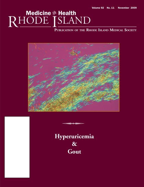
Cover: Monosodium urate crystals aspirated<br />
from the subcutaneous tissue of a patient with<br />
cutaneous manifestations of gout and viewed<br />
with a polarized light microscope, by Bernard<br />
Zimmermann, MD, and Peter Libbey, MD<br />
Medicine Health<br />
VOLUME 92 NO. 11 November 2009<br />
R HODE I SLAND<br />
PUBLICATION OF THE RHODE ISLAND MEDICAL SOCIETY<br />
COMMENTARIES<br />
350 Medication Trials: In an Imperfect World: <strong>Gout</strong> and Parkinson’s Disease<br />
Joseph H. Friedman, MD<br />
351 An Ailment for the Royally Nourished<br />
Stanley M. Aronson, MD<br />
CONTRIBUTIONS<br />
SPECIAL ISSUE: <strong>Hyperuricemia</strong> and <strong>Gout</strong><br />
Guest Editor: Bernard Zimmermann, MD<br />
352 Introduction: <strong>Hyperuricemia</strong> and <strong>Gout</strong><br />
Bernard Zimmermann, MD<br />
353 <strong>Medical</strong> Implications of <strong>Hyperuricemia</strong><br />
Larissa Sachs, MD, Kerri L. Batra, MD, and Bernard Zimmermann, MD<br />
356 Diagnosis and Management of Acute <strong>Gout</strong><br />
Nazli Conway, MD, and Stuart Schwartz, MD<br />
359 Approach To the Treatment of <strong>Hyperuricemia</strong><br />
Samuel H. Poon, MD, Harald A. Hall, MD, and Bernard Zimmermann, MD<br />
363 <strong>Gout</strong> In Women<br />
Jill McClory, MD, and Nuha Said, MD<br />
369 Treatment Failure <strong>Gout</strong><br />
Saman Ali, MD, and Edward V. Lally, MD<br />
COLUMNS<br />
372 IMAGES IN MEDICINE: Intranasal Mucosal Malignant Melanoma<br />
Robert Bagdasaryan, MD, and Mark Andreozzi, DO<br />
373 ADVANCES IN PHARMACOLOGY: Use of Angiotensin-Converting Enzyme Inhibitors/<br />
Angiotensin Receptor Blockers and Lipid-lowering Therapies Among <strong>Rhode</strong><br />
<strong>Island</strong> With Diabetes Enrolled In Medicare Part D Plans in 2006 and 2007<br />
Stephen Kogut, PhD, MBA, RPh, Aisling Caffrey, PhD, and Lynn Pezzullo, RPh<br />
377 GERIATRICS FOR THE PRACTICING PHYSICIAN: Asymptomatic Versus Symptomatic<br />
Urinary Tract Infections In Long-Term-Care-Facility Residents<br />
Porpon Rotjanapan, MD, and David Dosa, MD, MPH<br />
379 PHYSICIAN’S LEXICON: The Wanderings of the Vagus Nerve<br />
Stanley M. Aronson, MD<br />
380 HEALTH BY NUMBERS: Diabetes Prevention and Control: Progress Towards Healthy<br />
People 2010 Goals<br />
Annie Gjelsvik, PhD, Dona Goldman, RN, MPH, and Marilyn Moy, RN, MSW<br />
382 POINT OF VIEW: Prevention of Relapsing Mediocrity: How To Maintain Performance<br />
Improvement In Hospitals<br />
John S. Coldiron, MD, MPH<br />
385 November Heritage<br />
Medicine and Health/<strong>Rhode</strong> <strong>Island</strong> (USPS 464-820), a monthly publication, is owned and published by the <strong>Rhode</strong> <strong>Island</strong> <strong>Medical</strong> <strong>Society</strong>, 235<br />
Promenade St., Suite 500, Providence, RI 02908, Phone: (401) 331-3207. Single copies $5.00, individual subscriptions $50.00 per year, and $100<br />
per year for institutional subscriptions. Published articles represent opinions of the authors and do not necessarily reflect the official policy of the <strong>Rhode</strong> <strong>Island</strong><br />
<strong>Medical</strong> <strong>Society</strong>, unless clearly specified. Advertisements do not imply sponsorship or endorsement by the <strong>Rhode</strong> <strong>Island</strong> <strong>Medical</strong> <strong>Society</strong>. Periodicals postage<br />
paid at Providence, <strong>Rhode</strong> <strong>Island</strong>. ISSN 1086-5462. POSTMASTER: Send address changes to Medicine and Health/<strong>Rhode</strong> <strong>Island</strong>, 235 Promenade St.,<br />
Suite 500, Providence, RI 02908. Classified Information: RI <strong>Medical</strong> Journal Marketing Department, P.O. Box 91055, Johnston, RI 02919,<br />
phone: (401) 383-4711, fax: (401) 383-4477, e-mail: rimj@cox.net. Production/Layout Design: John Teehan, e-mail: jdteehan@sff.net.<br />
VOLUME 92 NO. 11 NOVEMBER 2009<br />
349
Commentaries<br />
350<br />
Medication Trials In an Imperfect World:<br />
<strong>Gout</strong> and Parkinson’s Disease<br />
This issue of Medicine & Health, <strong>Rhode</strong><br />
<strong>Island</strong> is the perfect venue to discuss a<br />
new, potentially useful treatment for slowing<br />
progression of Parkinson’s disease<br />
(PD), because it is based on increasing<br />
uric acid levels, hence putting people at<br />
risk for gout and possibly cardiovascular<br />
disease. It forces us to focus on several<br />
important clinical issues, including<br />
iatrogenesis, and how does one, both<br />
practically and ethically, recruit for a<br />
study that may cause significant complications<br />
in return for which the subject is<br />
rewarded with a clap on the back and an<br />
enhanced feeling of altruism but nothing<br />
else?<br />
The basic facts are straightforward.<br />
A few large studies of PD, performed for<br />
a variety of different reasons, have included,<br />
for safety purposes, uric acid levels.<br />
Analyses have shown, without conflict,<br />
that PD patients with higher uric<br />
acid levels progress more slowly than PD<br />
patients with lower levels. Studies comparing<br />
people with PD to those without<br />
PD have consistently shown that PD patients<br />
have lower uric acid levels. Uric<br />
acid is one of the body’s strongest antioxidants,<br />
and PD has been thought to<br />
result from excess oxidation within neurons.<br />
Thus elevated uric acid levels may<br />
be beneficial for people with PD. And<br />
although diet plays a role in uric acid levels,<br />
and diet may be altered in PD, experts<br />
believe that the differences in uric<br />
acid levels found in PD vs. controls cannot<br />
be explained by diet alone. These<br />
observations led to the idea of increasing<br />
uric acid serum levels to determine if this<br />
will slow PD progression. It must always<br />
be kept in mind that when one sees associations<br />
between potential cause and effect,<br />
that the connection, while robust,<br />
may not be what it seems. The connection<br />
between alcohol and lung cancer is<br />
strong, but is mediated via cigarettes, for<br />
example. Decreasing alcohol alone will<br />
not decrease lung cancer.<br />
MEDICINE & HEALTH/RHODE ISLAND<br />
<br />
The first step required to study a new<br />
drug to determine if it will slow disease<br />
progression is to perform a safety study, to<br />
prove to the Food and Drug Administration<br />
(FDA), which regulates testing of experimental<br />
medications, that this drug is<br />
safe to test in PD. Of course, this requires<br />
testing the drug in PD patients, but the<br />
structure of the study is quite different for<br />
a safety study than it is for an efficacy trial,<br />
for the goal of the preliminary study is to<br />
demonstrate that the drug is safe, not that<br />
it is therapeutically effective. The rationale<br />
for this is that safety studies require far fewer<br />
subjects, and generally are much shorter<br />
in duration so that fewer people are put<br />
at risk should the drug ultimately be<br />
shown to be unsafe.<br />
It is not easy to recruit for a study<br />
when the goal is safety, not efficacy. Most<br />
patients do not want a placebo; they want<br />
to feel better. Many of our efficacy studies<br />
promise the subjects that they will be<br />
able to take the experimental medicine<br />
once the placebo phase has ended on an<br />
open label basis. This is considered “fair”<br />
although it is an oddity of our testing system<br />
since the efficacy study is performed<br />
exactly to find out if the drug is, in fact,<br />
effective; so how, without knowing the<br />
results of the study, can we justify giving<br />
it to patients? In a safety study, we don’t<br />
even have any data, other than experimental,<br />
to indicate that the drug will be<br />
effective. In the case of a drug intended<br />
to slow disease progression, one doesn’t<br />
even feel better while taking the drug.<br />
Furthermore, in this particular study, we<br />
are even requiring a lumbar puncture to<br />
determine how well the study drug is altering<br />
urate levels within the cerebrospinal<br />
fluid, and, presumably, the brain.<br />
Would I enroll in this study if given<br />
the chance? I like to think so. I’m not sure<br />
I should be running a trial that I wouldn’t<br />
participate in as a subject. My own reservations<br />
have to do with the long-term<br />
safety of the drug. Will the FDA allow a<br />
drug that may induce gout and all its complications<br />
to be prescribed for people with<br />
PD? Will people with PD be willing to take<br />
a drug that may cause gout? At the initial<br />
meeting of site investigators, I asked the<br />
group who had made the observations<br />
about urate levels and PD, and who had<br />
designed this trial, if any of them had ever<br />
had a kidney stone. No one in the room<br />
had besides myself. Having had a few I<br />
can vouch for each one being memorably<br />
painful. Would I take a medication that<br />
might precipitate such a thing? Maybe, if<br />
it really slowed PD down a lot. What about<br />
cardiovascular disease? I’ve got that too.<br />
How keen am I to further increase my risk?<br />
Most patients don’t have a history of<br />
kidney stones, and if they did, they would<br />
not be urate stones, and the connection<br />
between cardiovascular disease and urate<br />
levels is no more solid than that between<br />
urate and PD.<br />
So the final decision rests, as it<br />
should, on clinical equipoise. Should we,<br />
or shouldn’t we? We simply have insufficient<br />
data to make a decision. When this<br />
happens, and the stakes are high, it is time<br />
for a study to answer the question. Unfortunately<br />
for those in the early studies,<br />
safety comes first. The later subjects have<br />
the benefit of helping determine if the<br />
drug actually works.<br />
If there were medals for altruism,<br />
those who volunteer for safety studies<br />
should get the gold ones. They truly reap<br />
no substantive gain.<br />
– JOSEPH H. FRIEDMAN, MD<br />
Disclosure of Financial<br />
Interests<br />
Joseph Friedman, MD, Consultant: Acadia<br />
Pharmacy, Ovation, Transoral; Grant Research<br />
Support: Cephalon, Teva, Novartis, Boehringer-<br />
Ingelheim, Sepracor, Glaxo; Speakers’ Bureau:<br />
Astra Zeneca, Teva, Novartis, Boehringer-<br />
Ingelheim, GlaxoAcadia, Sepracor, Glaxo Smith<br />
Kline, Neurogen, and EMD Serono.
An Ailment For the Royally Nourished<br />
<br />
A 59 yea- old British physician, in 1683, describes an attack of<br />
articular pain in his patient: “The victim goes to bed and<br />
sleeps in good health. About two o’clock in the morning he<br />
is awakened by a severe pain in the great toe, more rarely in<br />
the heel, ankle or instep. The pain is like that of a dislocation<br />
and yet the parts feel as though cold water were poured over<br />
them. Then follows chills and shiver and a little fever. The<br />
pain, at first moderate, becomes more intense. After a time<br />
this comes to full height accommodating itself to the bones<br />
and ligaments of the tarsus and metatarsus. Now it is a violent<br />
stretching and tearing of the ligaments – now it is a gnawing<br />
pain and now a pressure and tightening. So exquisite and<br />
lively meanwhile is the feeling of the part affected, that it<br />
cannot bear the weight of the bedclothes nor the jar of a<br />
person walking in the room.”<br />
Truly an authentic description of an age-old disorder called<br />
gout. The physician was Thomas Sydenham (1624 – 1689)<br />
and the patient was himself. When physicians describe diseases<br />
in their patients, the descriptions tend to be starkly objective<br />
and coldly impersonal with little of the patient’s inner reactions<br />
to the ailment. But when the physician is both victim and<br />
portrayer of the disease, the narration inevitably takes on dimension,<br />
greater accuracy and more benevolence.<br />
<strong>Gout</strong> was well known to the ancient Egyptians; and despite<br />
the passage of millennia, some of their mummified remains<br />
still contain toes with the characteristic anatomic changes<br />
of gout. The Classical-era Greeks were also familiar with this<br />
unique form of arthritis, calling the disorder podagra.<br />
This much was known about the predilection, pathology<br />
and prognosis of gout by the late 17 th Century, the dawn of<br />
scientific inquiry: It was a disease selectively but not exclusively<br />
of affluent society with a typical onset in late adult life. Men, to<br />
the exclusion of eunuchs, were its prime victims. Women became<br />
vulnerable to gout, but only beyond the menopause. <strong>Gout</strong><br />
seemed to choose its victims from amongst those who ate intemperately,<br />
especially a diet rich in dark meats; those who drank<br />
much, especially red wines such as port; and those who were<br />
obese and exercised little beyond their eccentricities. The British<br />
widely believed that gout was an inevitable retribution for<br />
the excesses of food, wine and debauchery.<br />
There is nothing timid or subtle about gout. It announces<br />
itself stridently; and if the walls are thin enough, even the<br />
neighbors will know when an attack of gout resumes. A powdery<br />
substance infiltrates the tissues near and within the affected<br />
joints. Occasionally this infiltrate accumulates to form a<br />
painful nodule, a calcareous concretion called a tophus. The<br />
Dutch scientist, Anton Leeuwenhoek (1632 - 1723) used his<br />
newly devised microscope to examine the gouty tophi and observed<br />
its white sediment to be composed of microscopic crystals.<br />
About two centuries latter the British physician, Archibald<br />
Garrod (1857 – 1936) demonstrated that gout represented a<br />
disorder of purine metabolism, sometimes hereditary, and was<br />
characterized by an excessive concentration of monosodium<br />
urates in the bloodstream (hyperuricemia) associated with an<br />
increased acidity of the circulating blood. Victims of gout often<br />
developed uric acid kidney stones, yet another source of<br />
exquisite pain.<br />
Many victims of gout in the wine-consuming population<br />
of the 18 th Century concurrently suffered from lead poisoning<br />
(saturnine gout) since many wines, then, were intentionally<br />
adulterated with sweetening agents such as lead acetate. The<br />
incidence of gout, as an inheritable disorder, is quite high in<br />
the indigenous populations of the Pacific region, particularly<br />
the Maori of New Zealand.<br />
<strong>Gout</strong> can be reproduced in experimental animals; but it is<br />
also encountered as a hereditary trait in some species of birds<br />
and reptiles. And paleontologists are quick to point out that<br />
some fossil remains of Tyrannosaurus rex, the great lizard of the<br />
age of dinosaurs, contained the bony changes seen in gout.<br />
<strong>Gout</strong> does not hide. Patients with gout tell the world of<br />
their affliction in their correspondence, diaries, autobiographies<br />
and even complaints to strangers on the streets. Amongst the<br />
founding fathers of this nation, the following had declared<br />
themselves to be the targets of gout: Benjamin Franklin, Thomas<br />
Jefferson, Alexander Hamilton, John Hancock, George<br />
Mason and John Jay. Historians have speculated that it may<br />
have required the anguish of gout to stir their strivings for independence.<br />
But when the biographies of the English leadership,<br />
on the other side of the Atlantic, are then examined, it<br />
appears that gout showed no political favoritism. <strong>Gout</strong> surfaced<br />
in William Pitt, Benjamin Disraeli, King George IV and<br />
Queen Anne. Other historic sufferers included John Calvin,<br />
John Milton, Isaac Newton, Samuel Johnson and even that<br />
champion of the proletariat, Karl Marx.<br />
Sydenham concluded: “<strong>Gout</strong>, unlike any other disease,<br />
kills more rich men than poor, more wise men than simple.<br />
Great kings, emperors, generals, admirals have all died of gout.”<br />
He might have added: “And countless commoners.”<br />
– STANLEY M. ARONSON, MD<br />
Disclosure of Financial Interests<br />
Stanley M. Aronson, MD, has no financial interests to<br />
disclose.<br />
CORRESPONDENCE<br />
e-mail: SMAMD@cox.net<br />
VOLUME 92 NO. 11 NOVEMBER 2009<br />
351
Introduction: <strong>Hyperuricemia</strong> and <strong>Gout</strong><br />
Bernard Zimmermann, MD<br />
<br />
The pathophysiology of gout is well<br />
understood, and effective treatments for<br />
acute gout and hyperuricemia leading to<br />
gout are widely available. Nonetheless,<br />
many patients suffer from severe tophaceous<br />
gouty arthritis, adverse effects of<br />
medications for gout, and inadequate<br />
treatment of hyperuricemia. The incidence<br />
of gout is rising in both men and<br />
women. The diagnosis and treatment of<br />
gout are challenging in patients with arthritis,<br />
renal and hepatic diseases. Acute<br />
gout is usually not difficult to treat, but<br />
evidence to guide therapeutic decisions<br />
for gout patients with complex medical<br />
problems is woefully lacking. Fortunately,<br />
new medications for treatment of gout<br />
and hyperuricemia are becoming available,<br />
and exciting new research is increasing<br />
our understanding of the role of hyperuricemia<br />
in the pathogenesis of hypertension<br />
and cardiovascular disease.<br />
In this issue of Medicine and Health/<br />
<strong>Rhode</strong> <strong>Island</strong> we discuss the epidemiology<br />
of gout in women, and review new<br />
data regarding the importance of hyperuricemia<br />
as a marker and perhaps a causative<br />
agent involved in the pathogenesis<br />
of the metabolic syndrome. We review<br />
the risks and benefits of various treatment<br />
options for acute gout and discuss the<br />
potential utility of febuxostat, the first<br />
new drug approved for treatment of hyperuricemia<br />
in 40 years. In addition, we<br />
describe ongoing studies of rasburicase<br />
and pegloticase which may offer dramatic<br />
improvement for patients with severe<br />
tophaceous gout.<br />
We hope to share the excitement in<br />
the rheumatology community that has<br />
been generated by the potential of new<br />
therapies and advances in our understanding<br />
of gout and hyperuricemia, and to<br />
improve the care of all patients with gout.<br />
Bernard Zimmermann, MD, is Director<br />
of the Division of Rheumatology at<br />
Roger Williams <strong>Medical</strong> Center, and Associate<br />
Professor of Medicine at the Boston<br />
University School of Medicine.<br />
Disclosure of Financial Interests<br />
The author has no financial interests<br />
to disclose.<br />
CORRESPONDENCE<br />
Bernard Zimmermann, MD<br />
Roger Williams <strong>Medical</strong> Center<br />
Division of Rheumatology<br />
825 Chalkstone Ave<br />
Providence, RI 02908<br />
e-mail: bzimmermann@rwmc.org<br />
352<br />
MEDICINE & HEALTH/RHODE ISLAND
<strong>Medical</strong> Implications of <strong>Hyperuricemia</strong><br />
Larissa Sachs, MD, Kerri L. Batra, MD, and Bernard Zimmermann, MD<br />
<br />
WHAT IS HYPERURICEMIA?<br />
Uric acid is an oxidation product of<br />
purine metabolism, which in primates (including<br />
humans) is largely eliminated by<br />
the kidney and the gut. Most non-primate<br />
mammals express uricase, an enzyme that<br />
converts serum uric acid into allantoin,<br />
which is easily excreted by the kidneys. Nonprimate<br />
mammals thus usually have low<br />
serum uric acid (SUA) levels (below 2 mg/<br />
dl), while primates have the potential to<br />
develop hyperuricemia, because they lack<br />
uricase. In humans, renal under excretion<br />
of uric acid is the cause of 90% of hyperuricemia,<br />
while 10% is due to overproduction<br />
of uric acid. Uric acid is more toxic to<br />
tissues than other purine metabolites such<br />
as xanthine, hypoxanthine and allantoin.<br />
<strong>Hyperuricemia</strong> is defined as a serum<br />
urate level greater than 6.0 mg/dl in<br />
women, and 7.0 mg/dl in men. Above<br />
this concentration, urate is supersaturated<br />
in body fluids, and is prone to crystallization<br />
and subsequent tissue deposition. The<br />
rising prevalence of hyperuricemia over<br />
the last several decades can be attributed<br />
to several factors. Westernization of diets<br />
and widespread use of high-fructose syrup<br />
may play a role in increasing SUA levels. 1<br />
Other factors that might be involved include<br />
increased lifespan and more common<br />
use of certain medications, including<br />
diuretics and cyclosporine. The prevalence<br />
of asymptomatic hyperuricemia in<br />
US is estimated to be 5-8 % among adult<br />
Caucasian men and it is even more common<br />
in some ethnic groups, such as<br />
Philippinos and Polynesians.<br />
While a few studies have noted a potential<br />
beneficial antioxidative effect of uric<br />
acid and even suggested a neuroprotective<br />
role, most studies link hyperuricemia with<br />
such co-morbidities as hypertension, renal<br />
disease, metabolic syndrome and cardiovascular<br />
disease (CVD).<br />
gout. The duration of each stage varies significantly<br />
among individuals. (Table 1)<br />
Fewer than one-third of individuals<br />
with asymptomatic hyperuricemia will develop<br />
gouty arthritis. The risk of developing<br />
gout increases with age and the degree<br />
of hyperuricemia. In the Normative<br />
Aging Study, the 5-year cumulative incidence<br />
of gout among those with a uric acid<br />
level between 7.0 and 8.0 mg/dl was 3%,<br />
compared to 22% in those with a uric acid<br />
level of 9.0 mg/dl. 2 (Figure 1)<br />
Acute gouty arthritis occurs when uric<br />
acid crystals interact with synovial phagocytes,<br />
which in turn activate neutrophils<br />
and initiate an inflammatory cascade. Urate<br />
crystals that serve as a trigger for an acute<br />
attack may derive from preformed synovial<br />
deposits or precipitate in the joint de novo.<br />
Clinically, acute gout presents with rapid<br />
onset of a painful, erythematous and swollen<br />
joint that may be accompanied by fever.<br />
Inflammation of the first metatarsophalangeal<br />
joint (also known as podagra), is the<br />
most characteristic presentation but other<br />
joints are often involved.<br />
Intercritical gout is the name given<br />
to the asymptomatic interval between<br />
acute attacks. In early gout, intercritical<br />
periods may last for years, but with progression<br />
of the disease the time between<br />
attacks tends to lessen.<br />
Chronic tophaceous gout is characterized<br />
by the development of tophi in and<br />
around the joints, which can cause destructive<br />
arthritis. (Figure 2) Tophi are commonly<br />
found in the soft tissues, including<br />
tendons, pinnae and subcutaneous fat.<br />
Tophi have been reported in such unusual<br />
locations as heart valves, spinal cord,<br />
sclerae, breast and even Cushing’s striae.<br />
Table 1. Stages of <strong>Hyperuricemia</strong> and <strong>Gout</strong>.<br />
Stage Duration Clinical Features<br />
Asymptomatic >10-15 years Asymptomatic<br />
hyperuricemia<br />
Acute gout 1-2 weeks Sudden onset of acute<br />
mono- or oligoarthritis<br />
(e.g., podagra)<br />
Intercritical gout From weeks Asymptomatic intervals between<br />
to years<br />
acute attacks<br />
Chronic tophaceous 10 or more years Development of tophi in and<br />
gout after the first episode around the joints and soft<br />
of acute gout tissues<br />
HYPERURICEMIA AND GOUT<br />
The most well known medical manifestation<br />
of hyperuricemia is gout. <strong>Gout</strong> is<br />
caused by deposition of uric acid crystals in<br />
and around the joints and has 4 stages: asymptomatic<br />
hyperuricemia, acute gout,<br />
intercritical gout, and chronic tophaceous<br />
Figure 1. Cumulative Incidence of <strong>Gout</strong>y Arthritis by Prior Serum Urate Levels.<br />
The numbers refer to the number of examination intervals for each group. Reprinted from<br />
American Journal of Medicine, 1987 March 82(3); Campion EW, Glynn RJ, DeLabry LO.<br />
Asymptomatic hyperuricemia. Risks and consequences in the Normative Aging Study.<br />
Pages: 421-6 Copyright 2009, with permission from Elsevier.<br />
VOLUME 92 NO. 11 NOVEMBER 2009<br />
353
354<br />
Figure 2. Severe tophaceous gout in the hands. Photograph by Harald A. Hall, MD<br />
MEDICINE & HEALTH/RHODE ISLAND<br />
HYPERURICEMIA AND HYPERTENSION<br />
Numerous studies have demonstrated<br />
an association between hyperuricemia and<br />
hypertension, and recent evidence even<br />
suggests there may be a causal relationship.<br />
This evidence was first noted in animal<br />
studies: in Sprague-Dawley rats, hyperuricemia<br />
can be induced by feeding<br />
with an uricase inhibitor. Mildly increased<br />
SUA is associated with development of<br />
hypertension in the rats within 3 weeks.<br />
The development of hypertension in the<br />
rats can be prevented by co-treatment<br />
with a uric acid lowering therapy such as<br />
allopurinol or a uricosuric agent. 3<br />
Upon pathologic evaluation, the<br />
hyperuricemic rats have lower levels of<br />
nitric oxide in the renal endothelium, suggesting<br />
increased renal vasoconstriction<br />
and activation of the renin-angiotensin<br />
system, leading to ischemic tissue damage.<br />
Uric acid crystal deposition is not seen in<br />
the kidneys of the hypertensive rats.<br />
In humans, a prospective study involving<br />
more then two thousand patients<br />
demonstrated that an increased SUA<br />
level predicts development of future hypertension<br />
independent of age, alcohol<br />
use or renal function. 4<br />
Further evidence of the association<br />
between hyperuricemia and hypertension<br />
comes from pediatric literature: 90% of<br />
adolescents with newly diagnosed hypertension<br />
are found to have hyperuricemia. 5<br />
In a double-blind placebo-controlled<br />
study involving 30 adolescents with hyperuricemia<br />
and hypertension, allopurinol<br />
therapy normalized blood pressure in 86%<br />
of patients compared to 3% of patients in<br />
a placebo group. 6 This suggests not only<br />
an early role of uric acid in the pathogenesis<br />
of primary hypertension, but also the<br />
possibility that early treatment of hyperuricemia<br />
may prevent the development of<br />
hypertension. Clearly, more clinical trials<br />
are needed to explore these issues.<br />
HYPERURICEMIA AND RENAL DISEASE<br />
Before uric acid lowering therapy was<br />
available, hyperuricemia was thought to be<br />
a cause of chronic kidney disease because<br />
of their frequent coexistence. However, in<br />
Epidemiologic<br />
evidence shows that<br />
there may be a<br />
connection between<br />
the rise of the use of<br />
high-fructose corn<br />
syrup, the increasing<br />
prevalence of<br />
metabolic syndrome,<br />
and the rapid<br />
increase in<br />
worldwide<br />
hyperuricemia<br />
the late 1970s the results of several epidemiologic<br />
studies made a direct causal relationship<br />
between elevated uric acid and<br />
renal impairment questionable. 7 It is clear<br />
that chronic kidney disease is associated<br />
with hyperuricemia. However, it is not clear<br />
whether renal impairment is due to a direct<br />
nephrotoxic effect of uric acid, or due<br />
to the conditions that are caused by hyperuricemia<br />
(e.g., hypertension).<br />
Most of the recent evidence for a direct<br />
pathogenic effect of hyperuricemia on<br />
the kidneys comes from animal studies, as<br />
noted above. In humans, epidemiologic<br />
studies demonstrate that hyperuricemia is<br />
associated with decline in kidney function. 8<br />
It has also been shown that allopurinol<br />
might slow this decline. In one prospective<br />
study patients with asymptomatic hyperuricemia<br />
treated with 300 mg of allopurinol<br />
showed significant improvement in glomerular<br />
filtration rate (GFR) after 3 months<br />
of the therapy. 9 Siu et al. reported that<br />
among the patients with chronic kidney<br />
disease treated with allopurinol, 16% progressed<br />
to end stage renal disease, compared<br />
to 46% in the control group. 10<br />
URIC ACID AND METABOLIC<br />
SYNDROME<br />
Metabolic syndrome is defined as a<br />
group of modifiable risk factors that are<br />
associated with an increased risk for CVD,<br />
type 2 diabetes mellitus, and mortality<br />
from CVD. The factors that define metabolic<br />
syndrome include systolic hypertension,<br />
hypertriglyceridemia, central obesity,<br />
and impaired glucose tolerance. <strong>Hyperuricemia</strong><br />
has been shown to be significantly<br />
more common in patients with metabolic<br />
syndrome than the normal population. 11<br />
In a recent population-based study all<br />
metabolic syndrome components correlated<br />
with elevated SUA level, with waist<br />
circumference being the strongest. 11<br />
It has been generally thought that hyperuricemia<br />
was a result of<br />
hyperinsulinemia in those with metabolic<br />
syndrome, as insulin decreases the renal excretion<br />
of uric acid. However, animal studies<br />
showed that the opposite scenario might<br />
be the case. Nakagawa et al. showed in fructose-fed<br />
rats with hyperuricemia, that treatment<br />
with allopurinol or benzbromarone<br />
(a uricosuric agent) prevented development<br />
of features of metabolic syndrome including<br />
hyperinsulinemia, systolic hypertension,<br />
hypertriglyceridemia, and weight gain. 1
It has been suggested that uric acid<br />
may cause metabolic syndrome by promoting<br />
a state of insulin resistance. It is<br />
well known that insulin stimulates glucose<br />
intake in skeletal muscle via increased<br />
blood flow to these tissues through a nitric<br />
oxide-dependent pathway. Uric acid<br />
decreases levels of nitric oxide and arterial<br />
dilatation and blocks the action of<br />
insulin, resulting in increased insulin resistance<br />
and hyperinsulinemia.<br />
One of the most interesting recent<br />
findings in hyperuricemia and gout concerns<br />
high-fructose corn syrup. Epidemiologic<br />
evidence shows that there may be a<br />
connection between the rise of the use of<br />
high-fructose corn syrup, the increasing<br />
prevalence of metabolic syndrome, and the<br />
rapid increase in worldwide hyperuricemia.<br />
Unlike glucose or other sugars, fructose rapidly<br />
increases uric acid production in humans<br />
and its consumption is associated with<br />
an increased incidence of gout. Animal<br />
studies have shown that when glucose and<br />
fructose-fed rats are compared, only fructose-fed<br />
animals developed metabolic syndrome<br />
as well as hyperuricemia. 1 Several<br />
large population-based studies have confirmed<br />
the correlation between increased<br />
fructose intake and hyperuricemia and<br />
metabolic syndrome in humans. 12<br />
HYPERURICEMIA AND<br />
CARDIOVASCULAR DISEASE (CVD)<br />
<strong>Hyperuricemia</strong> is frequently associated<br />
with CVD. However, conflicting data have<br />
caused controversy whether hyperuricemia<br />
is an independent risk factor for CVD or<br />
simply an indicator for co-morbidities that<br />
are frequently seen in patients with CVD<br />
(e.g., hypertension, insulin resistance).<br />
Analysis of data from the Framingham<br />
Heart study showed no association between<br />
hyperuricemia and cardiovascular outcomes.<br />
13 On the other hand, a study based<br />
on data from the first National Health and<br />
Nutrition Examination Survey<br />
(NHANES-1) did find an independent<br />
relationship between hyperuricemia and<br />
CVD, but only in women. 14 A meta-analysis<br />
of 21 prospective cohort studies by Baker<br />
et al. suggested a moderate and independent<br />
association between SUA and CVD<br />
in patients at high risk for CVD. 15 As for<br />
individuals with low risk for CVD, that correlation<br />
was not found to be consistent (only<br />
4 out of 6 studies demonstrated an independent<br />
link). It should be mentioned,<br />
however, that the studies of healthy individuals<br />
in which correlation between hyperuricemia<br />
and cardiovascular mortality<br />
was not found tended to have a low number<br />
of events per- person-years.<br />
<strong>Hyperuricemia</strong> has also been associated<br />
with peripheral vascular disease and<br />
all cause mortality. Baker et al. found that<br />
elevated level of SUA, independent of other<br />
co-morbidities, predicted worse outcomes<br />
after an acute stroke over 2 years. The association<br />
was so strong that it was suggested<br />
that in-house SUA level should be considered<br />
as a useful prognostic indicator in patients<br />
hospitalized for acute stroke.<br />
New data from the Chinese Cohort<br />
Study involving 93,393 participants (about<br />
half male, half female) demonstrated that<br />
hyperuricemia was an independent risk factor<br />
of mortality from all causes, total CVD<br />
and ischemic stroke. 16 This correlation was<br />
more significant in women than men. This<br />
study also found a linear relationship between<br />
SUA and all-cause and CVD mortality. In<br />
conclusion, hyperuricemia is clearly an important<br />
risk factor for not only for developing<br />
gout but also for other co-morbidities<br />
which are associated with cardiovascular and<br />
all-cause mortality. More clinical trials are<br />
needed to determine whether the treatment<br />
of asymptomatic hyperuricemia may reduce<br />
the incidence of severity of hypertension, cardiovascular<br />
and renal disease and related comorbid<br />
conditions.<br />
REFERENCES<br />
1. Nakagawa T, Hu H, et al. A causal role for uric<br />
acid in fructose-induced metabolic syndrome. Am<br />
J Physiol Renal Physiol 2006; 290:F625-31. Epub<br />
2005 Oct 18.<br />
2. Campion EW, Glynn RJ, DeLabry LO. Am J Med<br />
1987; 82:421-6.<br />
3. Mazzali M, Hughes J, et al. Elevated uric acid<br />
increases blood pressure in the rat by a novel crystal-independent<br />
mechanism. Hypertension<br />
2001;38:1101-6.<br />
4. Perlstein TS, Gumieniak O, et al. Uric acid and<br />
the development of hypertension. Hypertension<br />
2006; 48:1031-6. Epub 2006 Oct 23.<br />
5. Feig DI, Johnson RJ. <strong>Hyperuricemia</strong> in childhood<br />
primary hypertension. Hypertension<br />
2003;42:247-52. Epub 2003 Aug 4.<br />
6. Feig DI, Soletsky B, Johnson RJ. Effect of allopurinol<br />
on blood pressure of adolescents with<br />
newly diagnosed essential hypertension. JAMA<br />
2008;300:924-32.<br />
7. McLean L, Becker MA. The pathogenesis of gout.<br />
In Hochberg MC, editors. Rheumatology. Elsevier<br />
Limited; 2008. P.1824-5.<br />
8. Feig DI, Kang DH, Johnson RJ. Uric acid and<br />
cardiovascular risk. NEJM 2008; 359:1811-21.<br />
9. Kanbay M, Ozkara A, et al. Effect of treatment of<br />
hyperuricemia with allopurinol on blood pressure,<br />
creatinine clearance, and proteinuria in patients<br />
with normal renal functions. Int Urol<br />
Nephrol 2007;39:1227-33. Epub 2007 Aug 15.<br />
10. Siu YP, Leung KT, et al. Use of allopurinol in<br />
slowing the progression of renal disease through<br />
its ability to lower serum uric acid level. Am J<br />
Kidney Dis 2006;47:51-9.<br />
11. Puig JG, Martínez MA, et al. Serum urate, metabolic<br />
syndrome, and cardiovascular risk factors.<br />
Nucleosides Nucleotides Nucleic Acids 2008;<br />
27:620-3.<br />
12. Choi JW, Ford ES, et al. Sugar-sweetened soft<br />
drinks, diet soft drinks, and serum uric acid level.<br />
Arthritis Rheum 2008; 59:109-16.<br />
13. Abbott RD, Brand FN, et al. <strong>Gout</strong> and coronary<br />
heart disease. J Clin Epidemiol 1988;41:237-42<br />
14. Fang J, Alderman MH. Serum uric acid and cardiovascular<br />
mortality the NHANES I epidemiologic<br />
follow-up study, 1971-1992. National<br />
Health and Nutrition Examination Survey. JAMA<br />
2000; 283:2404-10.<br />
15. Baker JF, Krishnan E, et al.. Serum uric acid and<br />
cardiovascular disease. Am J Med 2005;118:816-<br />
26.<br />
16. Chen JH, Chuang SY, et al. Serum uric acid level<br />
as an independent risk factor for all-cause, cardiovascular,<br />
and ischemic stroke mortality. Arthritis<br />
Rheum 2009; 61:225-32.<br />
Larissa Sachs, MD, formerly a Fellow<br />
in Rheumatology at Roger Williams <strong>Medical</strong><br />
Center, is a rheumatologist with The<br />
Pinnacle Health Hospital in Harrisburg,<br />
Pennsylvania.<br />
Kerri Batra, MD, is a rheumatologist<br />
at <strong>Rhode</strong> <strong>Island</strong> Hospital, and Clinical<br />
Assistant Professor at the Warren Alpert<br />
School of Medicine of Brown University<br />
Bernard Zimmermann, MD, is Director<br />
of the Division of Rheumatology at<br />
Roger Williams <strong>Medical</strong> Center, and Associate<br />
Professor of Medicine at the Boston<br />
University School of Medicine.<br />
DISCLOSURE OF FINANCIAL<br />
INTERESTS<br />
The authors have no financial interests<br />
to disclose.<br />
CORRESPONDENCE<br />
Bernard Zimmermann, MD<br />
Roger Williams <strong>Medical</strong> Center<br />
Division of Rheumatology<br />
825 Chalkstone Ave<br />
Providence, RI 02908<br />
e-mail: bzimmermann@rwmc.org<br />
VOLUME 92 NO. 11 NOVEMBER 2009<br />
355
356<br />
Diagnosis and Management of Acute <strong>Gout</strong><br />
MEDICINE & HEALTH/RHODE ISLAND<br />
<br />
Nazli Conway, MD and Stuart Schwartz, MD<br />
Risk factors for the development of acute<br />
gout include hyperuricemia, increased age,<br />
and a family history of gout. Consumption<br />
of alcohol and purine-rich foods and medications<br />
such as thiazide diuretics, loop diuretics,<br />
and cyclosporine contribute to the<br />
development of hyperuricemia. Hypertension,<br />
diabetes, obesity, and chronic renal<br />
failure are often associated with gout.<br />
When gout is suspected, aspiration of the<br />
affected joint should be performed to confirm<br />
the presence of intracellular negatively<br />
birefringent needle-shaped crystals by compensated<br />
polarized light microscopy. However,<br />
in some situations this is not practical.<br />
The most recent European League Against<br />
Rheumatism report proposes ten key recommendations<br />
for the diagnosis of gout: 1<br />
(1) The rapid onset of severe pain and swelling<br />
(especially with overlying erythema)<br />
within 6 to 12 hours is highly suggestive of<br />
crystal inflammation. However, this is not<br />
specific for gout. (2) Classic podagra, involvement<br />
of the first metatarsophalangeal<br />
joint, has a high sensitivity and specificity,<br />
but is not definitive without crystal identification.<br />
(3) Definitive diagnosis is made by<br />
demonstration of monosodium urate<br />
(MSU) crystals in synovial fluid or tophus.<br />
(4) Synovial fluid from undiagnosed inflammatory<br />
arthritis should be routinely examined<br />
for the presence of monosodium urate<br />
crystals. (5) Identification of MSU crystals<br />
may be possible in intercritical periods (between<br />
attacks). (6) If septic arthritis is suspected,<br />
gram stain and culture of synovial<br />
fluid should be performed even if MSU<br />
crystals are present. (7) Serum uric acid levels<br />
alone cannot confirm or exclude gout,<br />
as patients with hyperuricemia may not<br />
develop gout, and uric acid may be normal<br />
during acute attacks. (8) In selected patients<br />
(those with a personal or family history of<br />
onset of gout before age 25, or patients with<br />
renal calculi), determination of renal uric<br />
acid excretion is useful for evaluation and<br />
management. (9) Radiographs are not<br />
helpful in confirming the diagnosis of early<br />
or acute gout, although they may be useful<br />
for differential diagnosis and may show<br />
characteristic changes in chronic gout. (10)<br />
Risk factors for gout and associated<br />
comorbidities should be assessed, including<br />
features of the metabolic syndrome<br />
(obesity, hyperglycemia, hyperlipidemia,<br />
and hypertension).<br />
The differential diagnosis for symptoms<br />
suggesting an acute gout attack includes<br />
acute pseudogout, septic arthritis,<br />
inflammatory arthritis, cellulitis, and<br />
trauma. Because these diagnoses may have<br />
similar presentations, it is important to establish<br />
a correct diagnosis, as the treatment<br />
implications are significant. For example,<br />
pseudogout, caused by calcium pyrophosphate<br />
crystals, should not be treated with<br />
urate-lowering therapy; and septic arthritis<br />
demands immediate attention with antibiotics<br />
and joint drainage.<br />
A definitive diagnosis<br />
of acute gout is<br />
made by detection of<br />
monosodium urate<br />
crystals in the<br />
synovial fluid of an<br />
inflamed joint.<br />
MEDICAL TREATMENT FOR ACUTE<br />
GOUT<br />
Colchicine<br />
Colchicine has been used both in the<br />
acute setting and in the management of<br />
chronic gout. It exerts anti-inflammatory<br />
effects through several mechanisms. Colchicine<br />
binds to tubulin causing anti-proliferative<br />
effects by arresting cell growth. It also<br />
inhibits phagocytic and cytokine secretory<br />
functions of leukocytes. Chemotactic responses<br />
of neutrophils to leukotriene B4, IL-<br />
8, and other cytokines are disrupted. At<br />
high concentrations, colchicine has recently<br />
been shown to inhibit urate crystal-induced<br />
activation of NALP3 inflammasome. 2 This<br />
protein complex cleaves caspase-1 which<br />
then activates interleukin 1-β, a pro-inflammatory<br />
cytokine felt to be central in the<br />
pathogenesis of gout.<br />
The optimal dosing of colchicine for<br />
treatment of acute gout remains controversial.<br />
In the only published randomized, placebo-controlled<br />
study of colchicine therapy,<br />
patients in the treatment group received oral<br />
colchicine 1 mg then 0.5 mg every 2 hours<br />
until a complete response or until side effects<br />
developed. 3 Of the 22 patients in the<br />
treatment group, 73% achieved greater than<br />
a 50% reduction in pain within 48 hours.<br />
However, gastrointestinal toxicity (diarrhea<br />
and/or vomiting) developed in 55% of the<br />
patients before the reduction in pain was<br />
achieved, and all patients experienced these<br />
adverse side effects at some point during the<br />
study. A randomized, controlled multicenter<br />
trial, presented in abstract form, compared<br />
high-dose colchicine (1.2 mg, then 0.6 mg<br />
hourly for six hours) versus low-dose colchicine<br />
(1.2 mg, then 0.6 mg in one hour) and<br />
showed equivalent efficacy and less gastrointestinal<br />
toxicity with the lower dose regimen.<br />
4 Recently, the European League Against<br />
Rheumatism issued consensus guidelines in<br />
favor of lower doses of colchicine to avoid<br />
unacceptable side effects while maintaining<br />
efficacy. 5 They recommend a maximum of<br />
three tablets of 0.5 mg in the first 24 hours.<br />
Overdose or chronic administration of<br />
colchicine may result in side effects including<br />
granulocytopenia, aplastic anemia, and<br />
reversible myopathic and neuropathic toxicity.<br />
6 Patients with abnormal renal function<br />
are at increased risk for developing neuromuscular<br />
toxicity by accumulation of<br />
toxic plasma levels of colchicine. This condition<br />
typically presents with proximal<br />
muscle weakness and elevated CPK, which<br />
can mimic polymyositis. Symptoms of<br />
colchicine myopathy resolve within three<br />
to four weeks after discontinuing the medication.<br />
Colchicine is contraindicated in<br />
patients on hemodialysis and should be used<br />
Table 1. Half-Life of Various NSAIDs. 12<br />
Short Half-Life (6 hours)<br />
Ibuprofen (2 hours)<br />
Nabumetone (24 hours)<br />
Diclofenac (1-2 hours)<br />
Naproxen (14 hours)<br />
Indomethacin (2.5 hours)<br />
Etodolac (7 hours)<br />
Ketoprofen (2 hours)<br />
Sulindac (13 hours)
Table 2. Recommended Corticosteroid Dose Based on Joint Size. 17<br />
Joint<br />
Intra-articular corticosteroid dose<br />
Knee<br />
40 mg triamcinolone<br />
Ankle<br />
30 mg triamcinolone<br />
Wrist<br />
30 mg triamcinolone<br />
Elbow<br />
30 mg triamcinolone<br />
Metacarpophalangeal joints<br />
10 mg triamcinolone<br />
Proximal interphalangeal joints 10 mg triamcinolone<br />
with caution in patients with renal insufficiency<br />
or hepatobiliary dysfunction. 7 Drug<br />
interactions have been reported with<br />
cyclosporine, statins, and macrolides.<br />
The FDA recently withdrew its approval<br />
for the use of intravenous colchicine. 2<br />
The most common side effect associated with<br />
IV colchicine is local extravasation of the drug<br />
which can lead to painful inflammation and<br />
necrosis of surrounding tissue. 8,9 Increased<br />
risk of systemic side effects such as renal and<br />
hepatic toxicity, bone marrow suppression,<br />
and congestive heart failure have also been<br />
reported with IV colchicine. 8 Fifty reports<br />
of adverse events, including 23 deaths, were<br />
submitted to the FDA through June 2007. 10<br />
Three of these deaths were attributed to an<br />
IV colchicine compounding error.<br />
Non-steroidal Anti-inflammatory<br />
Drugs (NSAIDs)<br />
NSAIDs are commonly used for treatment<br />
of acute gout. The anti-inflammatory<br />
effects of these medications occur<br />
mainly through inhibition of the cyclo-oxygenase<br />
enzyme which prevents the transformation<br />
of arachidonic acid to prostaglandins,<br />
particularly prostaglandin E2.<br />
Other NSAID mechanisms include inhibition<br />
of lipooxygenase with reduced generation<br />
of leukotriene B4 and inhibition of<br />
neutrophil activation and aggregation. 11<br />
Early administration and appropriate<br />
dosage of NSAIDs are more important in<br />
achieving a therapeutic response than the<br />
particular NSAID used. However,<br />
NSAIDs with short half-lives (less than six<br />
hours) reach steady-state levels more<br />
quickly than NSAIDs with long half-lives<br />
(more than six hours) and may be preferred<br />
in managing acute gout. (Table 1)<br />
Regardless of which NSAID is administered,<br />
large initial doses are recommended<br />
(indomethacin 150-200 mg/day, naproxen<br />
1000 mg/day, diclofenac 150 mg/day).<br />
Duration of treatment is generally 4-8 days<br />
which minimizes potential adverse side effects.<br />
Patients should be treated until symptoms<br />
resolve and then gradually tapered.<br />
Traditional NSAIDs inhibit both COX-<br />
1 and COX-2, with their main anti-inflammatory<br />
effects being via inhibition of COX-<br />
2 and most adverse effects by inhibition of<br />
COX-1. 13 While both selective and nonselective<br />
NSAIDs inhibit COX-2 equally, the<br />
selective COX-2 inhibitors spare inhibition<br />
of COX-1 therefore reducing gastrointestinal<br />
toxicity. Etoricoxib, a selective COX-2<br />
inhibitor, was shown to be as efficacious as<br />
indomethacin for treatment of acute gout<br />
with fewer gastrointestinal side effects. 14 This<br />
medication is not FDA-approved for use in<br />
the United States because of potential cardiovascular<br />
side effects. Celecoxib is the only<br />
approved selective COX-2 inhibitor available<br />
in the US; however, no trials of its use in<br />
gout have been published.<br />
The FDA recently<br />
withdrew its<br />
approval for the use<br />
of intravenous<br />
colchicine<br />
Relative contraindications to the use of<br />
NSAIDs include severe heart failure, peptic<br />
ulcer disease, gastrointestinal hemorrhage,<br />
aspirin-induced or NSAID-induced asthma<br />
and renal impairment. There is an increased<br />
risk of bleeding when NSAIDs are used concomitantly<br />
with warfarin.<br />
CORTICOSTEROIDS<br />
Systemic corticosteroids<br />
Systemic corticosteroids may be used<br />
for treating acute gout when NSAIDs and<br />
colchicine are contraindicated. Corticosteroids<br />
exert their anti-inflammatory effect<br />
by inhibition of pro-inflammatory cytokines<br />
(IL-1, IL-6, IL-8, and TNF-α) and<br />
upregulation of genes for lipocortin and<br />
vasocortin, which have anti-inflammatory<br />
effects by inhibiting phospholipase A2.<br />
A Cochrane review evaluated the efficacy<br />
and safety of systemic corticosteroids for<br />
the treatment of acute gout. 15 Only three<br />
studies involving a total of 74 patients were<br />
found that met the search criteria. In the<br />
first study, intramuscular (IM) injections of<br />
triamcinolone were compared to oral indomethacin.<br />
Intramuscular triamcinolone<br />
was compared to IM injections of ACTH<br />
in the second study. Oral prednisolone was<br />
compared to IM diclofenac combined with<br />
oral indomethacin in the third study. The<br />
results of these three studies show no clinically<br />
relevant differences between systemic<br />
corticosteroids and comparator drugs. No<br />
significant adverse side effects attributable<br />
to corticosteroids were found. This review<br />
concluded that systemic steroids are a safe<br />
and effective treatment for acute gout, but<br />
more studies are needed.<br />
One randomized, controlled trial published<br />
after the Cochrane review tested the<br />
equivalence of prednisolone and naproxen<br />
for treatment of monoarticular gout. 16<br />
One-hundred and twenty primary care<br />
patients with confirmed gout were randomly<br />
assigned to receive either prednisolone (35<br />
mg daily) or naproxen (500 mg twice daily)<br />
for five days. The primary outcome was<br />
pain. After 90 hours, the reduction in pain<br />
score was similar for both groups. Adverse<br />
effects were also comparable between both<br />
groups. This study suggests that corticosteroids<br />
are as effective and as safe as NSAIDs<br />
in the acute setting.<br />
Corticosteroids should be used with<br />
caution in patients with poorly controlled<br />
diabetes mellitus, hypertension, congestive<br />
heart failure, advanced coronary artery disease<br />
or severe infection. When using prednisone<br />
most practitioners start at a dose of<br />
20-40 mg per day and gradually taper and<br />
discontinue the drug over 10-14 days.<br />
Intraarticular corticosteroids<br />
Intraarticular corticosteroids have a role<br />
in the treatment of mono- or oligoarticular<br />
gout. 11 The absence of joint infection should<br />
be confirmed before injection of corticosteroid.<br />
The dose of steroid varies based on<br />
the size of the joint involved. (Table 2) Some<br />
potential (but rare) side effects of intra-articular<br />
corticosteroids include skin atrophy<br />
and septic arthritis. Systemic absorption does<br />
occur, but the clinical impact of this effect is<br />
mild and short-lived.<br />
ACTH<br />
Synthetic adrenocorticotropic hormone<br />
(ACTH) is another steroid preparation<br />
that has been useful in the acute set-<br />
VOLUME 92 NO. 11 NOVEMBER 2009<br />
357
358<br />
ting; however, it is not universally available.<br />
18 It is administered by intramuscular<br />
or intravenous injection of 40-80 IU.<br />
ACTH induces glucocorticoid release from<br />
the adrenal cortex. 11 Drawbacks include<br />
rebound attacks, mild hypokalemia, worsening<br />
of glycemic control, and fluid retention.<br />
Other features that render this a less<br />
attractive option are cost, inconvenience of<br />
parenteral administration, and dependence<br />
on the sensitivity of the adrenal cortex. 11<br />
Anakinra<br />
The inhibition of IL-1 has been investigated<br />
as a novel therapeutic option for the<br />
treatment of acute gout. Monosodium urate<br />
crystals activate toll-like receptors and the<br />
NALP3 “inflammasome complex” to release<br />
IL-1b from monocytes and synovial mononuclear<br />
cells. So, et al conducted a pilot study<br />
in which 10 patients with gout who were<br />
unable to tolerate conventional anti-inflammatory<br />
medication were given anakinra, an<br />
IL-1 inhibitor, daily at a dose of 100 mg subcutaneously<br />
for three days. 19 All 10 patients<br />
had a rapid response and no adverse side<br />
effects were noted. While these preliminary<br />
data are promising, these findings need to<br />
be confirmed in a larger, controlled study.<br />
COMMON PITFALLS IN THE<br />
MANAGEMENT OF ACUTE GOUT<br />
Patients taking uric acid lowering<br />
agents (allopurinol, febuxostat, or<br />
probenecid) should continue these medications<br />
during an acute attack. There are<br />
no data to suggest that a flare is exacerbated<br />
by continuing anti-hyperuricemics. An<br />
acute attack may, however, be precipitated<br />
during initiation or dose adjustment of<br />
urate lowering therapy if the patient is not<br />
also taking a prophylactic medication<br />
(colchicine or NSAIDs). This occurs as a<br />
result of fluctuations in serum uric acid levels<br />
that may trigger an acute attack.<br />
In one study, a retrospective chart review<br />
was performed to assess the treatment<br />
of acute gout in hospitalized patients. 20<br />
Only 25% of patients diagnosed with acute<br />
gout had arthrocentesis performed for crystal<br />
analysis, despite this being the gold standard.<br />
Combination anti-inflammatory<br />
agents (prednisone with colchicine,<br />
NSAIDs with colchicine, and steroid with<br />
NSAIDs) were used in over 50% of patients.<br />
There is a paucity of evidence to support<br />
such treatment, and this practice increases<br />
the risk of combined side effects.<br />
MEDICINE & HEALTH/RHODE ISLAND<br />
Over 80% of patients given colchicine or<br />
NSAIDs had renal failure. Renal failure<br />
increases the time needed for clearance of<br />
colchicine, thereby increasing the risk of<br />
toxic side effects. NSAIDs should be<br />
avoided in patients in this setting given their<br />
potential for further renal toxicity.<br />
Other pitfalls in management include<br />
delays in treatment, insufficient doses of<br />
medications, and premature termination of<br />
treatment. An appropriate dose of anti-inflammatory<br />
medication (NSAIDs, colchicine,<br />
or corticosteroids) should be given at<br />
the first onset of symptoms. Treatment should<br />
be continued until symptoms have resolved<br />
and then tapered for at least 2-3 days until<br />
all signs of inflammation are absent. 7<br />
SUMMARY<br />
A definitive diagnosis of acute gout is<br />
made by detection of monosodium urate<br />
crystals in the synovial fluid of an inflamed<br />
joint. However, when this is not feasible a<br />
clinical diagnosis can sometimes be made<br />
with reasonable accuracy. The mainstays<br />
of acute gout management are colchicine,<br />
NSAIDs, and systemic or intra-articular corticosteroids.<br />
NSAIDs are preferable to<br />
colchicine because of their more favorable<br />
side effect profile. Successful treatment occurs<br />
with the prompt initiation of high dose<br />
short half-life NSAIDS. Since many patients<br />
with gout have comorbidites that preclude<br />
the use of NSAIDS or colchicine, systemic<br />
corticosteroids are commonly used to<br />
treat acute gouty arthritis. Intra-articular<br />
injections are appropriate in the setting of<br />
mono- or oligoarticular involvement. Adequate<br />
duration of anti-inflammatory<br />
therapy and careful patient education are<br />
essential elements of successful therapy for<br />
acute gout. Evaluation and management<br />
of hyperuricemia should be undertaken<br />
when all symptoms of acute gout are resolved<br />
and the patient is stable on daily prophylaxis<br />
with NSAIDs or colchicine.<br />
REFERENCES<br />
1. Zhang W, Doherty M, et al. EULAR evidence based<br />
recommendations for gout. Part I: Diagnosis. Report of<br />
a task force of the standing committee for international<br />
clinical studies including therapeutics (ESCISIT). Ann<br />
Rheum Dis 2006; 65:1301-11.<br />
2. Terkeltaub RA. Colchicine update: 2008 [published<br />
online ahead of print October 28, 2008]. Semin Arthritis<br />
Rheum.<br />
3. Ahern MJ, Reid C, et al. Does colchicine work? Aust NZ<br />
J Med 1987; 17: 301-4.<br />
4. Terkeltaub R, Furst D, et al. Low dose (1.8 mg) vs high<br />
dose (4.8 mg) oral colchicine regimens in patients with<br />
acute gout flare in a large, multicenter, randomized,<br />
double-blind, placebo-controlled, parallel group study.<br />
Arthritis Rheum 2008; 58; (Abstract) (in press).<br />
5. Zhang W, Doherty M, et al. EULAR evidence based<br />
recommendations for gout. Part II: Management. Report<br />
of a task force of the EULAR Standing Committee<br />
for International Clinical Studies Including Therapeutics<br />
(ESCISIT). Ann Rheum Dis 2006; 65:1312-24.<br />
6. Kuncl RW, Duncan G, Watson D, et al. Colchicine<br />
myopathy and neuropathy. NEJM 1987; 316:1562-1568.<br />
7. Schumacher HR, Jr. The practical management of gout.<br />
Cleveland Clinic J Med 2008; 75 Suppl 5: S22-5.<br />
8. Emmerson BT. The management of gout. NEJM 1996;<br />
34:445-51.<br />
9. Wortmann RL. Treatment of acute gouty arthritis. Current<br />
Rheumatol Reports 2004; 6:235-9.<br />
10. US Food and Drug Administration Center for Drug<br />
Evaluation and Research. Questions and answers about<br />
FDA’s enforcement action against unapproved injectable<br />
colchicine products. http://www.fda.gov/cder/drug/<br />
unapproved_drugs/colchicine_qa.htm.<br />
11. Fam AG. Current therapy of acute microcrystalline arthritis<br />
and the role of corticosteroids. J Clin Rheumatol<br />
1997; 3:35-40.<br />
12. Carlo P. Non-steroidal anti-inflammatory drugs. In:<br />
Hochberg MC, Silman AJ, et al , editors. Rheumatology,<br />
Fourth Edition. Mosby Elsevier; 2008. p. 404<br />
13. Fam AG. Treating acute gouty arthritis with selective COX<br />
2 inhibitors. BMJ 2002; 325:980-1.<br />
14. Rubin BR, Burton R, et al. Efficacy and safety profile of<br />
treatment with etoricoxib 120 mg once daily compared<br />
with indomethacin 50 mg three times daily in acute<br />
gout. Arthritis Rheumatism 2004; 50:598-606.<br />
15. Janssens HJ, Lucassen PLBJ, et al. Systemic corticosteroids<br />
for acute gout (Review). Cochrane Database Systemic<br />
Rev 2008, Issue 2.<br />
16. Janssens HJ, Jansen M, et al. Use of oral prednisolone or<br />
naproxen for the treatment of gout arthritis. Lancet 2008;<br />
371:1854-60.<br />
17. Roberts Jr. WN. Intraarticular and soft tissue injections.<br />
Rose, BD, ed. UptoDate. Waltham, Mass: UpToDate,<br />
2009. Available at: http://www.utdol.com.<br />
18. Terkeltaub RA. <strong>Gout</strong>: recent advances and emerging<br />
therapies. Rheumatic Dis Clin 2008; 3.<br />
19. So A, De Smedt T, et al. A pilot study of IL-1 inhibition<br />
by anakinra in acute gout. Arthritis Research Therapy 2007,<br />
9:R28.<br />
20. Petersel D, Schlesinger N, et al. Treatment of acute gout<br />
in hospitalized patients. J Rheumatol 2007; 34:1566-8.<br />
Nazli R. Conway, MD, is a Fellow in Rheumatology<br />
at Roger Williams <strong>Medical</strong> Center.<br />
Stuart T. Schwartz, MD, is Clinical Associate<br />
Professor of Medicine at the Warren<br />
Alpert <strong>Medical</strong> School of Brown University.<br />
Disclosure of Financial Interests<br />
Nazli R. Conway, MD, has no financial<br />
interests to disclose.<br />
Stuart T. Schwartz, MD. Grant Research<br />
Support: Pfizer. Speaker’s Bureau:<br />
Forest, Cypress, Takeda<br />
Discussion of off-label use of medication:<br />
anakinra<br />
CORRESPONDENCE<br />
Stuart Schwartz, MD<br />
2 Dudley Street, Suite 370<br />
Providence, RI, 02905<br />
e-mail: sschwartz@lifespan.org
Approach To the Treatment of <strong>Hyperuricemia</strong><br />
Despite a sound understanding of the<br />
synthetic and metabolic pathways that<br />
control serum uric acid levels, clinicians<br />
have been limited to a few urate lowering<br />
agents and one urate synthesis inhibitor<br />
since the development of allopurinol<br />
in 1956. 1 Febuxostat (Uloric) became<br />
the second urate synthesis inhibitor when<br />
the Food and Drug Administration<br />
(FDA) approved it in early 2009. The<br />
role of febuxostat in the management of<br />
hyperuricemia and gout remains to be<br />
fully determined. This review will discuss<br />
the traditional agents used for the<br />
lowering of serum uric acid and address<br />
the potential benefits febuxostat may offer<br />
in clinical practice.<br />
SOURCES OF SERUM URATE<br />
Serum uric acid accumulates from<br />
the metabolism of purine nucleic acids<br />
which are derived either from cellular<br />
breakdown or directly from foods rich<br />
in purines such as red meats, beer, shellfish,<br />
and yeast extracts. 2 Catabolism of<br />
purines is mediated through a cascade of<br />
well regulated enzymes that includes<br />
phosphoribosyl pyrophosphate synthetase<br />
(PRPS), hypoxanthine-guanine<br />
phosphoribosyltransferase (HRPT), and<br />
xanthine oxidase (XO). 3 (Figure 1) A<br />
deficiency in HRPT or PRPS over-activity<br />
results in hyperuricemic syndromes like<br />
Lesch-Nyhan and Kelley-Seegmiller, with<br />
resultant gouty arthropathy in some patients.<br />
Unlike other animal species, humans<br />
and other primates do not express<br />
uricase, the enzyme which converts uric<br />
acid into the more soluble allantoin for<br />
excretion. Uric acid is therefore the end<br />
product in human purine catabolism and<br />
is ultimately excreted in urine and also,<br />
to a lesser proportion, in the stool. Drugs<br />
that inhibit xanthine oxidase and uricosuric<br />
agents that increase renal uric acid<br />
excretion are the cornerstone of therapy<br />
for hyperuricemia.<br />
ASYMPTOMATIC HYPERURICEMIA<br />
Uric acid exceeds its solubility in extracellular<br />
spaces such as the joint or soft<br />
tissue at a concentration of 6.8 mg/dL.<br />
Uric acid precipitates as monosodium<br />
<br />
Samuel H. Poon, MD, Harald A. Hall, MD, and Bernard Zimmermann, MD<br />
urate (MSU) crystals in these compartments<br />
but for varying reasons, does not<br />
always cause an inflammatory response.<br />
The risk of having symptomatic hyperuricemia<br />
is related to the degree of serum<br />
urate elevation. In an early study, Campion<br />
et al. demonstrated that in patients<br />
with serum urate levels less than 7.0mg/<br />
dL, the annual incidence of gouty arthritis<br />
was only 0.1% as compared to 4.9%<br />
in patients with serum rate greater than<br />
9.0 mg/dL. 4 However, after five years of<br />
follow-up even patients with serum urate<br />
greater than 9.0mg/dL only had a cumulative<br />
incidence of gouty arthritis of 22%,<br />
demonstrating that a large proportion of<br />
patients with increased serum uric acid<br />
remain unaffected by gout. While the<br />
extent of hyperuricemia is correlated with<br />
a higher risk for gouty arthritis, hyperuricemia<br />
for any individual can persists for<br />
years without symptoms. Therefore,<br />
empiric treatment of asymptomatic hyperuricemia<br />
is not advised.<br />
INDICATIONS FOR<br />
TREATMENT OF<br />
HYPERURICEMIA<br />
Pharmacologic<br />
reduction of hyperuricemia<br />
is generally<br />
required in patients<br />
with symptomatic<br />
disease.<br />
Most commonly,<br />
urate lowering<br />
agents are indicated<br />
for patients with<br />
more than one episode<br />
of acute gouty<br />
arthritis, all patients<br />
with tophaceous<br />
gout and chronic<br />
gouty arthritis, and<br />
patients with uric<br />
acid renal stones.<br />
Other situations that<br />
require the use of allopurinol<br />
include<br />
hyperuricemia associated<br />
with known<br />
inherited disorders<br />
like the Lesch-Nyhan<br />
Figure 1. Purine Metabolism Pathway.<br />
and Kelley-Seegmiller syndromes, or<br />
more frequently as prophylaxis before cytolytic<br />
therapy for cancer.<br />
TREATING HYPERURICEMIA:<br />
DIETARY APPROACH AND<br />
ADJUNCTIVE THERAPY<br />
Approximately 60% of patients who<br />
have experienced acute gout arthritis experience<br />
a repeat attack within one year,<br />
based on historical data. 5 Prospective data<br />
to guide physicians are lacking, but it may<br />
be beneficial to attempt lifestyle and dietary<br />
modification rather than to commit<br />
to lifelong therapy in patients with a<br />
single attack of gout. If this fails, antihyperuricemic<br />
therapy will be necessary.<br />
Patients should be educated on dietary<br />
and lifestyle modifications that can<br />
reduce serum urate levels. These patients<br />
should attempt to reduce purine-rich<br />
foods such as red or organ meats and<br />
shellfish, and they should avoid alcohol<br />
intake, especially beer. Weight loss and<br />
XO: xanthine oxidase; HPRT: hypoxanthine-guanine phosphoribosyltransferase; APRT:<br />
adenine phosphoribosyltransferase; PRPS: phosphoribosyl pyrophosphate synthetase.<br />
AMP: adenosine monophosphate; GMP: guanosine monophosphate; IMP: inosine<br />
monophosphate; ATP: adenosine triphosphate; R5P: ribose-5-phosphate.<br />
Adapted from Kassimatis et al, J Nephrol 2005; 18: 447-451. (3)<br />
VOLUME 92 NO. 11 NOVEMBER 2009<br />
359
360<br />
exercise have been also been shown to<br />
have a positive impact on urate reduction.<br />
6 From the clinician’s standpoint,<br />
loop diuretics or thiazide diuretics should<br />
be avoided when possible. Agents with<br />
mild uricosuric effects like losartan may<br />
be considered instead for patients with<br />
hypertension. Likewise, fenofibrate may<br />
be preferred for select patients with hypercholesterolemia<br />
and gout.<br />
WEIGHING THE DECISION TO TREAT<br />
HYPERURICEMIA<br />
The decision to initiate and commit<br />
to treatment with a long term serum<br />
urate lowering agent is not an easy one<br />
for patients. In a retrospective study of<br />
9,482 patients prescribed allopurinol for<br />
gout, only 65.9% filled their initial prescription;<br />
of these, 18% were fully compliant<br />
with the treatment regimen recommended<br />
by their primary providers. 7<br />
It is often difficult for clinicians to convince<br />
patients of the need to continue<br />
these medications during clinically quiescent<br />
periods, especially given the high<br />
medical costs incurred by patients with<br />
multiple medical conditions. 8<br />
Measurements of urine uric acid levels<br />
are no longer routinely performed during<br />
the workup of gout. Twenty-four hour<br />
urine uric acid and creatinine measurements<br />
continue to be useful in patients<br />
who are younger than 25 years old, those<br />
with strong family history of early onset<br />
gout, or patients with nephrolithiasis. 9 If<br />
urine uric acid is greater than 800mg per<br />
day, the patient is likely an over-producer<br />
of purines where hemolytic syndrome or<br />
lymphoproliferative disorders might be<br />
considered. If the patient has a uric acid<br />
clearance of less than 6ml per minute,<br />
then renal excretory defects such as Barter’s<br />
or Gitelman’s disease or lead nephropathy<br />
may be possible. 9 The urinary excretion<br />
of uric acid should be measured if<br />
uricosuric therapy is being considered, as<br />
these drugs will increase the possibility of<br />
renal stone formation in patients with high<br />
uric acid excretion.<br />
The treatment of acute gout is discussed<br />
elsewhere in this journal. It is<br />
important to note that during the<br />
“intercritical” period, when the patient<br />
is tapering off higher doses of non-steroidal<br />
anti-inflammatory drugs (NSAIDs)<br />
or oral prednisone, colchicine at 0.6 mg<br />
twice daily has been shown to be useful<br />
MEDICINE & HEALTH/RHODE ISLAND<br />
for preventing recurrent flares. Colchicine<br />
or NSAID prophylaxis should be<br />
given during the initiation of antihyperuricemic<br />
therapy and continued<br />
until the patient has been free of acute<br />
attacks for a prolonged period of time.<br />
URICOSURIC AGENTS<br />
Uricosuric agents are employed less<br />
frequently than allopurinol primarily<br />
because of the need for three times daily<br />
dosing and the requirement for measuring<br />
24-hour urinary uric acid before beginning<br />
therapy. Uricosurics continue to<br />
be useful in patients with allopurinol intolerance<br />
or hypersensitivity or in patients<br />
with known defects in renal excretion of<br />
uric acid.<br />
Probenecid is the primary uricosuric<br />
agent available in the United States.<br />
It blocks the renal proximal tubule exchanger<br />
URAT1, which reabsorbs uric<br />
acid. A study of forty patients with uncomplicated<br />
chronic gout receiving<br />
probenecid, or sulfinpyrazone versus allopurinol<br />
found the similar recurrence<br />
rates of acute gouty attacks, although serum<br />
urate level was lower in the allopurinol<br />
group. Other smaller trials suggest<br />
that uricosuric agents are just as efficacious<br />
as allopurinol in chronic gout patients<br />
without renal impairment. 8<br />
Initial dosing of probenecid is recommended<br />
to start at 0.5 grams per day<br />
to minimize flares, and then titrated to a<br />
typical dose of 0.5 grams twice a day to<br />
three times a day to a maximum dose of<br />
3 grams per day. The contraindications<br />
for uricosurics are patients who have any<br />
history of nephrolithiasis, patients with<br />
low urine output, or patients with reduced<br />
renal excretion (GFR less than<br />
60ml per minute). Significant drug interactions<br />
include the elevation of penicillin,<br />
salicylate, dapsone, heparin or<br />
zidovudine serum concentration by inhibition<br />
of renal tubular excretion. 2<br />
XANTHINE OXIDASE INHIBITORS<br />
Allopurinol<br />
Allopurinol and its metabolite,<br />
oxypurinol, competitively block xanthine<br />
oxidase conversion of hypoxanthine to<br />
xanthine and xanthine to uric acid. It is<br />
very effective in lowering serum urate levels<br />
and reducing tophus size as shown in<br />
a study of sixty-three patients who received<br />
allopurinol with or without concurrent<br />
bezbromarone, a uricosuric used<br />
in Europe and Asia. 10<br />
Allopurinol is generally well tolerated,<br />
with mild gastrointestinal intolerance<br />
being the most common side effect<br />
experienced. Allopurinol hypersensitiv-<br />
Table 1. Suggested Dosing of Allopurinol . Treatment should be initiated on a dose of<br />
allopurinol adjusted according to renal function. Then the dose of allopurinol should be<br />
titrated until serum uric acid is less than 6.0 mg/dL.<br />
CrCl: creatinine clearance; HD: hemodialysis.
ity syndrome (AHS) may occur during<br />
the first three months of therapy. It is<br />
estimated to occur in 0.1% of patients<br />
and has a mortality rate of as high as<br />
26%. 11,12 Signs and symptoms of AHS<br />
include severe exfoliative dermatitis progressing<br />
to toxic epidermal necrolysis, stomatitis,<br />
peripheral eosinophilia, leukocytosis,<br />
fever, hepatitis, and acute renal insufficiency.<br />
There exists no specific treatment<br />
for AHS other than discontinuation<br />
of allopurinol and supportive care,<br />
with the role of systemic steroids unsubstantiated.<br />
13<br />
Patients who develop an allergic reaction<br />
to allopurinol can be treated using<br />
an allopurinol desensitization protocol.<br />
Desensitization, however, should be<br />
attempted only in patients with mild to<br />
moderate reactions to allopurinol ranging<br />
from maculopapular rash, mild fever<br />
to peripheral eosinophilia without any<br />
liver or renal impairment. 11,14<br />
Initial allopurinol dose should be<br />
based on the patient’s GFR. A suggested<br />
starting dose according to creatinine<br />
clearance is provided in Table 1. 15 The<br />
starting dose, however, is often inadequate<br />
to reduce serum urate to target levels. In<br />
a study of 250 patients in New Zealand<br />
who were prescribed a fixed allopurinol<br />
dose based on renal dosing calculator,<br />
only 19% of the study cohort reached a<br />
target serum urate level of less than 6.0<br />
mg/dL. 16 Uric acid levels should be monitored<br />
at six to eight week intervals, and<br />
allopurinol increased until the serum uric<br />
acid is less than 6.0 mg/dL.<br />
Febuxostat<br />
Febuxostat is a non-purine inhibitor<br />
of xanthine oxidase. Because it is dissimilar<br />
from the structure of purines, it does<br />
not interfere with purine and pyrimidine<br />
metabolism. Furthermore, it is degraded<br />
by glucuronide formation and oxidation<br />
in the liver, with half of all febuxostat and<br />
its active metabolites excreted in the stool,<br />
with the other half in the urine. 17 Unlike<br />
allopurinol, febuxostat does not require<br />
renal dose adjustment. 18,19 The major reservation<br />
is its use in patients with underlying<br />
liver disease, current alcohol use, or<br />
history of alcohol abuse, but short term<br />
use of febuxostat 80mg daily for seven days<br />
was demonstrated to be safe in 8 patients<br />
with Child-Pugh A liver disease and 8 patients<br />
with Child-Pugh B disease. 17<br />
In the first phase III randomizeddouble-blind<br />
52-week multicenter trial<br />
of 762 patients with gout with serum<br />
urate levels greater than 8.0 mg/dL<br />
(FACT trial), febuxostat 80mg daily or<br />
febuxostat 120mg daily was compared<br />
with allopurinol 300mg daily. 18 The<br />
study population was composed primarily<br />
of Caucasian men with a mean age<br />
of 51 years. Patients with a creatinine<br />
greater than 1.5mg/dL, any history of<br />
alcohol abuse, or current alcohol use of<br />
more than fourteen drinks a week were<br />
excluded from the study. The authors<br />
found that a higher proportion of patients<br />
in the two febuxostat groups (53%<br />
& 62%) achieved serum urate levels of<br />
less than 6.0mg/dL than patients in allopurinol<br />
controls (21%). Patients receiving<br />
febuxostat also experienced a larger<br />
decrease in tophus size compared to patients<br />
on allopurinol. An important criticism<br />
is that patients given allopurinol<br />
were not allowed dose increases during<br />
the study. This arbitrary distinction underestimates<br />
the true efficacy of allopurinol<br />
in clinical practice.<br />
Adverse events were minor across all<br />
groups. Self limited reactions like diarrhea,<br />
headache, joint discomfort, and<br />
mild elevations in transaminase levels occurred<br />
in similar frequency in all groups.<br />
However, it was noted that significantly<br />
more patients in the febuxostat groups<br />
dropped out of the study and experienced<br />
more acute gout flares. An unexpected<br />
finding was four deaths in the<br />
febuxostat cohorts, as opposed to none<br />
in the allopurinol arm. The cause of<br />
death appeared unrelated to febuxostat<br />
and ranged from congestive heart failure,<br />
metastatic colon cancer complications,<br />
cardiac arrest, and anti-coagulation related<br />
diathesis.<br />
To address some of the concerns<br />
raised in the FACT trial, a 28-week, phase<br />
III randomized-double-blind placebo<br />
controlled trial was completed in 2008<br />
(APEX trial). 19 The same investigators<br />
studied 1,072 gout patients with serum<br />
urate greater than 8.0mg/dL. This study<br />
included patients with creatinine between<br />
1.5 mg/dL to 2.0mg/dL. The demographics<br />
of the study population were<br />
similar to the FACT trial, composed predominantly<br />
of Caucasian men. The authors<br />
looked at five study cohorts: patients<br />
either received febuxostat 80mg daily,<br />
120mg daily, 240mg daily, allopurinol<br />
dosed according to serum creatinine levels,<br />
or placebo. Similar to the FACT trial,<br />
the APEX trial found that febuxostat<br />
reached the primary endpoint of serum<br />
urate less than 6.0 mg/dL in higher proportion<br />
by the end of the study: 36% of<br />
patients on febuxostat 80mg; 52% of<br />
patients on febuxostat 120mg; 66% of<br />
patients on febuxostat 240mg; 10% of<br />
patients on allopurinol; 0% of patients<br />
on placebo. Similar results were seen in<br />
patients with renal impairments. They<br />
were unable to demonstrate a decrease<br />
in tophus size or number due to the short<br />
length of the study. The occurrence of<br />
minor and serious adverse reactions requiring<br />
hospitalization was similar across<br />
study group. Liver enzyme dysfunction<br />
was higher in patients receiving either<br />
febuxostat or allopurinol compared to<br />
placebo controls (19%-25% versus<br />
15%). Unlike the FACT trial, no deaths<br />
were reported during the study.<br />
Though the authors had intended<br />
to study febuxostat in patients with impaired<br />
renal function, only 40 patients<br />
recruited had creatinine between 1.5mg/<br />
dL to 2.0mg/dL. Additionally, patients<br />
with more impaired renal functions were<br />
excluded from this study, as in the FACT<br />
trial. Published data on febuxostat use<br />
patients with renal impairment or hepatic<br />
impairment continue to be lacking and<br />
will require ongoing post-market surveillance.<br />
CONCLUSION<br />
Management of hyperuricemia in<br />
the setting of recurrent or tophaceous<br />
gout has traditionally relied on the use of<br />
allopurinol. Uricosuric agents have<br />
proven utility but are beneficial in a limited<br />
number of situations. Febuxostat as<br />
described here and synthetic uricase as<br />
described elsewhere in this journal are<br />
much welcomed additions to the<br />
rheumatologist’s armamentarium. Experience<br />
with these agents is limited and<br />
their cost-effectiveness remains unstudied.<br />
Therefore, allopurinol will remain<br />
the first line agent for uric acid lowering<br />
therapy. In patients with clinical factors<br />
that preclude the use of probenecid and<br />
in those who had experienced allopurinol<br />
hypersensitivity, febuxostat may prove to<br />
be a valuable alternative.<br />
VOLUME 92 NO. 11 NOVEMBER 2009<br />
361
REFERENCES<br />
1. Robins RK. Potential purine antagonists. J Am<br />
Chem Soc 1956;78: 784-90.<br />
2. Hochberg MC, Silman AJ, et al. Clinical features<br />
of gout. Rheumatology, 4 th Ed.. Elsevier Limited,<br />
Philadelphia, 2008, pp. 1827-37.<br />
3. Kassimatis TI, Simmonds HA, et al. HPRT deficiency<br />
as the cause of ESRD in a 24-year-old<br />
patient. J Nephrol 2005; 18:447-51.<br />
4. Campion EW, Glynn RJ, DeLabry LO. Asymptomatic<br />
hyperuricemia. risks and consequences in<br />
the normative aging study. Am J Med 1987;<br />
82:421–6.<br />
5. Mandell, BF. Clinical manfestations of hyperuricemia<br />
and gout. Cleveland Clinic J Med<br />
2008;75(5): S5-8.<br />
6. Dessein PH, Shipton EA, et al. Beneficial effects<br />
of weight loss associated with moderate calorie/<br />
carbohydrate restriction, and increased proportional<br />
intake of protein and unsaturated fat on<br />
serum urate and lipoprotein levels in gout. Ann<br />
Rheum Dis 2000;59:539-43.<br />
7. Riedel A, Becker M. Compliance with allopurinol<br />
therapy among managed care enrollees with<br />
gout. J Rheumatol 2004; 31: 1575-81.<br />
8. Zhang W, Doherty M, et al. EULAR evidence<br />
based recommendations for gout. Part II: Management.<br />
Ann Rheum Dis 2006; 65; 1312-24.<br />
9. Zhang W, Doherty M, et al. EULAR evidence<br />
based recommendations for gout. Part I: Diagnosis.<br />
Ann Rheum Dis 2006; 65:1301–11.<br />
10. Perez-Ruiz F, Ruibal A. Effect of urate-lowering<br />
therapy on the velocity of size reduction of tophi<br />
in chronic gout. Arthritis & Rheum 2002;47:<br />
356-60.<br />
11. Fam AG, Lewtas J, et al. Desensitization to allopurinol<br />
in patients with gout and cutaneous<br />
reactions. Amer J Med 1992;93: 299-302.<br />
12. Singer J, Wallace S. The allopurinol hypersensitivity<br />
syndrome. Arthritis Rheum 1986;29: 82-7.<br />
13. Stamp, L, Dalbeth, N. Allopurinol dosing in<br />
renal impairment. Seminars in Dialysis 2007;<br />
20:391–5.<br />
14. Fam A, Dunne S, Paton TW. Efficacy and safety<br />
of desensitization to allopurinol following cutaneous<br />
reactions. Arthritis & Rheumatism 2001;<br />
44: 231–8.<br />
15. Hande KR, Noone RM, Stone WJ. Severe allopurinol<br />
toxicity. Am J Med. 1984 76:47-56.<br />
16. Dalbeth N, Gow P. Dose adjustment of allopurinol<br />
according to creatine clearance dose not provide<br />
adequate control of hyperuricemia in patients<br />
with gout. J Rheum 2006; 33: 1646-50.<br />
17. Khosravan R, Mayer M, Joseph-Ridge N.<br />
Febuxostat, a novel non-purine selective inhibitor<br />
of xanthine oxidase – effect of mild and moderate<br />
hepatic impairment on pharmacokinetics,<br />
pharmacodynamics, and safety. TAP Pharmaceutical<br />
Products Inc Study.<br />
18. Becker MA, Schumacher R, Joseph-Ridge N.<br />
Febuxostat compared with allopurinol in patients<br />
with hyperuricemia and gout. NEJM 2005;<br />
353:2450-61.<br />
19. Schumacher HR, Becker MA, Joseph-Ridge N.<br />
Effects of febuxostat versus allopurinol and placebo<br />
in reducing serum urate in subjects with<br />
hyperuricemia and gout. Arthritis & Rheumatism<br />
(Arthritis Care & Research) 2008;59:1540-8.<br />
Samuel H. Poon, MD, is a Fellow in<br />
Rheumatology at <strong>Rhode</strong> <strong>Island</strong> Hospital<br />
Harald A. Hall, MD, is a rheumatologist<br />
at Roger Williams <strong>Medical</strong> Center,<br />
and is Assistant Professor of Medicine at<br />
the Boston University School of Medicine.<br />
Bernard Zimmermann, MD, is Director<br />
of the Division of Rheumatology at<br />
Roger Williams <strong>Medical</strong> Center, and Associate<br />
Professor of Medicine at the Boston<br />
University School of Medicine.<br />
Disclosure of Financial Interests<br />
The authors have no financial interests<br />
to disclose.<br />
CORRESPONDENCE<br />
Bernard Zimmermann, MD<br />
Roger Williams <strong>Medical</strong> Center<br />
Division of Rheumatology<br />
825 Chalkstone Ave<br />
Providence, RI 02908<br />
e-mail: bzimmermann@rwmc.org<br />
362<br />
MEDICINE & HEALTH/RHODE ISLAND
<strong>Gout</strong> has long been associated with old<br />
men with red faces who imbibe heavy<br />
alcohol and eat rich meats. Because of<br />
this, it was dubbed “the disease of Kings.”<br />
These misconceptions persist, and many<br />
people continue to believe that gout is a<br />
condition that is self-inflicted by overindulgence,<br />
occurs only in men, with symptoms<br />
confined to the foot.<br />
<strong>Gout</strong> In Women<br />
Jill McClory, MD, and Nuha Said, MD<br />
<br />
WOMEN GET GOUT TOO<br />
<strong>Gout</strong> is not rare in women. Although<br />
women comprise approximately 5% of all<br />
gout patients, the incidence has risen.<br />
According to Arromdee, et al, the incidence<br />
of gout in men and women has<br />
doubled over the past 20 years. 1 Because<br />
it is principally a disease seen in men, gout<br />
is often misdiagnosed in women, and/or<br />
the diagnosis is often delayed.<br />
In this article we will discuss some<br />
of the epidemiological, clinical and treatment<br />
aspects that distinguish female<br />
from male gout patients.<br />
Women are older than men at the<br />
time of diagnosis of gout. In several studies,<br />
the mean age at diagnosis of gout was<br />
7-12 years greater in women than in<br />
men. 2-6 In patients older than 60 years<br />
with newly diagnosed gout, approximately<br />
half will be women. The incidence<br />
of females with gout peaks at age<br />
80 years and older.<br />
One important difference between<br />
women and men with gout is the change<br />
in urate levels that occurs in women after<br />
menopause. Serum urate concentrations<br />
in men average about 1mg/dl<br />
higher than those in adult women until<br />
after menopause, at which time the serum<br />
levels of uric acid in women approach<br />
or equal those in men. In 2008, Hak<br />
and Choi reviewed data from the Third<br />
National Health and Nutrition Examination<br />
Survey, and published their findings<br />
on menopause, postmenopausal hormone<br />
use, and serum uric acid levels in<br />
women. 7 They concluded that menopause<br />
was associated with higher serum<br />
uric acid levels, and that postmenopausal<br />
hormone replacement was associated<br />
with lower serum uric acid levels, suggesting<br />
that estrogen plays a key role in protecting<br />
women from hyperuricemia and<br />
gout.<br />
Pui et al found further evidence of<br />
the role of estrogens in regulating serum<br />
uric acid: they found early onset hyperuricemia<br />
and gout following hormone<br />
treatment given for the purpose of female<br />
to male gender reassignment. 8 Estrogen<br />
may enhance renal uric acid excretion.<br />
During gender reassignment, testosterone<br />
treatment likely dampens the effect<br />
of estrogen, and causes increased serum<br />
urate concentrations by reducing renal<br />
excretion of uric acid. It was also suggested<br />
that sex hormones may also influence<br />
the expression of acute gout through<br />
effects on the inflammatory response to<br />
monosodium urate crystals.<br />
…gout is often<br />
misdiagnosed in<br />
women<br />
Another important risk factor for<br />
gout in women is the use of diuretics.<br />
Both loop and thiazide diuretics increase<br />
serum uric acid. Meyers, et al., analyzing<br />
92 women with gout, found that 78%<br />
of them were receiving diuretic treatment.<br />
6 Although many of the women<br />
who were taking diuretics were postmenopausal,<br />
diuretic use appears to be an independent<br />
risk factor. Yu, et al found that<br />
18% of premenopausal women had “diuretic-induced<br />
acute gouty arthritis.” 9 In<br />
a case report Hayem et al. showed that<br />
diuretic abuse for the purpose of weight<br />
loss was implicated in three premenopausal<br />
women with tophaceous gout. 10<br />
Another case report described a 32-yearold<br />
woman with anorexia nervosa who<br />
developed tophaceous gout. This case was<br />
also attributed to diuretic abuse, as she<br />
had been taking furosemide to lose weight<br />
since age 18. 11<br />
Several studies have shown an increased<br />
incidence of renal insufficiency in<br />
women with gout. Renal insufficiency can<br />
reduce the serum uric acid excretion,<br />
thereby increasing risk for hyperuricemia<br />
and gout. 12 Puig, et al. found that more<br />
than 50% of women with gout had renal<br />
insufficiency, whereas approximately 11%<br />
of men with gout had renal insufficiency.<br />
This report described four of five premenopausal<br />
women with gout, none of<br />
whom were taking diuretics. Also, Yu’s<br />
study noted some form of renal disease in<br />
85% of premenopausal women with<br />
gout. 9 Many of the reports that showed<br />
higher prevalence of renal insufficiency<br />
in gouty women also showed a higher<br />
prevalence of hypertension and diuretic<br />
use.<br />
A widely observed association in female<br />
gout is the presence of pre-existing<br />
joint disease, in particular, osteoarthritis<br />
(OA). (Figure 1) Lally, et al. found preexisting<br />
joint disease in 70% of women<br />
and in only 37% of men. This paper<br />
described 6 women with tophi and/or<br />
acute gouty arthritis in association with<br />
Heberden’s or Bouchard’s nodes. 2 Similarly,<br />
Puig et al. reported 76% of women<br />
with gout had OA compared to 40% of<br />
men. 5 The latter study also found a relationship<br />
between nodal OA and monosodium<br />
urate crystal deposition in women<br />
with gout and nodal disease in the hands.<br />
The presentation of acute or subacute<br />
gout in the fingers of a woman with nodal<br />
OA may contribute to a delayed or in-<br />
Table 1. Differences between Women and Men with <strong>Gout</strong><br />
Women<br />
Men<br />
Age at presentation Older (50s-60s) Younger (40s)<br />
Renal insufficiency More (~28%) Less (~16%)<br />
Alcoholism Less More<br />
Family history More Less<br />
Pre-existing joint disease More (~50%) Less (~25%)<br />
Obesity Less More<br />
Diuretic use More (~30%) Less (~13%)<br />
VOLUME 92 NO. 11 NOVEMBER 2009<br />
363
364<br />
Figure 1: The hand of a 90-year-old woman with her first presentation of gout.<br />
Inflammation occurring in a joint with preexisting nodal osteoarthritis may obscure the<br />
correct diagnosis.<br />
MEDICINE & HEALTH/RHODE ISLAND<br />
correct diagnosis.<br />
Although environmental factors influence<br />
gouty arthritis, hereditary factors<br />
also play a role. Studies demonstrate an<br />
increased prevalence of gout in relatives<br />
of patients with gout. Serum uric acid<br />
levels are controlled by multiple genes in<br />
both genders. However, this seems to be<br />
more strongly associated with women. In<br />
one example, Yu found a higher familial<br />
influence in premenopausal gout patients,<br />
citing a positive family history of<br />
gout in 59% of premenopausal patients,<br />
versus 34% in postmenopausal ones. 9<br />
There also seems to be a relationship<br />
between serum glucose levels and uric<br />
acid levels. In the Third National Health<br />
and Nutrition Examination Survey, Choi<br />
and Ford observed that individuals with<br />
moderately elevated hemoglobin A1c levels<br />
(6.0-6.9), may be at higher risk of<br />
hyperuricemia and gout, particularly in<br />
women, however, higher HbA1c levels<br />
were associated with a lower risk of these<br />
conditions, particularly in men. 13 This<br />
observation corresponds to Chou’s report<br />
in 2001, which states that hyperuricemia<br />
in women was correlated with older age,<br />
higher fasting serum insulin levels, plasma<br />
glucose and hyperinsulinemia. 14 It is suggested<br />
that there is a uricosuric effect of<br />
glycosuria, which occurs when the blood<br />
glucose level is greater than 10 mmol/l.<br />
Several articles on risk factors for<br />
hyperuricemia and gout note the association<br />
of obesity and alcohol consumption;<br />
however, these associations are not<br />
as strong in women as they are in men.<br />
Puig reported the incidence of obesity<br />
seen in his gout population was approximately<br />
10% less in women, compared<br />
with the men in the study. And, in the<br />
comparison between women and men<br />
with gouty arthritis in Lally’s study, that<br />
less than 9% of the women had associated<br />
alcoholism, compared to 45% of<br />
men. 2,5<br />
CLINICAL MANIFESTATIONS<br />
The articular findings of gout are<br />
similar in men and women. Podagra is<br />
commonly the initial manifestation of<br />
gout in both genders. However, during<br />
the course of the disease, women tend to<br />
have more upper extremity joint involvement,<br />
and the onset of the gout attack is<br />
more often insidious in women. In some<br />
studies, women were more often afflicted<br />
with polyarticular gout. In fact, in an<br />
analysis of 92 women with gout by<br />
Meyers, et al, 70% of women presented<br />
with pauciarticular or polyarticular disease,<br />
and only 24% of those patients gave<br />
a history of preceding acute<br />
monoarticular attacks. 6 In some studies,<br />
women were shown to have higher incidence<br />
of tophi at presentation. Lally’s<br />
study, however, did not find a significant<br />
difference between tophi in men versus<br />
women at presentation. 2<br />
According to the data reported by<br />
Puig, et al. the mean serum uric acid levels<br />
in women (541µmol/L) with gout has<br />
been shown to be significantly higher<br />
than those seen in men (476µmol/L).<br />
This finding was supported by several<br />
other studies, including the studies by<br />
Lally and Meyers. 2,6 Puig also noted a<br />
lower mean urinary uric acid excretion<br />
in women with gout. This finding was<br />
independent of age, renal insufficiency,<br />
alcoholism or previous diuretic use. 5<br />
The diagnosis of gout should especially<br />
be considered in postmenopausal<br />
women, women who have associated comorbidities,<br />
women who are taking diuretics,<br />
women who have a positive family<br />
history, or women who present with<br />
an atypical pattern of inflammatory arthritis.<br />
Special attention should be given<br />
to women with nodal OA in order that<br />
coexisting gouty arthritis not be overlooked.<br />
TREATMENT CONSIDERATIONS<br />
Once the diagnosis of gout is made,<br />
treatment is basically the same in men and<br />
women. Recommendations are aimed at<br />
decreasing the modifiable risk factors, i.e.,<br />
avoid alcohol, discontinue diuretics if possible,<br />
maintain normal serum glucose levels<br />
and blood pressure, maintain ideal body<br />
weight and consume a diet low in red<br />
meat, fructose, and shell fish.<br />
The use of medications is often required<br />
for treatment of acute gout and<br />
maintenance of normal serum uric acid.<br />
The most commonly recommended<br />
therapies for acute gout include nonsteroidal<br />
anti-inflammatory drugs<br />
(NSAIDs), such as indomethacin or<br />
naproxsyn, intra-articular corticosteroid<br />
injections, oral colchicine, and oral corticosteroids.<br />
Uricosuric drugs such as<br />
probenecid, and the xanthine oxidase<br />
inhibitors allopurinol, and the newly<br />
FDA approved drug febuxostat are indicated<br />
for the control of hyperuricemia<br />
in gouty patients. The proper use of these<br />
medications is discussed elsewhere in this<br />
journal.<br />
Premenopausal women who are<br />
pregnant, or plan to become pregnant<br />
require special consideration. Colchicine<br />
and allopurinol are both classified as category<br />
C. Both enter the breast milk, and<br />
should be used with caution. NSAIDs<br />
are category C/D. They also enter the<br />
breast milk, and use is not recommended<br />
in females who are or may become pregnant.<br />
Sufficient data on febuxostat is not<br />
available for use in pregnant or lactating<br />
women, therefore, avoidance is recommended<br />
in that group. For acute flares<br />
of gout in pregnant or lactating women,<br />
prednisone may be the best option for<br />
treatment.
E S C L<br />
Established 1949<br />
East Side<br />
Clinical<br />
aboratory<br />
<strong>Rhode</strong> <strong>Island</strong>’s largest independent lab, servicing the<br />
<strong>Medical</strong>, Health Center & Long Term Care communities.<br />
• <strong>Rhode</strong> <strong>Island</strong>’s only ImmunoCap testing lab<br />
• Over 50 locations throughout RI and<br />
South Eastern Massachusetts<br />
• East Side Clinical Laboratory is a provider for all<br />
major insurances including United Healthcare, Tufts,<br />
and exclusive provider of outpatient laboratory<br />
services for Blue Chip Medicare.<br />
60<br />
Over<br />
years<br />
of service to<br />
the medical<br />
community.<br />
Visit our website www.esclab.com to view our locations throughout RI and South Eastern Massachusetts<br />
Administrative Offices and Main Laboratory | 10 Risho Avenue, East Providence, RI 02914
366<br />
MEDICINE & HEALTH/RHODE ISLAND
THE IMAGING INSTITUTE<br />
OPEN MRI • MEDICAL IMAGING<br />
• Offering both 1.5T High Field &<br />
Higher Field OPEN MRI Systems<br />
High Field MRI<br />
• Advanced CT with multi-slice<br />
technology, 3D reconstruction<br />
• Digital Ultrasound with enhanced<br />
3D/4D technology<br />
MRA<br />
• Digital Mammography with CAD<br />
(computer assisted diagnosis)<br />
CT • 3D CT<br />
CTA<br />
3D Ultrasound<br />
Digital Mammography<br />
Digital X-Ray & DEXA<br />
• Electronic <strong>Medical</strong> Record (EMR) Interfaces now available<br />
• Preauthorization Department for obtaining<br />
all insurance preauthorizations<br />
• Fellowship, sub-specialty trained radiologists<br />
• Friendly, efficient staff and convenient,<br />
beautiful office settings<br />
• Transportation Service for patients<br />
Higher Field OPEN MRI<br />
WARWICK<br />
250 Toll Gate Rd.<br />
TEL 401.921.2900<br />
CRANSTON<br />
1301 Reservoir Ave.<br />
TEL 401.490.0040<br />
CRANSTON<br />
1500 Pontiac Ave.<br />
TEL 401.228.7901<br />
N. PROVIDENCE<br />
1500 Mineral Spring<br />
TEL 401.533.9300<br />
E. PROVIDENCE<br />
450 Vets. Mem. Pkwy. # 8<br />
TEL 401.431.0080<br />
A Clearer Vision of Health <br />
theimaginginstitute.com
REFERENCES<br />
1. Arromdee E, Michet CJ, et al. Epidemiology of<br />
gout. J Rheumatol 2002; 29:2403-6.<br />
2. Lally EV, Ho Jr G, Kaplan SR. The clinical spectrum<br />
of gouty arthritis in women. Arch Intern<br />
Med 1986; 146:2221-5.<br />
3. De Souza AWS, Fernandes V, Ferrari AJL. Female<br />
gout. J Rheumatol 2005; 32:2186-8.<br />
4. Harrold LR, Yood RA, et al. Sex differences in<br />
gout epidemiology. Ann Rheum Dis 2006;<br />
65:1368-72.<br />
5. Puig JG, Michan AD, et al. Female gout. Arch<br />
Intern Med 1991; 151:726-32.<br />
6. Meyers OL, Monteagudo FS. A comparison of gout<br />
in men and women. S Afr Med J 1986; 70:721-3.<br />
7. Hak EA, Choi HK. Menopause, postmenopausal<br />
hormone use and serum uric acid levels in US<br />
women – The third national health and nutrition<br />
examination survey. Arthritis Research &<br />
Therapy 2008; 10:R116.<br />
8. Pui K, Waddell C, Dalbeth N. Early onset of<br />
hyperuricaemia and gout following treatment for<br />
female to male gender reassignment. Rheumatol<br />
(Oxford) 2008;;47:1840-1.<br />
9. Yu T. Some unusual features of gouty arthritis in<br />
females. Semin Arth Rheum 1977; 6:247-55.<br />
10. Hayem G, Delahousse M, et al. Female premenopausal<br />
tophaceous gout induced by long term<br />
diuretic abuse. J Rheumatol 1996; 23:2166-7.<br />
11. Nakazawa F, Ishihara H, Tanaka K. A case of female<br />
premenopausal tophaceous gout requiring surgical<br />
management. Mod Rheumatol 2004; 14:383-7.<br />
12. Rott KT, Agudelo CA. <strong>Gout</strong>. JAMA 2003;<br />
289:2857-60.<br />
13. Choi HK, Ford ES. Haemoglobin A1C, fasting<br />
glucose, serum C-peptide and insulin resistance<br />
in relation to serum uric acid levels-the third national<br />
health and nutrition examination survey.<br />
Rheumatol 2008; 47:713-7.<br />
14. Chou P, Lin KC, Tsai ST. Gender differences in<br />
the relationships of serum uric acid with fasting<br />
serum insulin and plasma glucose in patients without<br />
diabetes. J Rheumatol 2001; 28:571-6.<br />
Jill McClory, MD, formerly a Fellow<br />
in Rheumatology at <strong>Rhode</strong> <strong>Island</strong> Hospital,<br />
is a rheumatologist with Piedmont Rheumatology,<br />
PA, in Hickory, North Carolina.<br />
Nuha Said, MD, is a member of the<br />
academic rheumatology faculty at Roger<br />
Williams <strong>Medical</strong> Center.<br />
Disclosure of Financial Interests<br />
The authors have no financial interests<br />
to disclose.<br />
CORRESPONDENCE<br />
Nuha Said, MD<br />
Roger Williams <strong>Medical</strong> Center<br />
Division of Rheumatology<br />
825 Chalkstone Ave<br />
Providence, RI 02908<br />
e-mail: nuhasaid@yahoo.com<br />
368<br />
MEDICINE & HEALTH/RHODE ISLAND
Treatment Failure <strong>Gout</strong><br />
Saman Ali, MD, and Edward V. Lally, MD<br />
<br />
Treatment options for gout are well<br />
established and reasonably effective. 1<br />
They include anti-inflammatory agents<br />
such as non-steroidal anti-inflammatory<br />
drugs (NSAIDS), glucocorticoids and<br />
colchicine and urate-lowering therapies<br />
such as allopurinol and probenecid.<br />
However, despite the availability of effective<br />
urate-lowering therapy, there remains<br />
a subset of patients with gout who, despite<br />
aggressive therapy, have intractable<br />
disease, manifest as recurrent gout flares,<br />
chronic arthritis and progressive tophaceous<br />
deposits. These patients are referred<br />
to as having “refractory gout” or “treatment<br />
failure gout” (TFG). 1 The term<br />
“treatment failure gout” includes five ways<br />
by which patients may develop poorly<br />
controlled disease. These pertain mainly<br />
to the use of urate-lowering therapy and<br />
include delayed prescribing, inadequate<br />
dosage, intolerance, noncompliance and<br />
inadequate response in spite of generally<br />
acceptable dosages. 2 (Table 1) The new<br />
xanthine oxidase inhibitor febuxostat<br />
(Uloric) has not been studied in TFG,<br />
but may ultimately prove useful for this<br />
indication. Febuxostat is discussed elsewhere<br />
in this journal.<br />
Regardless of the pathway, all patients<br />
with treatment failure gout are<br />
unable to reduce and maintain serum<br />
urate below the therapeutic target of 6<br />
mg/dl. 2 An estimated 100,000 to<br />
300,000 of the nearly 3 million cases of<br />
gout in the US are not adequately managed<br />
with current therapies. 3 Additionally,<br />
many patients with gout have significant<br />
and multiple co-morbidities that<br />
preclude the use of those therapies.<br />
Allopurinol, the most common treatment<br />
used to lower serum urate levels, is<br />
generally regarded as safe and effective. 3<br />
It lowers serum urate by inhibiting the<br />
purine nucleotide pathway enzyme xanthine<br />
oxidase. However, about 20% of<br />
patients receiving allopurinol report side<br />
effects and about 5% discontinue the<br />
medication due to intolerance. 4 Allopurinol<br />
may also be contraindicated because<br />
of potential adverse drug interaction<br />
with azathioprine. 5 Moreover, the presence<br />
of kidney disease may preclude adequate<br />
dosing because allopurinol-related<br />
toxicity is increased in the presence of significant<br />
renal impairment. Even though<br />
severe toxicity is rare, inadequate dosing<br />
of allopurinol to achieve target serum<br />
urate level
370<br />
Table 1. Mechanisms of Treatment Failure <strong>Gout</strong><br />
1.Physician failure to diagnose and treat gout<br />
2.Inadequate dosing of urate-lowering therapy<br />
3.Allergy or intolerance to urate-lowering therapy<br />
4.Inadequate response to adequate dosing of urate-lowering therapy<br />
5.Lack of compliance with prescribed therapy<br />
MEDICINE & HEALTH/RHODE ISLAND<br />
malignancies. A recombinant Aspergillus<br />
flavus uricase, known as rasburicase, became<br />
available in the US in 2002 and<br />
has been approved by the Food and Drug<br />
Administration (FDA) for use in patients<br />
for the prevention of tumor lysis syndrome<br />
after chemotherapy. 8,9 For this<br />
indication, the recommended dosage is<br />
0.20 mg/kg per day for 5-7 days administered<br />
intravenously (IV). It has been<br />
demonstrated to be superior to allopurinol<br />
in the control of serum urate in a<br />
randomized trial of pediatric and adult<br />
patients at risk for tumor lysis syndrome. 10<br />
Although the FDA has not approved<br />
rasburicase for the management of gout,<br />
a number of case reports and small series<br />
have described its use in patients with<br />
severe tophaceous gout.<br />
A 56-year-old woman with chronic<br />
renal insufficiency and severe recurrent<br />
gouty arthritis who had an allopurinol<br />
hypersensitivity reaction was treated successfully<br />
with 10 IV infusions of<br />
rasburicase over 16 months. Rasburicase<br />
treatment was well tolerated without allergic<br />
side effects, but she did have gout<br />
flares during the first three infusions.<br />
After the sixth infusion, the patient had<br />
dramatic regression of hand synovitis,<br />
resolution of gouty tophi and restoration<br />
of functional capacity to both hands. Her<br />
renal function remained stable. 11<br />
Another case was reported of a 33-<br />
year-old renal transplant patient with recurrent<br />
gout attacks and an allergy to allopurinol.<br />
She had also had surgery for<br />
recurrent tophi. She was begun on<br />
rasburicase infusions at a dose of 0.15<br />
mg/kg IV monthly. Rasburicase therapy<br />
was well tolerated and produced no adverse<br />
effects other than occasional gout<br />
flares which resolved with a decrease in<br />
the dose. During the 3 years of treatment<br />
the patient experienced resolution of gout<br />
attacks; the size of tophi decreased substantially<br />
and her functional status improved.<br />
12<br />
In a retrospective study of 10 patients,<br />
the short term safety and efficacy<br />
of rasburicase 0.2 mg/kg in monthly vs.<br />
daily dosing schedules was compared in<br />
patients with renal failure and tophaceous<br />
gout, not adequately treated by allopurinol.<br />
The 5 patients receiving a daily infusion<br />
for 5 days had rapid but not sustained<br />
reduction in serum urate concentration<br />
and did not have any reduction<br />
in tophus size. Fifty percent of the patients<br />
in the group receiving monthly infusions<br />
for 6 months had reduction in<br />
tophus size. Serum urate was dramatically<br />
decreased with maximal decline at 7 days.<br />
Eight of ten patients experienced gout<br />
flares that were treated with colchicine<br />
and NSAIDs. Two patients also had hypersensitivity<br />
reactions to the medication.<br />
13 Rasburicase has a short half-life (18<br />
hours) requiring repeated infusions. It is<br />
known to be antigenic. This raises the<br />
concern for hypersensitivity and decreased<br />
efficacy with repeated administration.<br />
Studies have demonstrated that<br />
time to detection of antibodies ranges<br />
from 1-6 weeks after administration and<br />
that the presence of antibodies is not associated<br />
with severe side effects. One of<br />
the byproducts of urate breakdown by<br />
uricase is hydrogen peroxide. Therefore<br />
G6PD deficiency contraindicates treatment<br />
with rasburicase because of risk of<br />
hemolysis. Other side effects include fever,<br />
respiratory distress, sepsis, neutropenia<br />
and mucositis. 14<br />
Although the results from these reports<br />
appear promising, data regarding<br />
optimal dosing and interval between infusions<br />
are lacking due to the absence of<br />
larger clinical studies of rasburicase in<br />
gout.<br />
PEGLOTICASE<br />
Pegloticase is a genetically engineered,<br />
recombinant polyethylene glycol<br />
(PEG)-conjugated mammalian uricase. A<br />
pegylated form of uricase has been formulated<br />
with the potential for reduced<br />
immunogenicity and a longer half-life.<br />
Pegloticase has been studied extensively<br />
to evaluate both efficacy and safety in<br />
treatment failure gout. It is undergoing<br />
clinical trials in human subjects.<br />
The results of two phase I clinical<br />
trials involving subcutaneous and IV infusions<br />
of PEG-conjugated uricase<br />
(pegloticase) demonstrated that this agent<br />
rapidly lowered and maintained serum<br />
urate levels at
SURGICAL THERAPY FOR GOUT<br />
The use of surgical intervention for<br />
gout dates back to the time of<br />
Hippocrates, when relief from severe<br />
pain was provided by burning the painful<br />
tophus with crude flax. Before the<br />
introduction of effective urate lowering<br />
therapy in the management of gout, surgery<br />
was frequently used for cosmetic reasons<br />
or for removal of large deposits of<br />
sodium urate. 18<br />
Tophi are characteristically deposited<br />
in articular and periarticular structures,<br />
and have a predilection for avascular structures.<br />
Nerves, blood vessels and muscles<br />
are not usually involved. Straub et al condensed<br />
and reclassified the earlier indications<br />
of Linton and Talbot for surgery in<br />
gout into four main categories: 1) functional:<br />
excision to permit wearing of shoes<br />
and clothing, restoration of motion, and<br />
stabilization of joints; 2) symptomatic: control<br />
of drainage and infection, reduction<br />
of pain and decompression of nerves; 3)<br />
cosmetic restoration and 4) metabolic:<br />
decrease of total body urate. 19<br />
The role of surgery for gout is now<br />
generally limited to the complications of<br />
tophaceous disease which include infection,<br />
nerve compression due to mass effect<br />
of the tophus, joint deformity and<br />
intractable pain. Tophaceous gout may<br />
compress peripheral nerves, the cauda<br />
equina or the spinal cord in which case<br />
prompt surgical intervention is needed<br />
to prevent permanent neurological impairment.<br />
In a retrospective analysis of 45 patients<br />
who underwent surgery for gouty<br />
tophi, sepsis control in infected or ulcerated<br />
tophi was the main indication for<br />
surgery (51%), followed by mechanical<br />
problems caused by foot, elbow and hand<br />
tophi (27%). Four percent of patients<br />
underwent tophus surgery mainly for<br />
pain control. 20<br />
Recurrence of tophi is unpredictable.<br />
In the series of Straub, 36 procedures<br />
were performed and tophi recurred<br />
in only 3 cases. However, few<br />
clinical trials address the long term efficacy<br />
of surgery in the management of<br />
tophaceous disease.<br />
It is universally recommended that<br />
before surgery is considered for the treatment<br />
of gout, optimal urate-lowering<br />
medical therapy should be employed to<br />
reduce the size of tophi.<br />
REFERENCES<br />
1. Fels E, Sunday JS. Refractory gout. Current Opinion<br />
Rheumatol 2008; 20:198-202.<br />
2. Sundy JS, Hershfield MS. Uricase and other novel<br />
agents for the management of patients with treatment<br />
failure gout. Curr Rheumatol Rep 2007; 9:<br />
258-64.<br />
3. Edwards NL. Treatment-failure gout. Arthritis<br />
Rheum 2008; 58: 2587-90.<br />
4. Stamp LK, O’Donnell JL, Chapman PT. Emerging<br />
therapies in the long-term management of<br />
hyperuricemia and gout. Intern Med J 2007 Apr;<br />
37: 258-66.<br />
5. Bardin T. Current management of gout in patients<br />
unresponsive or allergic to allopurinol. Joint<br />
Bone Spine 2004; 71: 481-5.<br />
6. Chohan S, Becker MA. Update on emerging<br />
urate-lowering therapies. Curr Opin Rheumatol<br />
2009; 21: 143-9.<br />
7. Becker MA, Schumacher HR, et al. Quality of life<br />
and disability in patients with treatment-failure<br />
gout. J Rheumatol 2009; 36: 1041-8.<br />
8. Bomalaski JS, Clark MA. Serum uric acid-lowering<br />
therapies. Curr Rheumatol Rep 2004; 6:240-7.<br />
9. Schumacher HR, Chen LX. Newer therapeutic<br />
approaches. Rheum Dis Clin North Am 2006; 32:<br />
235-44.<br />
10. Goldman SC, Holcenberg JS, et al. Blood 2001;<br />
97:2998-3003.<br />
11. Richette P, Bardin T. Successful treatment with<br />
rasburicase of a tophaceous gout in a patient allergic<br />
to allopurinol. Nat Clin Pract Rheumatol 2006;<br />
2:338-42.<br />
12. Vogt B. Urate oxidase (rasburicase) for treatment<br />
of severe tophacheous gout. Nephrol Dial Transplant<br />
2005:20: 431-3.<br />
13. Richette P, Briere C, et al Rasburicase for tophaceous<br />
gout not treatable with allopurinol. J<br />
Rheumatol 2007; 34: 2093-8.<br />
14. Pui C. Rasburicase. Expert Opin Pharmacother<br />
2002; 3: 433-42<br />
15. Ganson NJ, Kelly SJ, et al Control of hyperuricemia<br />
in subjects with refractory gout, and induction<br />
of antibody against poly (ethylene<br />
glycol)(PEG), in a phase I trial of subcutaneous<br />
PEGylated urate oxidase. Arthritis Res Ther<br />
2006;8(1):R12.<br />
16. Sundy JS, Ganson NJ et al. Pharmacokinetics and<br />
pharmacodynamics of intravenous PEGylated recombinant<br />
mammalian urate oxidase in patients with refractory<br />
gout. Arthritis Rheum 2007; 56: 1021-8.<br />
17. Sundy JS, Becker MA, et al. Reduction of plasma<br />
urate levels following treatment with multiple<br />
doses of pegloticase (polyethylene glycol-conjugated<br />
uricase) in patients with treatment-failure<br />
gout. Arthritis Rheum 2008; 58: 2882-91.<br />
18. Casagrande PA. Surgery for tophaceous gout. Seminars<br />
Arthritis Rheumatism 1971; 1: 262-73.<br />
19. Straub LR, Smith JW, et al The surgery of gout in<br />
the upper extremity. J Bone Joint Surg Am 1961;<br />
43:731-74.<br />
20. Kumar S, Gow P. A survey of indications, results<br />
and complications of surgery for tophaceous gout.<br />
N Z Med J 2002; 115:U109.<br />
Saman Ali, MD, is a Fellow in Rheumatology<br />
at Roger Williams <strong>Medical</strong> Center.<br />
Edward V. Lally, MD, is Director of<br />
the Division of Rheumatology at <strong>Rhode</strong> <strong>Island</strong><br />
Hospital and Professor of Medicine<br />
at the Warren Alpert <strong>Medical</strong> School of<br />
Brown University.<br />
Disclosure of Financial Interests<br />
Saman Ali, MD, has no financial interests<br />
to disclose.<br />
Edward V. Lally, MD. Consultant:<br />
Abbott Laboratories, Gilead Science.<br />
Discussion of off-label usage of product:<br />
pegloticase, rasburicase<br />
CORRESPONDENCE<br />
Edward V. Lally, MD<br />
2 Dudley Street, Suite 370<br />
Providence, RI, 02905<br />
e-mail: elally@lifespan.org<br />
VOLUME 92 NO. 11 NOVEMBER 2009<br />
371
Images In Medicine<br />
Intranasal Mucosal Malignant Melanoma<br />
Robert Bagdasaryan, MD, Mark Andreozzi, DO<br />
A 84-year old woman with left-sided pansinusitis had a<br />
large mass in the left nasal cavity which was removed with<br />
endoscopic surgery. Histologically, the tumor demonstrated<br />
highly abnormal malignant cells with increased<br />
mitotic activity without necrosis. Immunohistochemically,<br />
malignant cells express Pan-Melanoma Marker and S-100<br />
protein. All others, including organ-specific markers are<br />
not expressed.<br />
Fortunately, primary malignant mucosal melanoma<br />
of the head and neck is rare. Approximately 1% of all<br />
melanomas are mucosal melanomas, and ~55% of them<br />
involve mucous membranes of head and neck. 1<br />
In sinonasal melanoma, ~57% patients present with<br />
symptoms of obstruction, followed by epistaxis (52%).<br />
In the late stages of disease, rhinorrhea, epiphora, proptosis,<br />
facial pain, and swelling may occur.<br />
Once mucosal melanoma is discovered, the main<br />
goal is to gain local control at the primary site. If local<br />
control is achieved, patients have a better survival and a<br />
decreased chance of developing distant metastasis. 2<br />
Drug regimens for treating mucosal melanoma are<br />
still in the trial phase.<br />
Gene therapy is also a possible treatment modality.<br />
Mucosal melanoma is a rare disease that even with<br />
aggressive resection and treatment still has a poor 5-year<br />
survival.<br />
H&E, high power view.<br />
REFERENCES:<br />
1. Mucosal melanoma. J La State Med Soc 2005; 157:147-51.<br />
2. Malignant mucosal melanoma of head and neck. Cancer 1997,<br />
80:1373-86.<br />
Robert Bagdasaryan, MD, is an attending pathologist,<br />
Kent County Memorial Hospital.<br />
Mark Andreozzi, DO, is an otolaryngologist, Cranston,<br />
<strong>Rhode</strong> <strong>Island</strong>.<br />
Pan-Melanoma marker expression.<br />
Disclosure of Financial Interests<br />
The authors have no financial interests to disclose.<br />
CORRESPONDENCE<br />
Robert Bagdasaryan, M.D.<br />
E-mail: robbagdasaryan@yahoo.com<br />
372<br />
MEDICINE & HEALTH/RHODE ISLAND<br />
S-100 protein expression.
ADVANCES IN PHARMACOLOGY<br />
Use of Angiotensin-Converting Enzyme Inhibitors/Angiotensin<br />
Receptor Blockers and Lipid-lowering Therapies among <strong>Rhode</strong><br />
<strong>Island</strong>ers with Diabetes Enrolled in Medicare Part D Plans in<br />
2006 and 2007<br />
Stephen Kogut, PhD, MBA, RPh, Aisling Caffrey, PhD, and Lynn Pezzullo, RPh<br />
DISCLAIMER<br />
The analyses upon which this publication is based were performed<br />
under Contract HHSM-500-2006-RI, ‘‘Utilization<br />
and Quality Control Peer Review Organization for the State<br />
of <strong>Rhode</strong> <strong>Island</strong>,’’ sponsored by CMS, Department of Health<br />
and Human Services. The contents of this publication do not<br />
necessarily reflect the views or policies of the Department of<br />
Health and Human Services, nor does mention of trade names,<br />
commercial products, or organizations imply endorsement by<br />
the U.S. government. The authors assume full responsibility for<br />
the accuracy and completeness of the ideas presented. This article<br />
is a direct result of the Health Care Quality Improvement<br />
Program initiated by CMS, which has encouraged identification<br />
of quality improvement projects derived from analysis of<br />
patterns of care and therefore required no special funding on<br />
the part of this contractor. Feedback to the authors concerning<br />
the issues presented is welcomed.<br />
WITH THE INTRODUCTION OF MEDICARE PART D IN 2006, MEDICARE<br />
Quality Improvement Organizations (QIOs) were directed<br />
to collaborate with Medicare Part D prescription drug plan<br />
providers to improve the safety, efficiency and effectiveness of<br />
prescription drug use. 1 Quality Partners of <strong>Rhode</strong> <strong>Island</strong>, the<br />
Medicare-contracted QIO for <strong>Rhode</strong> <strong>Island</strong>, initiated an effort<br />
to improve the quality of medication use among that state’s<br />
Medicare beneficiaries with diabetes mellitus. Selecting diabetes<br />
presented a logical starting point, because epidemiologic<br />
studies have described the under-use of medications. 2-7 Additionally,<br />
patients with diabetes can be identified from pharmacy<br />
claims data with an acceptable level of specificity.<br />
This initiative sought to increase the use of lipid-lowering<br />
and angiotensin-directed drug therapies (i.e. angiotensin-converting<br />
enzyme inhibitors (ACEIs) and angiotensin receptor<br />
blockers (ARBs)), from the perspective of both prescribing and<br />
patient adherence. When this study was initiated, the American<br />
Diabetes Association (ADA) recommended that all senior<br />
patients with diabetes be treated with statin therapy to achieve<br />
cholesterol reduction, regardless of baseline LDL level. 8 The<br />
ADA also recommended that ACEI/ARB therapy be prescribed<br />
for patients having both diabetes and hypertension, a<br />
group which comprised “the majority of people with diabetes”.<br />
8 The ADA noted that “ACE inhibitors have been shown<br />
to improve cardiovascular outcomes in high–cardiovascular risk<br />
patients with or without hypertension.”<br />
Quality Partners collaborated with the University of <strong>Rhode</strong><br />
<strong>Island</strong> College of Pharmacy, provider groups, and several Medicare<br />
Part D drug plans in the state. The initiative included academic<br />
detailing by a clinical pharmacist, presentations at physician<br />
group meetings, dissemination of educational materials,<br />
and targeted letters presenting physician-level prescribing rates<br />
for ACEI/ARB and lipid-lowering therapies. The audience<br />
learned the utilization rates of ACEI/ARB and lipid-lowering<br />
therapies among Part D plan enrollees having diabetes, and<br />
the significant differences in the rates of use of these therapies<br />
among sub-groups. This report presents that data.<br />
METHODS<br />
We conducted cross-sectional analyses of dispensings of<br />
ACEI/ARB and lipid-lowering drugs for two 6-month periods:<br />
January 1 - June 30th, 2006; and January 1 - June 30th,<br />
2007. Pharmacy data were provided by several but not all<br />
Medicare Part D prescription drug plans operating in <strong>Rhode</strong><br />
<strong>Island</strong> during 2006 and 2007. The pharmacy data included<br />
patient age and gender; a prescriber identifier (DEA number);<br />
the drug name and quantity, and the date of dispensing. For<br />
2007, information describing gender was missing for approximately<br />
one-third of patients and was imputed based on the<br />
percentage of patients in the population for which gender was<br />
documented.<br />
Among patients receiving medications for diabetes, we<br />
determined the percentage of patients who were dispensed an<br />
ACEI/ARB and a lipid-lowering drug therapy at least once<br />
during a 6-month period. Lipid-lowering therapies included<br />
both statin and non-statin medications.<br />
We classified age as 50-64 years, 65-74 years, or 75 years of<br />
age or older. We hypothesized that medication utilization would<br />
be higher among patients with coronary artery disease, and we<br />
used the proxy of receiving a prescription for a nitrate-containing<br />
product to identify this comorbidity. We also sought to determine<br />
if ACEI/ARB and lipid-lowering therapy utilization rates<br />
were higher among patients receiving care from one of the physician<br />
group practices that were participating in this initiative,<br />
VOLUME 92 NO. 11 NOVEMBER 2009<br />
373
374<br />
Table 1. Patient and Provider Characteristics<br />
among a Sample of Diabetic Medicare Part D<br />
Plan Enrollees in <strong>Rhode</strong> <strong>Island</strong>, 2006 and 2007<br />
Characteristics January 1 – January 1 –<br />
June 30, 2006 June 30, 2007<br />
N = 5,009 N = 7,331<br />
n (%) n (%)<br />
Age (years)<br />
50-64 621 (12.4) 916 (12.5)<br />
65-74 2,080 (41.5) 3,108 (42.4)<br />
75 + 2,308 (46.1) 3,307 (45.1)<br />
Gender<br />
Female 2,989 (59.7) 4,390 (59.9)<br />
Male 2,020 (40.3) 2,940 (40.1)<br />
Coronary artery<br />
disease<br />
(receiving nitrates)<br />
Yes 461 (9.2) 635 (8.7)<br />
No 4,548 (90.8) 6,696 (91.3)<br />
Physician affiliation<br />
Group practice<br />
collaborator 2,795 (55.8) 4,083 (55.7)<br />
Other 2,214 (44.2) 3,248 (44.3)<br />
Endocrinologist care<br />
Yes 708 (14.1) 1,071 (14.6)<br />
No 4,301 (85.9) 6,260 (85.4)<br />
and thus compared rates of use among patients receiving prescriptions<br />
from physicians affiliated with these groups versus patients<br />
receiving prescriptions from physicians not affiliated with<br />
these groups. ACEI/ARB and lipid-lowering therapy rates were<br />
assessed among patients receiving at least one dispensing for any<br />
medication from an endocrinologist as compared to patients who<br />
did not receive prescriptions from an endocrinologist.<br />
Within-group comparisons for each year were conducted,<br />
and reported here as the frequency and percent of use of ACEI/<br />
ARB and lipid-lowering medications. For these analyses we assessed<br />
the statistical significance of group differences using the<br />
chi-square test.<br />
MEDICINE & HEALTH/RHODE ISLAND<br />
Table 2. Use of ACEI/ARB and Lipid-lowering<br />
Medications among a Sample of Diabetic<br />
Medicare Part D Plan Enrollees in <strong>Rhode</strong><br />
<strong>Island</strong>, 2006 and 2007<br />
January 1 – January 1 –<br />
June 30, 2006 June 30, 2007<br />
N = 5,009 N = 7,331<br />
n (%) n (%)<br />
ACEI/ARB 3,478 (69.4) 5,123 (69.9)<br />
Lipid therapy 3,165 (63.2) 4,796 (65.4)<br />
Abbreviation: ACEI/ARB = angiotensin-converting enzyme inhibitors and<br />
angiotensin receptor blockers<br />
RESULTS<br />
Part D pharmacy data were provided for nearly 38,000<br />
Medicare beneficiaries, representing approximately 40% of the<br />
population of beneficiaries enrolled in Medicare drug plans in<br />
<strong>Rhode</strong> <strong>Island</strong> during 2006 and 2007. For 2006, we identified<br />
5,009 patients who received medications for diabetes; in 2007,<br />
7,331 such patients.<br />
Table 1 presents descriptive statistics for the patients. Most<br />
were older: roughly 12% were younger than 65 years. Approximately<br />
9% of patients received dispensings for nitratecontaining<br />
medications. Slightly more than half of patients<br />
(56%) received medications from a practitioner affiliated with<br />
one of the collaborating group practices. Roughly 15% of patients<br />
received medication from an endocrinologist.<br />
Table 2 presents overall rates of use of ACEI/ ARB and<br />
lipid-lowering therapies for the two periods. For the analysis<br />
using 2006 data, 69% of beneficiaries received at least one<br />
dispensing for an ACEI or ARB medication, and 63% of patients<br />
received at least one dispensing for a lipid-lowering drug.<br />
In the following year, 70% of patients received at least one<br />
dispensing for an ACEI or ARB medication, and 65% received<br />
at least one dispensing for a lipid-lowering drug. The increased<br />
utilization of lipid-lowering therapy observed between 2006<br />
and 2007 was statistically significant (p
Table 3. Use of ACEI/ARB and Lipid-lowering<br />
Medications According to Patient and Provider<br />
Characteristics, January 1 – June 30, 2007<br />
(N = 7,331)<br />
Characteristic ACEI/ARB Lipid therapy<br />
n (%) n (%)<br />
Overall percentage: 5,123 (69.9) 4,796 (65.4)<br />
Age (years)<br />
50-64 611 (66.7) 591 (64.5)<br />
65-74 2,280 (73.4) 2,190 (70.5)<br />
75 + 2,232 (67.5) 2,015 (60.9)<br />
Gender<br />
Female 3,236 (73.7) 3,090 (70.4)<br />
Male 2,063 (70.2) 1,840 (62.6)<br />
Coronary artery disease<br />
(receiving nitrates)<br />
Yes 473 (74.5) 488 (76.9)<br />
No 4,650 (69.4) 4,308 (64.3)<br />
Physician affiliation<br />
Group practice<br />
collaborator 2,930 (71.8) 2,769 (67.8)<br />
Other 2,193 (67.5) 2,027 (62.4)<br />
Endocrinologist care<br />
Yes 770 (71.9) 774 (72.3)<br />
No 4,353 (69.5) 4,022 (64.3)<br />
Abbreviation: ACEI/ARB = angiotensin-converting enzyme inhibitors and<br />
angiotensin receptor blockers<br />
p < 0.05 for all comparisons except by endocrinologist care among patients<br />
receiving ACEI/ARB therapy<br />
Caution should be applied in comparing our results with<br />
other research, given differing data sources, populations, and<br />
methodologies. Yet it is evident that in our study, ACEI/ARB<br />
and lipid-lowering medications were frequently prescribed<br />
among diabetic Medicare beneficiaries, and at higher rates than<br />
reported elsewhere.<br />
Patients in the 65-74 year old age group received both<br />
medication classes more frequently than those in the younger<br />
and older age categories. Perhaps this indicates a greater prevalence<br />
of severe illness among the oldest patients, such as advanced<br />
renal disease and other conditions where lipid management<br />
was a lesser priority. Yet the less frequent utilization of<br />
lipid-lowering medications among the oldest patients perhaps<br />
reflects a lack of clinical aggressiveness that may not be justified.<br />
9 The lower frequency of use of these medications among<br />
younger patients is also of concern. Physicians may be less inclined<br />
to prescribe these medications among younger, healthier<br />
diabetic patients. Perhaps this results from younger patients’<br />
poorer adherence in refilling medications. Regardless of the<br />
cause, our analyses point out that younger diabetic patients<br />
received these medications less frequently.<br />
To determine if these medications were more likely to be<br />
received among patients having coronary disease, we used a<br />
proxy of receiving nitrate-containing prescriptions to identify<br />
such patients. While this method is a poorly sensitive means for<br />
identifying patients with coronary disease, we believed it to be<br />
sufficiently specific, and would provide some evidence of<br />
known-groups validity. The results revealed that patients receiving<br />
prescriptions for nitrate-containing medications also<br />
more frequently received ACEI/ARB and lipid-lowering medications.<br />
Among users of nitrate-containing products, approximately<br />
1 in 4 did not receive a dispensing for a lipid-lowering<br />
medication during the study timeframe.<br />
This project entailed collaboration with local physician<br />
group practices. We sought to determine if ACEI/ARB and<br />
lipid-lowering drug utilization rates were higher among physicians<br />
affiliated with these practices, compared with overall rates<br />
among physicians not affiliated with these practices. We found<br />
that rates of use of these medications were higher for patients<br />
receiving prescriptions from physicians affiliated with one of<br />
the collaborating group practices<br />
As a final sub-group analysis, we determined if rates of<br />
medication use were higher when a patient was receiving care<br />
from an endocrinologist. Indeed, patients receiving medications<br />
from an endocrinologist more frequently filled prescriptions<br />
for ACEI/ARB and lipid-lowering therapies, with a substantial<br />
and statistically significant difference in the use of lipidlowering<br />
therapies (72% versus 64%, p
Fourth, to mitigate the potential effect of being in the Part<br />
D coverage gap (i.e. the “donut hole”), we analyzed pharmacy<br />
claims data from the first 6 months of the year, before most<br />
patients would have fallen into that gap.<br />
CONCLUSION<br />
This report describes rates of use of two clinically important<br />
drug therapies for patients having diabetes mellitus. While<br />
a majority of patients were receiving these medications, approximately<br />
30% of patients did not fill prescriptions for an ACEI/<br />
ARB therapy, while 35% of patients did not receive a dispensing<br />
for a lipid-lowering medication. We note that this may reflect<br />
either failure to prescribe, or the failure of patient persistence<br />
in refilling medication. We cannot ascertain which cause<br />
may have contributed the most to our findings.<br />
Patients received these medications more frequently if they<br />
were age 65-74 years (compared with younger and older<br />
groups), and also if they were female. Patients dispensed medications<br />
prescribed by a physician affiliated with one of the collaborating<br />
provider groups more frequently received these<br />
therapies. Lipid-lowering medications were more frequently<br />
utilized by patients under the care of an endocrinologist.<br />
We hope these findings may help identify patients who<br />
should be receiving these therapies.<br />
Gateway Healthcare is a non-profit behavioral health<br />
care organization that provides a wide array of services to<br />
adults, children and families in <strong>Rhode</strong> <strong>Island</strong>. We provide<br />
services to people of all cultural and economic backgrounds,<br />
who may not otherwise have access to the services they<br />
need. GHI has grown to include more than 41 locations<br />
across <strong>Rhode</strong> <strong>Island</strong>, including Pawtucket, Central Falls,<br />
Johnston, Cranston, Middletown, Lincoln, Smithfield,<br />
Woonsocket and West Greenwich. Gateway’s tenure in<br />
the <strong>Rhode</strong> <strong>Island</strong> community boasts an annual clientele of<br />
over 14,000 men, women and children with over $3 million<br />
dollars given in free care each year. Together with our staff<br />
of over 700, we continue to expand our services to reflect<br />
the needs of the clients and communities we serve.<br />
We are currently seeking both full time and part time<br />
PSYCHIATRISTS<br />
To join our medical staff in Adult Services. Must be Adult<br />
boarded or board eligible. On-call required for two months<br />
per year. Benefits include malpractice insurance, four<br />
weeks vacation, CME time of one week as well as competitive<br />
health, dental and other benefits.<br />
Please send current CV as well as references,<br />
salary history and cover letter to:<br />
Maura Goodwin, VP HR<br />
Gateway Heathcare, Inc.<br />
249 Roosevelt Avenue,<br />
Suite 205, Pawtucket, RI 02860<br />
Fax 401/722-2250<br />
Email: hr@gatewayhealth.org<br />
Or apply online at<br />
www.gatewayhealth.org and<br />
click on the employment tab.<br />
Stephen Kogut, PhD, MBA, RPh, is Associate Professor of Pharmacy<br />
Practice, College of Pharmacy, University of <strong>Rhode</strong> <strong>Island</strong>.<br />
Aisling Caffrey, PhD, a graduate of the Program in<br />
Pharmacoepidemiology and Pharmaceconomics, College of Pharmacy,<br />
University of <strong>Rhode</strong> <strong>Island</strong>, is with the Infectious Diseases Research<br />
Program, Providence Veterans Affairs <strong>Medical</strong> Center.<br />
Lynn Pezzullo, RPh, is Senior Program Administrator, Quality<br />
Partners of <strong>Rhode</strong> <strong>Island</strong>.<br />
Disclosure of Financial Interests<br />
The authors have no financial interests to disclose.<br />
REFERENCES<br />
1. Schulke DG, Krantzberg E, Grant J. Introduction: Medicare quality improvement<br />
organizations—activities and partnerships. J Manag Care Pharm<br />
2007;13:S3-6.<br />
2. Segars LW, Lea AR. Assessing prescriptions for statins in ambulatory diabetic<br />
patients in the United States. Clin Ther 2008;30:2159-66.<br />
3. Tjia J, Briesacher BA. Prescription drug benefits and use of guideline recommended<br />
medications by elderly Medicare beneficiaries with diabetes mellitus.<br />
J Am Geriatr Soc 2008;56:1879-86.<br />
4. Winkelmayer WC, Fischer MA, et al. Underuse of ACE inhibitors and angiotensin<br />
II receptor blockers in elderly patients with diabetes. Am J Kidney Dis<br />
2005;46:1080-7.<br />
5. Yan AT, Yan RT, et al. Contemporary management of dyslipidemia in high-risk<br />
patients: targets still not met. Am J Med 2006;119:676-83.<br />
6. Rosen AB. Indications for and utilization of ACE inhibitors in older individuals<br />
with diabetes. J Gen Intern Med 2006;21:315-9.<br />
7. Toth PP, Zarotsky V,et al. Dyslipidemia treatment of patients with diabetes<br />
mellitus in a US managed care plan. Cardiovasc Diabetol 2009;8:26.<br />
8. Standards of medical care in diabetes—2006. Diabetes Care 2006;29 Suppl<br />
1:S4-42.<br />
9. Ko DT, Mamdani M, Alter DA. Lipid-lowering therapy with statins in highrisk<br />
elderly patients. JAMA 2004;291:1864-70.<br />
10. Third Report of the National Cholesterol Education Program (NCEP) Expert<br />
Panel on Detection, Evaluation, and Treatment of High Blood Cholesterol in<br />
Adults (Adult Treatment Panel III) final report. Circulation 2002;106:3143-<br />
421.<br />
11. Snow V, Aronson MD, et al. Lipid control in the management of type 2<br />
diabetes mellitus. Ann Intern Med 2004;140:644-9.<br />
12. Benner JS, Glynn RJ, et al. Long-term persistence in use of statin therapy in<br />
elderly patients. JAMA 2002;288:455-61.<br />
13. McDonald HP, Garg AX, Haynes RB. Interventions to enhance patient adherence<br />
to medication prescriptions. JAMA 2002;288:2868-79.<br />
CORRESPONDENCE<br />
Stephen Kogut, PhD, MBA, RPh<br />
41 Lower College Road<br />
College of Pharmacy<br />
University of <strong>Rhode</strong> <strong>Island</strong><br />
Kingston, RI 02881<br />
phone: (401) 874-5370<br />
e-mail: Kogut@URI.edu<br />
376<br />
MEDICINE & HEALTH/RHODE ISLAND
THE WARREN ALPERT MEDICAL SCHOOL<br />
OF BROWN UNIVERSITY<br />
Division of Geriatrics<br />
Department of Medicine<br />
GERIATRICS FOR THE<br />
PRACTICING PHYSICIAN<br />
Asymptomatic Versus Symptomatic Urinary Tract Infections<br />
In Long-Term-Care-Facility Residents<br />
Porpon Rotjanapan, MD, and David Dosa, MD, MPH<br />
Quality Partners of RI<br />
EDITED BY ANA TUYA FULTON, MD<br />
You are in your office seeing patients when a nurse calls<br />
from the nursing home to report the urinalysis results of Mrs. X.<br />
Mrs. X, 75 years-old, with mild Alzheimer’s dementia, was admitted<br />
to the facility for rehabilitation and low back pain control<br />
after two falls. Her medical history also includes hypertension,<br />
hyperlipidemia, chronic obstructive pulmonary disease<br />
(COPD), osteoporosis, and two new vertebral compression fractures.<br />
Yesterday the nursing staff noticed foul smelling urine and<br />
contacted your covering physician for an order to obtain a clean<br />
catch urine. Currently, Mr.s X is afebrile, without any specific<br />
complaints. She takes oxycodone and acetaminophen for pain<br />
control, donepezil for her Alzheimer’s, a bisphosphonate for her<br />
osteoporosis, and an albuterol inhaler for her COPD. She has<br />
no medication allergies. The urinalysis reveals 20-30 WBC/ml,<br />
5-10 RBC /ml, and numerous bacteria. A urine culture has been<br />
sent but results are unavailable. The nurse asks whether you would<br />
like to start antibiotics.<br />
Urinary tract infections (UTIs) are the most common<br />
bacterial infection in older populations, both in the community<br />
and in the nursing home (NH). 1 UTIs are the most common<br />
reason for antimicrobial prescriptions in NHs, and are<br />
responsible for the initiation of 20-60% of systemic antimicrobial<br />
courses in long term care residents. 2 Fifty percent of<br />
female and 40% of male residents have been reported to have<br />
a UTI at one time or another. Nursing home residents are at<br />
particular risk for UTIs. Risk factors for infection in this population<br />
include immobility, which leads to incomplete bladder<br />
emptying; poor hygiene, which favors bacterial growth; incontinence;<br />
and age-associated physiological changes, such<br />
as a decline in the effectiveness of immune function, loss of<br />
estrogen effect on genitourinary mucosa, and changes in colonizing<br />
flora. 3-5<br />
Despite the high prevalence of UTIs in the NH, most<br />
patients are clinically asymptomatic. Numerous organizations<br />
(e.g., the American <strong>Medical</strong> Director’s Association, The Infectious<br />
Disease <strong>Society</strong> of America) have advocated against<br />
the treatment of asymptomatic bacteriuria for a number of<br />
reasons. First, the presence of asymptomatic bacteriuria in<br />
the older patient - including the diabetic - does not predict<br />
future UTI or mortality. 2,6,7 Furthermore, treatment of asymptomatic<br />
bacteriuria does not prevent recolonization or<br />
reduce the risk of developing symptomatic UTI. On the other<br />
hand, unnecessary treatment of asymptomatic UTIs has been<br />
correlated with increased resistance in colonizing bacteria.<br />
Are there criteria that I can use to determine if my patient<br />
has asymptomatic or symptomatic bacteriuria?<br />
The Department of Health and Human Services and the<br />
Centers for Medicare and Medicaid Services issue yearly guidelines<br />
for NHs that state that only residents meeting the McGeer<br />
Criteria should be treated for UTI. 8,9 Urine culture results are<br />
not required by these criteria to make a decision on empirical<br />
coverage. The McGeer criteria for NH residents without an<br />
indwelling catheter state that 3 of the following criteria must<br />
be met to identify a UTI: (1) a temperature of 38 C (100.4 F)<br />
or higher; (2) new or increased burning sensation on urination,<br />
frequency of urination, or urgency of urination; (3) new<br />
flank or suprapubic pain or tenderness; (4) change in character<br />
of urine; and (5) worsening of mental or functional status.<br />
Other guidelines for UTIs proposed by Loeb et al. can also be<br />
useful as minimum criteria necessary for empirical antibiotic<br />
therapy. 10 For nursing home residents without an indwelling<br />
catheter, the Loeb criteria recommend empirical coverage in<br />
the setting of: acute dysuria alone or fever (a temperature of<br />
greater than 37.9 C (100 F) or an increase of 1.5 C (2.4 F)<br />
above baseline temperature) plus at least 1 of the following<br />
symptoms: new or worsening urgency or frequency of urination,<br />
suprapubic pain, gross hematuria, costovertebral angle<br />
tenderness, or urinary incontinence. 11,12<br />
You decide not to start antimicrobial therapy for Mrs. X,<br />
since her clinical presentation does not meet either the McGeer or<br />
Loeb criteria. Three months later, Mrs. X develops dysuria and a<br />
fever to 101. Examination reveals tachycardia and suprapubic<br />
tenderness. Her urinalysis reveals 30 WBC/ml, 3-5 RBC /ml, 1+<br />
squamous epithelial cells. Given her overt symptoms, you decide to<br />
start empiric antibiotic,s pending results from a urine culture.<br />
Your patient has no known drug allergies. Which antibiotic is the<br />
best option?<br />
EMPIRICAL TREATMENT FOR SYMPTOMATIC UTIS<br />
The treatment of a lower tract UTI can usually be managed<br />
in the NH. Choices for empiric regimens should always<br />
be made with the knowledge of the individual nursing home’s<br />
antibiogram and resistance pattern, and resistance patterns of<br />
the usual colonizing flora. In most nursing homes, the medical<br />
director leads the infection control team and assembles an<br />
antibiogram for distribution to other clinicians practicing there.<br />
If one is not available, there are several general recommendations.<br />
VOLUME 92 NO. 11 NOVEMBER 2009<br />
377
378<br />
In the setting of an acute uncomplicated bacterial cystitis<br />
in an otherwise healthy adult nonpregnant woman, current<br />
recommendations suggest that a 3-day course of a recommended<br />
antibiotic is as effective as the same antimicrobial<br />
given for a longer duration. 13,14 Trimethoprim-<br />
Sulfamethoxazole (TMP-SMZ) for 3 days is generally considered<br />
first line therapy in non-sulfa allergic patients. Other<br />
antibiotics that have been shown to be as equally effective as<br />
TMP-SMZ for empiric therapy include: Trimethoprim alone,<br />
and renally excreted members of the fluoroquinolone family<br />
(e.g., ofloxacin, ciprofloxacin, levofloxacin). Although empirical<br />
therapy with a quinolone or other broad-spectrum antimicrobial<br />
may be appropriate in selected clinical presentations,<br />
the universal application of empirical therapy with a<br />
given agent should be discouraged, given the developing resistance<br />
to these drugs and the overall expense related to prescribing<br />
these medications compared with less costly alternatives.<br />
6,7,14,15<br />
Other drugs to consider empirically include Nitrofurantoin<br />
and fosfomycin, both of which may become more useful<br />
in the future as resistance to TMP-SMZ and trimethoprim<br />
alone increases. Nevertheless, nitrofurantoin should not be prescribed<br />
for patients with a creatinine clearance < 60 ml/min.<br />
14,15<br />
Oral amoxicillin-clavulanate may be used as an alternate<br />
agent, and is especially useful when a patient has a polymicrobial<br />
infection with both susceptible gram-negative rods and<br />
Enterococcus spp. Finally, several oral second-and third-generation<br />
cephalosporins, such as cefuroxime axetil, cefixime,<br />
ceftibuten, and cefpodoxime, may be used as alternate therapies<br />
in the management of UTI when patients cannot tolerate<br />
first-line therapies or have organisms with resistance to firstline<br />
agents. 6,7<br />
There are many antibiotics that should be avoided for empiric<br />
therapy. For example, when given for 3 days, B-lactam<br />
penicillins and cephalosporins as a group are less effective than<br />
the previously described drugs. Amoxicillin and ampicillin<br />
should not be used empirically because many community-acquired<br />
and nursing home-acquired strains of E coli produce B-<br />
lactamase, which renders these agents inactive. Additionally,<br />
numerous studies over the years have shown that B-lactam antibiotics,<br />
such as penicillins and cephalosporins are not as efficacious<br />
in curing cystitis or eradicating uropathogens from their<br />
perineal reservoirs. 6,7<br />
When an oral agent cannot be used, IM ceftriaxone,<br />
cefotaxime, or another injectable cephalosporin is appropriate.<br />
The duration of treatment for older women with uncomplicated<br />
cystitis can be 3 days. Seven to 10 days of treatment<br />
should be prescribed for: 6,7<br />
• Women with more than 1 week of symptoms<br />
prior to diagnosis<br />
• Women with structural or functional abnormalities<br />
of the urinary tract<br />
• Infection caused by S saprophyticus<br />
• Men<br />
Double strength TMP-SMZ is started on Mrs. X. At 48 hours,<br />
she feels much better and final urine culture report reveals E coli<br />
that is sensitive to TMP-SMZ, ciprofloxacin, levofloxacin,<br />
MEDICINE & HEALTH/RHODE ISLAND<br />
amoxicillin/clavulanic acid, nitrofurantoin, and ceftriaxone. You<br />
opt not to change her previous antibiotics. After the three-day<br />
course of TMZ-SMZ, MRs X’s family raises a concern as to whether<br />
she should repeat urine tests to confirm success of treatment.<br />
Routine follow-up, including urine culture, is generally<br />
unnecessary after treatment for cystitis, unless symptoms do<br />
not abate. 16<br />
A review of Mrs. X’s medical records reveals that this is<br />
her 3 rd UTI in 6 months. The nurse asks you if you would like<br />
to consider prophylactic antibiotics to prevent future infections.<br />
Prophylaxis should not be initiated until the eradication<br />
of active infection is confirmed by a negative urine culture at<br />
least one to two weeks after treatment is discontinued. Continuous<br />
prophylaxis, typically with medication taken once<br />
daily at bedtime, is an option for women who have had two<br />
or more symptomatic infections during one 6-month period<br />
or three or more such infections over a 12- month period. 16<br />
Acceptable choices for prophylaxis include single strength<br />
TMP-SMZ ½ tablet a night or three times weekly;<br />
Trimethoprim 100 mg nightly; or Nitrofurantoin<br />
macrocrystals 50-100 mg nightly. 16<br />
The TMP-SMZ prophylaxis is initiated after a negative urine<br />
culture at two weeks confirms sterile urine.<br />
Summary of recommendations:<br />
1. Only patients meeting the McGeer or Loeb criteria<br />
should be treated for UTI based on currently<br />
accepted guidelines.<br />
2. Screening for and treatment of asymptomatic bacteriuria<br />
is not recommended for diabetic women,<br />
elderly, institutionalized patients, and catheterized<br />
patients while the catheter remains in situ<br />
3. Most patients with consistent symptoms and a<br />
positive dipstick test can be treated without the<br />
need to obtain a urine culture unless any of the<br />
factors associated with an upper tract or complicated<br />
infection is present<br />
4. Cultures are warranted to identify usual or resistant<br />
organisms in women whose symptoms either<br />
do not abate or recur within two to four weeks<br />
after the completion of treatment<br />
5. Prophylaxis might be warranted for two or more<br />
symptomatic infections during one 6-month period<br />
or three or more such infections over a 12-<br />
month period<br />
REFERENCES<br />
1. Yoshikawa TT. Epidemiology and unique aspects of aging and infectious diseases.<br />
Clin Infect Dis 2000;30:931-3.<br />
2. Schweizer AK. Managing urinary tract infections in nursing homes. Pharm<br />
World Sci 2005;27: 159-65.<br />
3. Nicolle LE. Urinary tract infections in long-term-care facilities. Infection Control<br />
Hospital Epidemiol 2001; 22: 167-75.<br />
4. Whippo CC. Bacteriuria and urinary incontinence in aged female nursing<br />
home residents. J Advance Nurs 1989;14:287-225.
5. Strausbaugh LJ. Emerging health care- associated infections in the geriatric<br />
population. Emerging Infectious Dis 2001;7:268-71.<br />
6. Davey P. Management of suspected bacterial urinary tract infection in adults, A<br />
national clinical guidelines, Scottish Intercollegiate Guidelines Network, July 2006<br />
7. Treatment Guidelines from The <strong>Medical</strong> Letter 2007; 5 (57).<br />
8. McGeer A, Campbell B, et al. Definitions of infection for surveillance in longterm<br />
care facilities. Am J Infect Control 1991;19:1-7.<br />
9. Centers for Medicare & Medicaid Services. State Operations Manual. Appendix<br />
PP, Section 483.25(d); 2005. Publication #100-07. http://cms.hhs.gov/<br />
manuals/Downloads/som107ap_pp_guidelines_ltcf.pdf.<br />
10. Loeb M, Bentley DW, et al. Development of minimum criteria for the initiation<br />
of antibiotics in residents of long-term care facilities. Infect Control Hosp<br />
Epidemiol 2001;22:120-4.<br />
11. Juthani-Mehta M. Asymptomatic bacteriuria and urinary tract infection in<br />
older adults. Clinics Geriatric Med 2007; 23:585-94.<br />
12. Juthani-Mehta M. Nursing home practitioner survey of diagnostic criteria for<br />
urinary tract infections. J Amer Geriatrics Soc 2005; 53:1989-90.<br />
13. Warren JW. Guidelines for antimicrobial treatment of uncomplicated acute bacterial<br />
cystitis and acute pyelonephritis in women. Clin Infectious Dis 1999; 29:745-58.<br />
14. Hummers-Pradier E. Management of urinary tract infections in female general<br />
practice patients. Fam Practice 2005; 22:71-7.<br />
15. Nicolle LE. Resistant pathogens in urinary tract infections. J Amer Geriatrics<br />
Soc .2002;50S230-5.<br />
16. Fihn SD. Acute uncomplicated urinary tract infection. NEJM 2003;349:259-66.<br />
Porpon Rotjanapan, MD, is an Infectious Disease Fellow at<br />
the University of Iowa Hospitals and Clinics.<br />
David Dosa, MD, is Assistant Professor of Medicine and<br />
Community Health, The Warren Alpert School of Medicine.<br />
Disclosure of Financial Interests<br />
The authors have no financial interests to disclose.<br />
9SOW-RI-GERIATRICS-112009<br />
THE ANALYSES UPON WHICH THIS PUBLICATION IS BASED were performed<br />
under Contract Number 500-02-RI02, funded by the<br />
Centers for Medicare & Medicaid Services, an agency of the<br />
U.S. Department of Health and Human Services. The content<br />
of this publication does not necessarily reflect the views or policies<br />
of the Department of Health and Human Services, nor does<br />
mention of trade names, commercial products, or organizations<br />
imply endorsement by the U.S. Government. The author assumes<br />
full responsibility for the accuracy and completeness of<br />
the ideas presented.<br />
The vagus nerve, sometimes called the<br />
pneumogastric nerve, is the tenth of<br />
twelve paired nerves emanating from the<br />
primate brain stem and are collectively<br />
called the cranial nerves. It is the longest<br />
and most complex of the cranial nerves.<br />
It emerges from the medulla oblongata,<br />
between the olivary nucleus and the inferior<br />
cerebellar peduncle; it then exits<br />
the infratentorial space through the jugular<br />
foramen, courses caudally through the<br />
carotid sheath and finally distributes its<br />
roots to structures in the neck, thorax<br />
and, via the diaphragm, the abdomen.<br />
About 80% of its fibers are sensory but it<br />
does innervate numerous muscles including<br />
those of the larynx and also carries<br />
parasympathetic fibers.<br />
The phrase, vagus nerve, is derived<br />
from the Latin, nervus vagus, meaning<br />
wandering nerve. A number of other<br />
English words are also descended from<br />
the Latin, vagus, all reflecting the sense<br />
of wandering or impermanence. Thus,<br />
one encounters the word, vagabond, (a<br />
Physician’s Lexicon<br />
The Wanderings of the Vagus Nerve<br />
<br />
person leading a wandering, nomadic life<br />
and sometimes thought to be shiftless,<br />
irresponsible and without a permanent<br />
home); the word, vagrant, (a person with<br />
neither home not visible means of support,<br />
a wanderer); the word, vague,<br />
(something not clearly perceived or understood,<br />
something imprecise); and extravagant<br />
(spending too much; wandering<br />
beyond the bounds of fiscal reason<br />
and prudence). And the words reverie<br />
and rave are distantly related.<br />
Still further English words trace back<br />
to the Latin, vagus. A vade mecum (literally,<br />
in Latin, “go with me”) defines working<br />
or instructional manuals in various<br />
occupations and avocations (in more modern<br />
vernacular, “how-to” books). Even the<br />
slang, vamoose (directly from the Spanish,<br />
vamos, meaning let us go; previously<br />
from the Latin, vadere, meaning to go, and<br />
ultimately from the Latin, vagus.)<br />
The word, wander, however, is<br />
purely Germanic (through various north<br />
Teutonic permutations including Old<br />
English) with not a trace of Latin or<br />
Greek. The word, vandal, derives from<br />
it; and through a reverse linguistic migration<br />
then turns up in Latin as<br />
Vandalus, the name that the Romans bestowed<br />
upon the pagan Germanic tribes<br />
that ravaged Spain and Gaul particularly<br />
during the Fifth Century.<br />
The vagus nerve has been a sturdy<br />
mainstay within textbooks on neuroanatomy<br />
for centuries. No medical student<br />
could possibly consider promotion<br />
to the clinical years without knowing the<br />
distribution and sensory/motor responsibilities<br />
of this important cranial nerve.<br />
And now, with the increasing employment<br />
of the vagus nerve stimulator<br />
(VNS) as a therapeutic adjunct, the nerve<br />
has assumed even greater practical importance.<br />
– STANLEY M. ARONSON, MD<br />
VOLUME 92 NO. 11 NOVEMBER 2009<br />
379
RHODE ISLAND DEPARTMENT OF HEALTH • DAVID GIFFORD, MD, MPH, DIRECTOR OF HEALTH<br />
EDITED BY SAMARA VINER-BROWN, MS<br />
380<br />
MEDICINE & HEALTH/RHODE ISLAND<br />
Diabetes Prevention and Control:<br />
Progress Towards Healthy People 2010 Goals<br />
Annie Gjelsvik, PhD, Dona Goldman, RN, MPH, and Marilyn Moy, RN, MSW<br />
Diabetes is the seventh leading cause of death in the United<br />
States. People with diabetes have twice the risk of death of people<br />
without diabetes of the same age, 1 and complications from the<br />
disease can diminish the quality of life. However, people with<br />
diabetes can take steps to control the disease and minimize the<br />
risks of complications. 1 <strong>Rhode</strong> <strong>Island</strong> tracks several Healthy People<br />
2010 goals (HP2010) goals for diabetes. 2 The RI Diabetes Prevention<br />
and Control Program is preparing to lead the Statewide<br />
Diabetes Health System (SDHS) in strategic planning for the<br />
next five years. Prevalence estimates and progress towards clinical<br />
preventive services and mortality goals are presented here.<br />
METHODS<br />
RI Behavioral Risk Factor Surveillance System (RI<br />
BRFSS) data were used to obtain prevalence estimates, track<br />
clinical preventive services and obtain denominator data for<br />
mortality rates. 3 Methodology of the BRFSS is described elsewhere.<br />
4 In order to increase the statistical reliability of the estimates<br />
three years of data (2006, 2007 and 2008) were combined<br />
for prevalence estimates among subgroups.<br />
RI mortality data were obtained using the Office of Vital<br />
Records of the <strong>Rhode</strong> <strong>Island</strong> Department of Health death certificate<br />
data. Diabetes (ICD-10 E10-E14) was coded if it was<br />
mentioned on the death certificate as either the underlying or<br />
a contributing cause of death. A cardiovascular death had cardiovascular<br />
disease (ICD 10 I00-I78) listed as the underlying<br />
cause and diabetes as any other listed cause. Denominators were<br />
obtained from the 2007 US census population estimates. 5 Census<br />
denominators were adjusted to produce estimates of the<br />
number of diabetics through weighting with three-year age-,<br />
race-, and sex- specific average prevalence rates for diabetes<br />
from the RI BRFSS.<br />
Prevalence and death rates were age-adjusted to the 2000<br />
United States Standard Population using age groups 0-44, 45-64<br />
and 65+ for general population estimates and age groups 18-44,<br />
45-64 and 65+ for diabetic population estimates. To allow comparisons<br />
between groups age-adjusted estimates are reported.<br />
US age-adjusted estimates for clinical preventive services<br />
were downloaded from the Centers for Disease Prevention<br />
and Control (CDC) Division of Diabetes Translation’s Diabetes<br />
Data and Trends webpage. 6 US age-adjusted estimates for<br />
diabetes related mortality were downloaded using CDC’s<br />
WONDER system. 7<br />
RESULTS<br />
The increase in diabetes among adults in RI closely resembled<br />
the nation’s trend. In 2007, an estimated 7.2% of<br />
<strong>Rhode</strong> <strong>Island</strong> adults aged 18 years or older had diagnosed diabetes<br />
compared to 8.0% of United States adults. 8 The proportion<br />
of adults in RI with diabetes rose to 11% when the approximately<br />
30,000 adults with diabetes but who were undiagnosed<br />
were included with the known 60,000 cases. 9<br />
Older adults had a high prevalence of diagnosed diabetes<br />
compared to younger adults (3% age 18-44, 9% age 45-64<br />
and 17% age 65+). Men had a higher prevalence compared<br />
to women (8.1% compared to 6.5%). In <strong>Rhode</strong> <strong>Island</strong> the<br />
prevalence of diagnosed diabetes was highest among Black, non-<br />
Hispanic adults (15.7%) and Hispanic adults (11.3%) compared<br />
to White non-Hispanic adults (6.7%) and adults of other<br />
or multiracial identity (6.9%). People who preferred to speak<br />
Spanish had a higher prevalence (14.4%) compared to those<br />
whose preferred language was English (7.1%).<br />
Table 1: Prevalence of diagnosed diabetes among<br />
civilian, non-institutionalized adults age 18+<br />
Population Age-adjusted 95%<br />
Group prevalence Confidence<br />
estimate Interval<br />
All 7.2 6.8, 7.7<br />
Age Group<br />
18-44 2.6 2.0, 3.2<br />
45-64 9.2 8.3, 10.1<br />
65+ 16.9 15.7, 18.3<br />
Sex<br />
Male 8.1 7.3, 8.9<br />
Female 6.5 5.9, 7.0<br />
Race/Ethnicity<br />
White, Non-Hispanic 6.7 6.2, 7.2<br />
Black, Non-Hispanic 15.7 11.7, 19.7<br />
Hispanic 11.3 8.7, 13.9<br />
Other/multi-racial 6.9 4.5, 9.3<br />
Preferred Language<br />
English 7.1 6.6, 7.6<br />
Spanish 14.4 10.5, 18.3<br />
Income<br />
Less than $25,000 11.4 9.9, 13.0<br />
$25,000 - $74,999 7.3 6.5, 8.1<br />
$75,000+ 5.2 4.4, 6.0<br />
Education<br />
Less than High School 11.9 9.9, 14.0<br />
High School Graduate 7.5 6.6, 8.5<br />
At least Some College 6.4 5.8, 7.0<br />
Insurance Status<br />
Medicare 16.2 12.0, 20.5<br />
Private 6.6 5.9, 7.2<br />
Fee for Service Medicaid 16.4 11.4, 21.5<br />
Uninsured 4.4 1.5, 7.3<br />
RIte Care 8.9 0.0, 19.9<br />
Other 9.4 7.3, 11.4
Table 2: Healthy People 2010 Diabetes Goals for RI adults age 18+<br />
Clinical Preventive HP 2010 2007 US 2007 RI 2007 RI<br />
Services Goal Goal (Age- (Crude) (Age-<br />
Adjusted)<br />
Adjusted)<br />
At Least 2 A1c 50% 69.6% 73.1 73.1<br />
Tests in Past Year (67.7, 78.6) (66.3, 80.0)<br />
Annual Dilated 75% 66.3% 81.8 78.7<br />
Eye Exam (77.6, 86.0) (73.1, 84.3)<br />
Annual Foot 75% 69.4% 74.9 75.1<br />
Exam (70.0, 79.9) (68.9, 81.4)<br />
Attended Diabetes 60% 57.7% 46.8 45.5<br />
Outpatient Education (41.0, 52.5) (38.3, 52.7)<br />
Ever<br />
Ever had 60% 38.9% 59.0 47.7<br />
Pneumococcal (18-64) (53.3, 64.7) (40.8, 54.6)<br />
Vaccine 90%<br />
(65+)<br />
Annual Influenza 60% 51.3% 69.4 62.3<br />
Vaccine (18-64) (64.0, 74.7) (55.4, 69.3)<br />
90%<br />
(65+)<br />
Mortality Goal HP 2010 2006 US 2007 RI 2007 RI<br />
Goal (Age- (Crude) (Age-<br />
Adjusted)<br />
Adjusted)<br />
Reduce diabetes-related 78 per 62 15.5 78.9<br />
deaths among people 10,000 (15.0, 16.0) (75.1, 82.7)<br />
with diabetes<br />
adults with<br />
diabetes<br />
Reduce deaths from 30.9 deaths 20.2 58.7 29.0<br />
cardiovascular disease per 10,000 (55.6, 61.8) (26.8, 31.1)<br />
in people with diabetes. adults with<br />
diabetes.<br />
HEALTHY PEOPLE 2010 GOALS<br />
RI met or exceeded the HP2010 goals<br />
for adults with diabetes having at least two<br />
A1C test in the past year, having an annual<br />
dilated eye exam, and for having an annual<br />
foot exam. (Table 2) In addition, while RI<br />
has not yet met the HP2010 goal for having<br />
ever had a pneumococal vaccine or having<br />
had an annual influenza vaccine, RI was<br />
substantially higher than the national averages<br />
for these clinical preventive services (and<br />
for the age group 45-64 RI has met the<br />
HP2010 goal for annual influenza vaccine).<br />
Only in ever having attended diabetes outpatient<br />
education has RI not surpassed the<br />
national average or met the HP2010 goal.<br />
RI reduced diabetes-related deaths from<br />
86.3 in 1999 to 78.9 per 10,000 people with<br />
diabetes in 2007. This met the HP2010 goal<br />
but was higher than the national rate. Cardiovascular<br />
disease is a major cause of death<br />
among persons with diabetes. Adults with<br />
diabetes have heart disease rates about two to<br />
four times higher than adults without heart<br />
disease. 1 RI reduced deaths due to cardiovascular<br />
disease among persons with diabetes<br />
from 50.5 in 1999 to 29.0 per 10,000<br />
people with diabetes in 2007. This exceeded<br />
the HP2010 goal but was higher than the<br />
national rate. Perhaps due to the increasing<br />
prevalence of diabetes, diabetes-related deaths<br />
among the general population rose from 6.4<br />
in 1999 to 7.8 per 10,000 general population,<br />
failing to meet the HP2010 goal and<br />
remaining higher than the US average.<br />
Reduce the diabetes 4.5 deaths 7.4 8.8 7.8<br />
death rate. per 10,000 (8.5, 9.1) (7.5, 8.2)<br />
population.<br />
Nationally, low income populations have a diabetes prevalence<br />
of up to two times higher compared to wealthy populations.<br />
10 In RI 11.4% of those with an income less than<br />
$25,000, 7.3% of those with an income between $25,000 and<br />
$75,000, and 6.9% of those with and income greater than<br />
$75,000 had diagnosed diabetes. A similar trend was seen in<br />
education: those without a high school education had the highest<br />
prevalence of diagnosed diabetes (11.9%) compared to high<br />
school graduates (7.5%) or people who had attended college<br />
(6.4%). In addition, prevalence of diabetes differed by insurance<br />
status. The highest prevalence was among those with<br />
Medicare (16.2%) and Fee-for-Service-Medicaid (16.4%) compared<br />
to those with RIte Care (8.9%), private insurance (6.6%),<br />
other insurance (9.4%), or the uninsured (4.4%). It should be<br />
noted, however, that those without insurance were less like to<br />
be screened for diabetes (only 40% screened in the past three<br />
years compared to greater than 57% for all other groups).<br />
DISCUSSION<br />
While RI met or exceeded five HP2010<br />
goals, more work remains for the SDHS, to<br />
reduce the prevalence of diabetes and meet<br />
patients’ needs for clinical preventive services. 11<br />
Acknowledgement: We thank Kathy Taylor from the Center for<br />
Health Data and Analysis for providing death certificate data.<br />
This work supported in part by the <strong>Rhode</strong> <strong>Island</strong> Collaborative<br />
Chronic Disease, Health Promotion, and Surveillance<br />
Award (1U58DP001988-01)<br />
REFERENCES<br />
1. Centers for Disease Control and Prevention. National diabetes fact sheet. Atlanta,<br />
GA: US Department of Health and Human Services, CDC, 2008.<br />
2. US Department of Health and Human Services. Healthy People 2010. 2nd ed.<br />
With Understanding and Improving Health and Objectives for Improving Health.<br />
2 vols. Washington, DC: US Government Printing Office, November 2000.<br />
3. <strong>Rhode</strong> <strong>Island</strong> Behavioral Risk Factor Surveillance System, [2006-2008] Center<br />
for Health Data and Analysis, <strong>Rhode</strong> <strong>Island</strong> Department of Health, and<br />
supported in part by the National Center for Chronic Disease Prevention<br />
and Health Promotion Programs, Centers for Disease Control and Prevention,<br />
Cooperative Agreement (2003 – 2007), 5U58DP122791 (2007 –<br />
2008).<br />
VOLUME 92 NO. 11 NOVEMBER 2009<br />
381
4. Centers for Disease Control and Prevention. CDC’s Behavioral Risk Factor<br />
Surveillance System Website. [http://www.cdc.gov/brfss/]<br />
5. http://www.census.gov/popest/states/asrh/files/SC-EST2008-AGESEX-<br />
RES.csv<br />
6. ` http://www.cdc.gov/diabetes/statistics/preventive_national.htm accessed on<br />
9/8/09<br />
7. http://wonder.cdc.gov/data2010/<br />
8. Behavioral Risk Factor Surveillance System: Prevalence Data. Centers for Disease<br />
Control and Prevention. (2007). http://apps.nccd.cdc.gov/brfss/<br />
list.asp?cat=DB&yr=2007&qkey=1363&state=All.<br />
9. Prevalence of Diabetes and Impaired Fasting Glucose in Adults — United<br />
States, 1999–2000. MMWR 2003;52:833-837.<br />
10. Rabi DM, et al. Association of socio-economic status with diabetes prevalence<br />
and utilization of diabetes care. BMC Health Services Research. 2006;<br />
6:124.<br />
11. Kondilis B, Lindenmaye J, Goldman G. Diabetes: An ppidemic of a chronic<br />
disease. Health by Numbers. RI Department of Health (April 2002) 4: 4.<br />
Annie Gjelsvik PhD, is the Epidemiologist for the RI Diabetes<br />
Prevention and Control Program. She is an Assistant Professor<br />
(Research) at the Warren Alpert <strong>Medical</strong> School at Brown University.<br />
Dona Goldman, RN, MPH, is the lead for the RI Chronic<br />
Care and Disease Management Team and Director of the Diabetes<br />
Prevention and Control Program.<br />
Marilyn Gurney Moy, RN, MSW, is the Program Coordinator<br />
of the RI Diabetes Prevention and Control Program.<br />
Disclosure of Financial Interests<br />
The authors have no financial interests to disclose.<br />
382<br />
Point of View<br />
Prevention of Relapsing Mediocrity:<br />
How to Maintain Performance Improvement in Hospitals<br />
Anyone experienced in performance<br />
measurement and improvement has felt<br />
the frustration of maintaining high performance<br />
levels. Complex systems that<br />
we frequently rely on in hospitals are subject<br />
to breakdown through distracting<br />
forces such as changing priorities, staff<br />
turnover without adequate training,<br />
shortcutting due to excessive workload,<br />
etc. These systems that are codified as<br />
policies, procedures and processes seem<br />
subject to unforgiving degradation.<br />
This predictable deterioration can be referred<br />
to as “relapsing mediocrity”. How<br />
can one maintain high levels of performance?<br />
IMPLEMENTATION ISSUES<br />
It is a basic tenet that processes and<br />
procedures should be simple and unambiguous<br />
in design. Complexity begets errors<br />
through the possibilities of poor<br />
handoffs, misapplication, etc. Secondly,<br />
complex processes are high maintenance<br />
systems that require more resources to<br />
keep them functioning at the top level.<br />
Managers often do not recognize, or<br />
overlook, this second reality. Resources<br />
for the ongoing orientation of new employees,<br />
refresher training for existing<br />
staff, performance measurement of sufficient<br />
frequency to be meaningful, periodic<br />
feedback reports at both the group<br />
and individual level, and, if necessary,<br />
MEDICINE & HEALTH/RHODE ISLAND<br />
John S. Coldiron, MD, MPH<br />
revision and retraining must be anticipated<br />
in order to avoid relapse (deterioration<br />
of performance). A myriad of<br />
once-touted creative initiatives that have<br />
fallen to mediocre levels because of lost<br />
leadership or shifted resource priority can<br />
be cited. While organizations can, with<br />
fanfare, implement data-based “best practices,”<br />
it can be difficult to sustain those<br />
initiatives. 1<br />
The evidence is that reimbursement<br />
concerns have long had a higher priority<br />
than the quality of care, including patient<br />
safety. Concern for the accuracy<br />
of financial data in the hospital information<br />
system has exceeded the concern<br />
for accurate clinical data at the individual<br />
provider level. The funds to purchase<br />
software and consulting for fiscal<br />
services have exceeded those available for<br />
risk management and quality management.<br />
Quality managers’ failure to<br />
maintain many of the past improvements<br />
in performance has not helped to forward<br />
the argument for resources. Staffing<br />
levels of the typical medical staff office<br />
and quality management and risk<br />
management programs are usually very<br />
small compared to the expectations demanded<br />
by even the basic requirements<br />
of the Joint Commission. The reason<br />
seems related to the focus on short term<br />
“return-on-investment” priority in resource<br />
allocation. Organizations may be<br />
reluctant to implement or sustain improved<br />
care practices unless they can<br />
project a financial benefit. 2 Clinical<br />
outcomes have only recently become a<br />
consideration in this decision-making<br />
process. There are and will be increasing<br />
financial consequences of quality<br />
problems that will work to shift this balance.<br />
3<br />
HOLDING THE LEVEL OF<br />
PERFORMANCE<br />
What can be done now?<br />
Most important is the establishment<br />
of genuine organizational support.<br />
If there is not commitment and advocacy<br />
within senior management that includes<br />
willingness to create the proper<br />
organizational structure, develop and<br />
enforce the necessary policies and procedures<br />
and provide adequate resources,<br />
it will not be possible to sustain and improve<br />
the level of performance within<br />
the organization. “Proper organizational<br />
structure” is an organizational chart that<br />
groups the departments that are key to<br />
execution of the collection, performance<br />
monitoring, training for performance<br />
improvement, performance measurement,<br />
quality, risk management/patient<br />
safety, physician credentialing and profiling<br />
and all related reporting into one<br />
administrative division. The “necessary”<br />
policies and procedures must be writ-
ten to ensure coordinated, uninterrupted flow of information<br />
and data that enables accurate monitoring, measurement<br />
and reporting, and unambiguous delineation of accountability<br />
for performance of each function. “Adequate resources,” at<br />
a minimum, means sufficient software and programming to<br />
relieve the staff of the mountains of paperwork involved in<br />
aggregating data from dissimilar and non-communicating<br />
databases to produce non-standardized, individualized reports<br />
to countless committees, managers and other publics. It also<br />
means providing funds for a dedicated position with Staff<br />
responsibility for oversight of the key aspects of performance<br />
measurement and improvement within the organization. This<br />
position should be viewed as similar to an internal auditor<br />
but with an expert level of familiarity with the tools of quality<br />
improvement and quality management. The authority to hold<br />
any group or individual at any level within the organization<br />
accountable for compliance with the established Performance<br />
Improvement Plan should be granted. Accountability and<br />
reporting should be to a top manager and/or the top Quality<br />
oversight body.<br />
The next most important activity is the provision of feedback,<br />
formatted to be useful to the target audience. This usually<br />
requires multiple revisions to reach a report that conveys<br />
the intended information to a large majority of the users. This<br />
is the reason for the data collection and analysis and performance<br />
measurement. Without the feedback of results, the<br />
collection and analysis activities are wasted.<br />
OTHER CONSIDERATIONS<br />
As one confronts new issues requiring improvement, the<br />
process designed is likely to include new work for someone. It<br />
may be a simple item for documentation. But, how long will it<br />
really take? How inconvenient is it within the existing workflow<br />
of the provider? Is it truly “simple” to do or is it going to place<br />
additional stress on individuals who already struggle to meet all<br />
the demands placed on their time? Will this “last straw” force a<br />
decision to trade one task for another or as a last resort, since<br />
documentation is audited, to supply false documentation? Don’t<br />
forget the relationship between workload and performance as<br />
one approaches capacity for added duties. It may require a<br />
work analysis prior to process implementation.<br />
Prudent direct observation of how a process is actually<br />
being performed is often dropped from consideration since it<br />
can embarrass the observer and is perceived to demean the<br />
observed. None-the-less, this is an important function within<br />
the performance management program. Familiar examples are<br />
anonymous observation of hand-washing and use of a disclosing<br />
compound to measure housekeeping effectiveness. Some<br />
activities, such as pre-operative protocols to prevent wrong site<br />
surgery, are too important to leave any doubt as to whether<br />
there is 100% compliance. Documentation is not the same as<br />
witnessing it happen.<br />
Don’t forget the characteristics of memory that should influence<br />
planning for training and retraining. The simple rule<br />
is, the amount learned depends on the time spent learning. 4<br />
This, of course, is impacted by the quality of the teaching provided<br />
and how well accelerated learning techniques can be<br />
applied. Also to be incorporated in the planning is the reality<br />
that there is a rapid loss of material that has been learned. The<br />
frequency of retraining is modified by the complexity of the<br />
process design, the critical rating of the process or procedure,<br />
and the frequency the task is performed.<br />
CONCLUSION<br />
No professional wishes to affiliate with an institution that<br />
is not committed to achieving and maintaining a high level of<br />
performance. Mediocre performance demeans the provider<br />
and inevitably results in patient harm and wasted resources.<br />
Prevention of relapse in performance levels requires planning,<br />
persistence and a reward structure that promotes performance<br />
advances. Inattention will result in frustration and rework that<br />
competes with new initiatives. The energy devoted to maintaining<br />
improvement levels should be valued as equally satisfying<br />
and beneficial to patients as efforts given to new improvement<br />
projects. It requires both to produce a high level of performance<br />
throughout the institution.<br />
REFERENCES<br />
1. Beck C, et al. Sustaining a best-care practice in a nursing home. J Healthcare<br />
Quality 2005;27:5-16.<br />
2. Leatherman S, et al. The business case for quality. Health Affairs 2003; 22:17-<br />
30.<br />
3. Milstein A. Ending extra payment for “never events.” NEJM 2009;360:2388-<br />
90.<br />
4. Ebbinghaus H. Memory: A Contribution to Experimental Psychology. New<br />
York City: Teachers College, Columbia University; 1913.<br />
John S. Coldiron, MD, MPH, is Vice President, <strong>Medical</strong><br />
Management Insight Health Solutions.<br />
Disclosure of Financial Interests<br />
The author has no financial interests to disclose.<br />
CORRESPONDENCE<br />
John S. Coldiron, MD, MPH<br />
Insight Health Solutions<br />
2377 Pawtucket Avenue<br />
East Providence, RI 02914<br />
phone: (866) 743-9481<br />
e-mail: jcoldiron@insighthealthsolutions.com<br />
VOLUME 92 NO. 11 NOVEMBER 2009<br />
383
The Official Organ of the <strong>Rhode</strong> <strong>Island</strong> <strong>Medical</strong> <strong>Society</strong><br />
Issued Monthly under the direction of the Publications Committee<br />
VOLUME 1 PER YEAR $2.00<br />
NUMBER 1 PROVIDENCE, R.I., JANUARY, 1917 SINGLE COPY, 25 CENTS<br />
<br />
FIFTY YEARS AGO - NOVEMBER 1959<br />
Captain George Calvy, MC, NSN, Commanding Officer,<br />
the Naval <strong>Medical</strong> Field Research Lab, Camp Lejeune, presented<br />
the following talk at the Interim Meeting of the <strong>Rhode</strong><br />
<strong>Island</strong> <strong>Medical</strong> <strong>Society</strong>, September 1959: “Staphylococcal Pulmonary<br />
Infections.” He reflected on his personal experience<br />
in a large hospital, seeing more than 40 cases of antibioticresistant<br />
staphylococcal pneumonia, “principally due to a hospital<br />
acquired strain, diagnosed and treated over 20 years.”<br />
Seven members of his staff were infected. He found that adding<br />
ristocetin to the medications was effective.<br />
Joseph S. Kars, MD, Kenneth B. Nanian, MD, Lester L.<br />
Vargard, MD, and Frank Merlino, MD, all from <strong>Rhode</strong> <strong>Island</strong><br />
Hospital, discussed six cases in “Combined Left Ventricular<br />
and Suprasternal Percutaneous Puncture in Assessment of Mitral<br />
and Aortic Valve Disease.”<br />
Seebert J. Goldowsky, MD, in “The Hospital at Portsmouth<br />
Grove,” recounted the history of this Civil War Hospital<br />
(called Lovell General) at Portsmouth Grove Avenue on<br />
Aquidneck <strong>Island</strong>. In July 1862 two reporters noted: “The<br />
stern realities of war are now brought to our own doors.” Ships<br />
brought 1724 sick and wounded patients, casualties from the<br />
Army of the Potomac, who had been evacuated from the hospital<br />
at Yorktown, PA. A reporter noted that 1800 citizens,<br />
including physicians, greeted the patients. At the time the<br />
<strong>Medical</strong> Director at Yorktown was Dr. Francis L. Wheaton,<br />
son of Dr. Levi Wheaton (Brown <strong>Medical</strong> School 1826).<br />
An Editorial, “Quackery on Television,” railed against the<br />
“outpouring of tawdry and misleading advertising by the patent<br />
medicine industry…There are products for ‘tired blood,’ to<br />
quench burning fires in the stomach and to still the hammers<br />
that pound and the lightning that flashes inside the cranium.<br />
Diagrams show drugs that go ‘round and round and come out<br />
here…’” The Editorial contrasted the surge of misleading advertising<br />
with the industry’s efforts to stop rigged quiz shows.<br />
TWENTY-FIVE YEARS AGO – NOVEMBER 1984<br />
Sarah C. Aronson, Kemi Nakabashi, Michael Siegel, William<br />
Q. Sturner, MD, and Stanley M. Aronson, MD, in “Traffic<br />
Fatalities in <strong>Rhode</strong> <strong>Island</strong>: Part IV. The Pedestrian Victim,”<br />
recounted results from a statistical analysis of the 173 pedestrian<br />
deaths recorded by the <strong>Medical</strong> Examiner’s Office 1977-<br />
1982 (22.6% of the registered 766 traffic fatalities). Pedestrian<br />
fatalities occurred more frequently on weekends, especially<br />
Saturdays. Victims had blood alcohol levels at > .06 gm<br />
per cent in one-third of the cases. Most fatalities happened<br />
after dark.<br />
A.A. Savastano, MD, Chair, <strong>Rhode</strong> <strong>Island</strong> Board of <strong>Medical</strong><br />
Review (established 1976 by the General Assembly) described<br />
the make-up: 9 members appointed by the Governor<br />
“for not more than 2 consecutive 3-year terms.” The nine comprised<br />
5 physicians, 1 hospital administrator, 2 public sector<br />
members, and the Department of Health Director ex officio.<br />
As of 1982, the board had heard 73 allegations (24 related to<br />
insurance company repeals, 16 to JUA malpractice cases, 15<br />
to allegations of unprofessional conduct).<br />
John DiOrio, MD, in “Short-Course Antibiotic Prophylaxis<br />
in First-Trimester Abortion,” noted: “Complications were<br />
acceptably low and consistent with previous studies.” The study<br />
followed 478 patients at a local outpatient ambulatory facility<br />
from October 1982 to January 1983. A study group received<br />
500 mg tetracycline at the time of the procedure, 500 mg 6<br />
hours later. A standard group received 250 mg at the onset,<br />
then 250 mg every 6 hours for 4 days.<br />
384<br />
MEDICINE & HEALTH/RHODE ISLAND
The Name of Choice in MRI<br />
Open MRI<br />
of New England, Inc.<br />
‘OASIS’ 1.2 Tesla open-sided scanner<br />
• High Field Open-Sided and<br />
Short-Bore Systems<br />
• Fast appointments and reports<br />
• Insurance authorization services,<br />
physician web portal and EMR<br />
system interfaces<br />
ADVANCED<br />
Radiology, Inc.<br />
• ‘Multislice’ CT systems<br />
• Digital xray, bone density<br />
and ultrasound<br />
• Insurance authorization services,<br />
physician web portal and EMR<br />
system interfaces<br />
Multislice CT system<br />
525 Broad St • Cumberland<br />
T 725-OPEN (6736) F 726-2536<br />
501 Great Road • North Smithfield<br />
T 766-3900 F 766-3906<br />
1002 Waterman Ave • East Providence<br />
T 431-5200 F 431-5205<br />
335 Centerville Rd • Warwick<br />
T 732-3205 • F 732-3276<br />
148 West River St • Providence<br />
T 621-5800 F 621-8300<br />
101 Airport Rd • Westerly<br />
T 315-0095 F 315-0092
When’s the last time your insurance<br />
company paid you?<br />
We’ve declared more than $386 million in dividends for our<br />
policyholders since 1975. That includes $14 million in dividends<br />
paid in the past year. When you become a NORCAL Mutual<br />
policyholder you own a piece of one of the nation’s top medical<br />
Our passion protects<br />
your practice<br />
liability insurers.<br />
Call RIMS Insurance Brokerage Corporation at 401.272.1050<br />
to purchase your NORCAL Mutual coverage.<br />
NORCAL Mutual is proud to be endorsed<br />
by the <strong>Rhode</strong> <strong>Island</strong> <strong>Medical</strong> <strong>Society</strong> as<br />
the preferred professional liability insurer<br />
for its members.