Microbe Hunter Microbe Hunter - MicrobeHunter.com
Microbe Hunter Microbe Hunter - MicrobeHunter.com
Microbe Hunter Microbe Hunter - MicrobeHunter.com

Create successful ePaper yourself
Turn your PDF publications into a flip-book with our unique Google optimized e-Paper software.
IMAGE PROCESSING<br />
Exposure control<br />
Sometimes it is impossible to show both bright and dark areas of a micrograph correctly<br />
exposed. HDR imaging <strong>com</strong>bines micrographs of different exposures to obtain a single,<br />
correctly exposed picture. This article shows three ways on how to do this.<br />
Oliver Kim<br />
Some microscopic specimens can<br />
have a large difference in bright<br />
and dark areas. Insects and other<br />
arthropods, for example, have a dark<br />
chitin exoskeleton which does not allow<br />
much light to go through. In order to see<br />
structural details of the insect's body, it<br />
is necessary to turn the microscope's<br />
lamp up and it is also necessary to open<br />
the condenser aperture diaphragm. This,<br />
however, may cause other parts of the<br />
specimens, such as the thinner legs, to<br />
be<strong>com</strong>e overexposed. In extreme cases,<br />
the finer structures are not visible at all,<br />
as they are <strong>com</strong>pletely flooded by light.<br />
Often contrast adjustment using digital<br />
image processing is not a solution.<br />
One may consider to use the “levels”<br />
tool of image editing software to make<br />
the dark areas of the image brighter and<br />
the bright areas darker. This only works<br />
to a point, however. Image information<br />
which is not there in the first place can<br />
not be recovered this way (Figure 3).<br />
Many microscopic cameras are not<br />
able to capture the full range of brightness.<br />
Some pixels of the digital camera<br />
may go into saturation (in the bright<br />
parts), and are thus not capable of capturing<br />
the image information in these<br />
areas. Other parts of the camera’s sensor<br />
receive so little light that even increasing<br />
the brightness in these areas<br />
will not reveal much image detail (Figure<br />
3 illustrates this).<br />
This issue can be over<strong>com</strong>e to a<br />
certain extent by using digital SLR cameras<br />
that have the capability of recording<br />
image in the RAW format. This<br />
format is capable of storing a far greater<br />
color depth than JPG images, and it may<br />
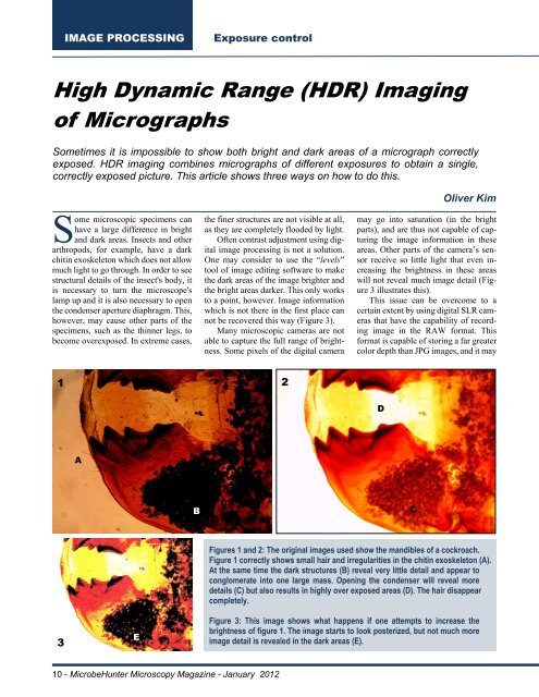
1 2<br />
D<br />
A<br />
B<br />
C<br />
3<br />
E<br />
Figures 1 and 2: The original images used show the mandibles of a cockroach.<br />
Figure 1 correctly shows small hair and irregularities in the chitin exoskeleton (A).<br />
At the same time the dark structures (B) reveal very little detail and appear to<br />
conglomerate into one large mass. Opening the condenser will reveal more<br />
details (C) but also results in highly over exposed areas (D). The hair disappear<br />
<strong>com</strong>pletely.<br />
Figure 3: This image shows what happens if one attempts to increase the<br />
brightness of figure 1. The image starts to look posterized, but not much more<br />
image detail is revealed in the dark areas (E).<br />
10 - <strong>Microbe</strong><strong>Hunter</strong> Microscopy Magazine - January 2012