Ahmed Azab *, Adel Hanafy *, Alaa Essa, Khaled Aref, Hesham ...
Ahmed Azab *, Adel Hanafy *, Alaa Essa, Khaled Aref, Hesham ...
Ahmed Azab *, Adel Hanafy *, Alaa Essa, Khaled Aref, Hesham ...

Create successful ePaper yourself
Turn your PDF publications into a flip-book with our unique Google optimized e-Paper software.
Menoufiya Medical Journal 189 Surgical Aspects in Management<br />
Vol. 21 No . 2 July 2008 <strong>Ahmed</strong> <strong>Azab</strong> et al.,<br />
SURGICAL ASPECTS IN MANAGEMENT OF DORSOLUMBAR<br />
JUNCTION & UPPER LUMBAR DISC LESIONS<br />
<strong>Ahmed</strong> <strong>Azab</strong> *, <strong>Adel</strong> <strong>Hanafy</strong> *, <strong>Alaa</strong> <strong>Essa</strong> $ , <strong>Khaled</strong> <strong>Aref</strong> $ ,<br />
<strong>Hesham</strong> Abo El Enin $ and Mohamed Fahmy $<br />
INTRODUCTION:<br />
*Minoufiya University & $ Alexanderia University<br />
Abstract:<br />
Background: Approximately 5% of lumbar disc herniations occur at the upper levels of the lumbar<br />
spine & dorsolumbar Junction (D12-L1, L1-2, L2-3, L3-4 levels) although the incidence reported<br />
in the literature varies from < 1% to 10.4%. This wide discrepancy in the incidence is partly the<br />
result of non standardized terminology regarding what constitutes an upper lumbar disc herniation.<br />
Some authors report upper lumbar discs as only D12-L1, L1-2, L2-3 & another as L1-2, L2-3.<br />
Aim of the work: This study aims at the evaluation of the clinical and radiological condition of the<br />
upper lumbar disc prolapse & the dorsolumbar junction disc and to evaluate the adequet surgical<br />
approach posteriorly either just posterior laminectomy only or transpedicular approach. PATIENTS<br />
AND METHODS: This prospective study included thirty patients with herniated discs at the upper<br />
lumbar & the dorsolumbar junction operated during the last two years. The posterior surgical<br />
approach done for the patients included: Decompressive laminectomy alone in patient with<br />
calcified discs with stenotic canal. Unilateral facetectomy with or without partial or total pedicle<br />
removal. Discectomy will be done for selected cases. Instrumental fusion was done when needed<br />
according to preoperativc radiological assessment and the planned surgical procedures. Results:<br />
Operative procedures: 60% of cases were treated with laminectomy uniletral facetectomy and<br />
discectomy at the involved level. 30% of cases were treated with laminectomy without discectomy<br />
due to hard spondylotic bar. In 3 cases constituting 10% at all cases, laminectomy, bilateral<br />
facetectomy, pediclectomy and discectomy was done followed by transpedicular fixation.<br />
Postoperatively 24 cases (80%) showed complete relief of their preoperative symptoms while 6<br />
cases (20%) showed partial relief of their preoperative. Discussion: All the patients were<br />
subjected to the posterior surgical approach as the most commonly used approach. Also because<br />
the patients of our study suffered not only isolated upper L.D.P but also, they were associated with<br />
lower lumbar canal abnormalities including the concomitant lower lumbar canal stenosis and lower<br />
lumbar disc prolapses, so the surgery was not directed only to the upper L.D.P, but also to the<br />
associated pathology, which are commonly treated with the posterior surgical approach.<br />
Disc pathology involving the proximal levels of<br />
the lumbar spine & dorsolumbar Junction have<br />
not been appreciated as well before the clinical<br />
use of magnetic resonance imaging (MRI) scan.<br />
Review of literature showed that most lumbar<br />
disc herniations are found in the lower levels.<br />
Disc herniation at D12-L1, L1-2, L2-3 levels<br />
were reported in less than 3% of the cases, of 88<br />
lumbar disc herniations reported by love et al<br />
two were found in D12-L1 & one in L1-2 & two<br />
in L 2-3. Scott et al reported that approximately<br />
MMJ ( July 2008)Vol. 21 N0:2:P 189-198<br />
5% of lumbar disc herniations occur at the upper<br />
levels of the lumbar spine & dorsolumbar<br />
Junction (D12-L1, L1-2, L2-3, L3-4 levels)<br />
although the incidence reported in the literature<br />
varies from < 1% to 10.4%. This wide<br />
discrepancy in the incidence is partly the result<br />
of non standardized terminology regarding what<br />
constitutes an upper lumbar disc herniation.<br />
Some authors report upper lumbar discs as only<br />
D12-L1, L1-2, L2-3 & another as L1-2, L2-3 (1).<br />
Those reporting an incidence closer to 1 to 2% of<br />
all herniated lumbar discs tended not to include<br />
the L3-4 the L3-4 level in their definition (2).
Menoufiya Medical Journal 190 Surgical Aspects in Management<br />
Vol. 21 No . 2 July 2008 <strong>Ahmed</strong> <strong>Azab</strong> et al.,<br />
The clinical presentation of disc proloapse at the<br />
level of upper lumbar spine & dorsolumbar<br />
Junction varies from; Low back pain which is<br />
usually dull aching pain , may be sever and<br />
causing abdominal pain. Pain exacerbates with<br />
coughing or valsalva maneuver as it increases the<br />
intra thecal pressure, Radicular pain in the<br />
dermatomal sensory distribution at the<br />
compressed root can be the only pain or<br />
associated with low back pain, Cauda equine<br />
compression syndrome due to central mechanical<br />
compression of the cauda equina, Neurogenic<br />
claudications as limited walking tolerance, back<br />
pain, much like patients with lower lumbar canal<br />
stenosis, Myelopathic manifestations,<br />
myelopathy with long tract signs, gait<br />
disturbance, weakness or spasticity, Conus<br />
medullaries compression resulting in paraparesis<br />
& even paraplegia with bowel & bladder<br />
dysfunction (3).<br />
Diagnostic investigations is very important for<br />
surgical decision such as plain radiography<br />
which can indirectly suggest pathology such as<br />
decreased disc space. The CT scan is one of the<br />
imaging modalities used for diagnosis of<br />
herniated lumbar disc in addition to its<br />
important role in detection of spinal canal<br />
stenosis. MRI gives excellent visualization of<br />
root anatomy, disc herniation and fragments<br />
including foraminal and far lateral pathologies (4).<br />
AIM OF THE WORK:<br />
This study aims at the evaluation of the clinical<br />
and radiological condition of the upper lumbar<br />
disc prolapse & the dorsolumbar junction disc<br />
and to evaluate the adequet surgical approach<br />
posteriorly either just posterior laminectomy<br />
only or transpedicular approach.<br />
PATIENTS AND METHODS:<br />
This prospective study included thirty patients<br />
with herniated discs at the upper lumbar & the<br />
dorsolumbar junction operated during the last<br />
two years. All patients were subjected to the<br />
following:<br />
Complete history including the inquirements<br />
about the age, sex, the onset, duration and nature<br />
of the complaints, the history of previous<br />
operations, also any associated diseases in<br />
addition to inquirement about available<br />
radiological investigations.<br />
General examination To assess the conditions<br />
of the chest, heart, abdomen and lower limbs,<br />
which is important to determine whether the<br />
patient is fit for surgery or not.<br />
Neurological examination including: Evaluation<br />
of patient's gait. Back examination for<br />
paravertebral spasm, limitation of movement, the<br />
lumbosacral angle and curve, any tenderness in<br />
the back and scar of any previous operations.<br />
Femoral stretch test is tested in both lower limbs.<br />
Straight leg raising test is tested in both lower<br />
limbs and its angle is recorded. Reflex changes is<br />
examined and recording any absence or<br />
diminution. Testing any dermatomal loss.<br />
Routine laboratory investigations including:<br />
Complete blood count. Liver function tests.<br />
Renal function tests. Bleeding and coagulation<br />
profile. Random blood sugar.<br />
Radiological investigations including: Plain X<br />
rays of the dorsolumbar and lumbosacral spine<br />
with Anteroposterior and lateral films, Stress<br />
films, Oblique films. MRI of the dorsolumbar<br />
and lumbosacral spine. CT scan was available<br />
with 50% of the patients on admission.<br />
The clinical indications for surgery were:<br />
Progressive neurological deficits. Pain not<br />
responding to conservative measures.<br />
The posterior surgical approach done for the<br />
patients included:<br />
The patient is under general anesthesia in a<br />
prone position. Midline back incision and<br />
incision of the fascia and then retraction of the<br />
muscles. Intraoperative fluoroscopy used when<br />
needed for levelling. Decompressive<br />
laminectomy alone in patient with calcified discs<br />
with stenotic canal. Unilateral facetectomy with<br />
or without partial or total pedicle removal.
Menoufiya Medical Journal 191 Surgical Aspects in Management<br />
Vol. 21 No . 2 July 2008 <strong>Ahmed</strong> <strong>Azab</strong> et al.,<br />
Discectomy will be done for selected cases.<br />
Instrumental fusion was done when needed<br />
according to preoperativc radiological<br />
assessment and the planned surgical procedures.<br />
Postoperative follow up clinically and<br />
radiographically;<br />
Clinically; as regard the improvement of<br />
symptoms and signs and occurrence of<br />
complications including neurological deficits and<br />
infection.<br />
Picture( 1 ) MRI LSS showing D12-L1disc prolapse<br />
Picture( 2 ) MRI LSS showing L1-L2 disc prolapse<br />
Radiologically; by pain X-ray films of<br />
dorsolumbar and lumbosacral spine at one month<br />
and 6 months postoperatively to assess the<br />
development of segmental instability.<br />
Postoperative MRI L.S.S will be done when<br />
needed if patient deteriorated postoperatively.
Menoufiya Medical Journal 192 Surgical Aspects in Management<br />
Vol. 21 No . 2 July 2008 <strong>Ahmed</strong> <strong>Azab</strong> et al.,<br />
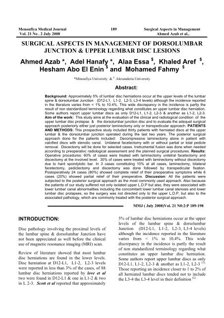
Picture( 3 ) MRI LSS showing L2-L3 disc prolapse.<br />
Picture(4) Post operative Plain x ray LSS after L2-L3 discectomy & fixation
Menoufiya Medical Journal 193 Surgical Aspects in Management<br />
Vol. 21 No . 2 July 2008 <strong>Ahmed</strong> <strong>Azab</strong> et al.,<br />
RESULTS:<br />
Table (I): Sex distribution: Males were more affected than females. Males (80%), females (20%).<br />
Gender No. of patients Percentage<br />
Males 24 80%<br />
Females 6 20%<br />
Total 30 100%<br />
Table (2): Age distribution: Most of patients affected were in the age group from 40-49 ys.<br />
Age group (years) No. of patients Percentage<br />
20-29 1 3.3%<br />
30-39 9 30%<br />
40-49 12 40%<br />
50-59 7 23.3%<br />
60-69 1 3.3%<br />
Total 30 100%
Menoufiya Medical Journal 194 Surgical Aspects in Management<br />
Vol. 21 No . 2 July 2008 <strong>Ahmed</strong> <strong>Azab</strong> et al.,<br />
Table (3): Clinical picture: Low back pain was the main complaint followed by the crural and<br />
anterior thigh pain. The most dominant clinical sign was the positive femoral stretch test (70%),<br />
followed by motor weakness in (60%). The straight leg raising test (SLRT) was positive in 30%<br />
only with concomitant lower lumber disc affection:<br />
Symptoms No. of patients Percentage<br />
Low back pain (L.B.P) 30 100%<br />
Anterior thigh pain 27 90%<br />
Sensory changes 15 50%<br />
Claudication 12 40%<br />
Sphincteric disturbances 3 10%<br />
Signs<br />
Back signs 15 50%<br />
Positive femoral stretch test 21 70%<br />
Positive SLRT 9 30%<br />
Reflex changes 18 60%<br />
Sensory changes 15 50%<br />
Motor weakness 18 60%<br />
Table (4): The level of disc prolapse, the most frequent level affected was the L2-3 level,<br />
followed by Ll-2 level and lastly D12-L1 level.<br />
Level No of patients Percentage<br />
D12-L1 3 10%<br />
L1-L2 9 30%<br />
L2-L3 18 60%<br />
Total 30 100%
Menoufiya Medical Journal 195 Surgical Aspects in Management<br />
Vol. 21 No . 2 July 2008 <strong>Ahmed</strong> <strong>Azab</strong> et al.,<br />
Table(5): MRI and CT findings:<br />
MRI and CT findings No. of patients Percentage<br />
Diffuse annulus disc bulge 24 80%<br />
Central hard disc protrusion 6 20%<br />
Indentation ofthecal sac 30 100%<br />
Encroachment on lateral recess and nerve root exit<br />
foraminae<br />
24 80%<br />
Caudal migration of disc prolapse 3 10%<br />
Lumbar spondylo degenerative changes 18 60%<br />
Concomitant lower lumbar disc prolapse 9 30%<br />
Concomitant lower lumbar canal stenosis 18 60%<br />
Sagittal stenosis 18 60%<br />
Table (6) Operative procedures: 60% of cases were treated with laminectomy uniletral<br />
facetectomy and discectomy at the involved level. 30% of cases were treated with laminectomy<br />
without discectomy due to hard spondylotic bar. In 3 cases constituting 10% at all cases,<br />
laminectomy, bilateral facetectomy, pediclectomy and discectomy was done followed by<br />
transpedicular fixation.<br />
Operative procedures<br />
No. of<br />
patients<br />
Percentage<br />
Laminectomy without discectomy at involved level. 9 30%<br />
Laminectomy, unilateral facetectomy with discectomy at<br />
involved level<br />
Laminectomy, bilateral facetectomy with pediclectomy<br />
discectomy and transpedicular fixation.<br />
18 60%<br />
3 10%
Menoufiya Medical Journal 196 Surgical Aspects in Management<br />
Vol. 21 No . 2 July 2008 <strong>Ahmed</strong> <strong>Azab</strong> et al.,<br />
Table (7) Complications: One patient was complicated with dural tear and developed<br />
postoperative C.S.F. leakage. One patient developed post operative spondylodiscitis. One<br />
patient developed cauda equina syndrome due to post operative haematoma<br />
Complications No. of patients Percentage<br />
C.S.F. leakage 1 3.3%<br />
Spndylodiscitis 1 3.3%<br />
Cauda equine syndrome 1 3.3%<br />
Table (8) Operative outcome: Postoperatively 24 cases (80%) showed complete relief of their<br />
preoperative symptoms while 6 cases (20%) showed partial relief of their preoperative.<br />
Postoperative sequel No. of patients Percentage<br />
Complete relief 24 80%<br />
Partial relief 6 20%<br />
DISCUSSION:<br />
Approximately 5% of lumbar disc herniations<br />
occur at upper levels of the lumbar spine (L1-<br />
2,L2-3,L3-4), although the incidence reported in<br />
the literature varies from < l-10.4% (5 ' 6 ' 7) This<br />
wide discrepancy in incidence is partly the result<br />
of non standardized terminology regarding what<br />
constitutes an "upper" lumbar disc herniation -<br />
some authors report upper lumbar discs as only<br />
L1-2and L2-3 (1) . Those reporting an incidence<br />
closer to 1-2% of all herniated lumbar discs did<br />
not include the L3-4 level in their definition (2).<br />
In the present study, the disc herniation at the<br />
level of L3-4 was not included among our cases<br />
of upper lumbar disc herniations, so the disc<br />
prolapses at D12-L1, L1-2 and L2-3 were the<br />
target of the present study.<br />
Similarly, Scott P. Sanderson et al in 2004, in<br />
their study about the unique characteristics of<br />
upper lumbar disc herniations, reported that disc<br />
herniations at L3-4 are different in their patient<br />
population and surgical outcome from those at<br />
L1-2 and L2-3 levels, and also reported that disc<br />
herniations at L3-4 are more similar to those<br />
occurring at L4-5 and L5-S1in their surgical<br />
outcome and patient population (8).<br />
The lower incidence of upper L.D.P. may be<br />
attributed to the decreased motion of the upper<br />
lumbar spine compared with the lower lumbar<br />
spine that may lead to less spondylosis, less disc<br />
degeneration and thus fewer herniated discs (9).<br />
In the present study, the MRI of L.S.S. was<br />
regarded as the most important and main<br />
investigation done for all patients and surgery<br />
was decided up on it.<br />
Similarly, Albert et al in 1993, recommended the<br />
MRI as the main diagnostic tool in the work-up<br />
of patients with upper L.D.P (10) Also, Mark<br />
Weidenbaum et al in 2002 reported that MRI has<br />
become the imaging procedure of choice in
Menoufiya Medical Journal 197 Surgical Aspects in Management<br />
Vol. 21 No . 2 July 2008 <strong>Ahmed</strong> <strong>Azab</strong> et al.,<br />
evaluating disc pathology, neural structures and<br />
muscloligamentous components and also gives<br />
excellent visualization of root anatomy, disc<br />
herniation and other conditions that may mimic it<br />
such as synovial cyst, neurofibroma or perineural<br />
cyst (11).<br />
All the patients were subjected to the posterior<br />
surgical approach as the most commonly used<br />
approach. Also because the patients of our study<br />
suffered not only isolated upper L.D.P but also,<br />
they were associated with lower lumbar canal<br />
abnormalities including the concomitant lower<br />
lumbar canal stenosis and lower lumbar disc<br />
prolapses, so the surgery was not directed only to<br />
the upper L.D.P, but also to the associated<br />
pathology, which are commonly treated with the<br />
posterior surgical approach. Intraoperatively, in<br />
18 cases constituting (60%), there was a soft disc<br />
bulge encroaching upon the nerve root exit<br />
foramine, while in 3 cases constituting (10%)<br />
there was a sequestrated disc prolpase with<br />
unilateral encroachment on the nerve root exit<br />
foramina. But in 9 cases constituting 30% of all<br />
cases in the present study, there was a hard<br />
spondylotic bar with a lateral recess stenosis and<br />
so, the surgical procedure done for this group<br />
was just a laminectomy and foraminotomy at the<br />
involved level without discectomy. While in 18<br />
cases constituting 60% of all cases in the present<br />
study were subjected to laminectomy (including<br />
formal and hemilaminectomy) unilateral<br />
facetectomy and discectomy at the involved<br />
level. Three cases with disc prolapses at D12-L1<br />
had an additional surgical procedures aiming to<br />
gain a lateral access to the disc prolapse with<br />
avoidance of manipulation to mobilize the<br />
sensitive neural structures at the upper lumbar<br />
levels including the conus medullaris and cauda<br />
equina. These additional surgical procedures<br />
included bilateral facetectomy with pedicle<br />
removal which necessitated an additional<br />
transpedicular fixation of one level above and<br />
one level below the involved level with<br />
instrumentation to avoid the late instability. So,<br />
the instrumental fusion was done for these<br />
patient depending only on one indication that<br />
was the extensive facetectomy to avoid the late<br />
instability.<br />
However, Scott P. Sanderson et al in 2004 in his<br />
retrospective study about patients with upper<br />
L.D.P, he mentioned that the fusion in patients<br />
with upper L.D.P treated surgically was done if<br />
the patient had incapacitating back pain, had<br />
preexisting spondylolisthesis or required such<br />
extensive facet removal to access the disc that<br />
predisposed the patient to instability (8).<br />
In addition, to the surgical intervention at the<br />
involved level, patients who suffered<br />
concomitant lower lumbar canal stenosis were<br />
subjected to decompressive laminectomy at<br />
lower lumbar levels, those patients were 18 cases<br />
constituting 60% of all cases of the present<br />
study. While patients who suffered concomitant<br />
disc herniation at lower lumbar level were 9<br />
cases: 5 cases of them were associated with disc<br />
prolapse at L4/5 level while 2 cases were<br />
associated with disc prolapse at L3/4 level and<br />
another two cases at L5/S1 level, and those<br />
patients were subjected to concomitant surgical<br />
intervention at the affected level in the form of<br />
laminectomy and discectomy in addition to<br />
surgical intervention at the upper lumbar disc<br />
prolapse.<br />
Regarding the postoperative sequelae of the<br />
patients operated in the present study, 24 patients<br />
(80%) showed complete relief of symptoms<br />
postoperatively, while 20% of patients showed<br />
partial relief of their complaints and those<br />
patients were found to have significant<br />
neurological deficits preoperatively mainly the<br />
motor weakness and sphincteric disturbances.<br />
Among this group of patients that showed partial<br />
relief of symptoms postoperatively, one patient<br />
was complicated with unintended durotomy<br />
intraoperatively and developed C.S.F. leak<br />
postoperatively which was managed in the ward<br />
conservatively till the leakage stopped and<br />
patient was discharged with relief of preoperative<br />
symptoms. Another patient developed<br />
postoperative spondylodiscitis and was treated<br />
with conservative measure and showed relief of<br />
preoperative symptoms after a period of one
Menoufiya Medical Journal 198 Surgical Aspects in Management<br />
Vol. 21 No . 2 July 2008 <strong>Ahmed</strong> <strong>Azab</strong> et al.,<br />
month. One patient was complicated with cauda<br />
equina syndrome postoperatively, MRI was done<br />
that showed postoperative extradural hematoma<br />
compressing the thecal sac, this patient needed<br />
another immediate intervention for evacuation of<br />
the hematoma and showed rapid recovery<br />
immediately after evacuation of the hematoma.<br />
REFERENCES<br />
1- Su H, Zucherman J, Shea W. High lumbar<br />
disc herniation, incidence and etiology. Spine.<br />
1990;15:679-82.<br />
2- Bosacco Sj, Berman AT, Raisis LW,<br />
Zamarin RI. High lumbar disc herniation case<br />
reports. Orthopedics. 1989;12:275-8.<br />
3-Lee DY, Kim HS, Lee SH. Multi access for<br />
diagnosis of missed upper lumbar disc<br />
herniation. J Korean Neurosurgery Soc.<br />
2005;38:144-6.<br />
4- Abrishamkar S, Mansour BA, Arti H. The<br />
effectiveness of computed tomography scans<br />
versus magnetic resonance imaging for decision<br />
making in patients with low back pain and<br />
radicular leg pain. Journal of Reserch in Medical<br />
Sciences. Nov & Dec 2006;11 (6):351-4.<br />
5- Aronson HA, Dunsmore RH. Herniated<br />
lumbar discs. J Bone Joint Surg.1993;45-A:311-7.<br />
6- Greenberg M et al. Lumbar spoinal stenosis,<br />
Handbook of Neurosurgery. 6 th ed. New York:<br />
Thieme;2006;289-369.<br />
7- Albert TJ, Baiderston RA, Heller JG,<br />
Herkowitz HN, Garfin SR, Tomany K, et al.<br />
Upper lumbar disc herniations. J Spinal Disord.<br />
1993;6:351-9.<br />
8- Sanderson SP, Houten J, Errico T,<br />
Forshaw D, Bauman J, Cooper PR. The<br />
unique characteristics of upper lumbar disc<br />
herniation. Neurosurgery. 2004; 55:385-9.<br />
9- Fontnesi G, Tartalgia I, Cavazutti A,<br />
Giancecchi F. Prolapse intervertebral disc at the<br />
upper lumbar level. Ital J Ortop Traumatol.<br />
1987;13:501-7.<br />
10- Albert TJ, Baiderston RA,Heller JG,<br />
Herkowitz HN, Garfin SR, Tomany K, et al.<br />
Upper lumbar disc herniations. J Spinal Disord.<br />
1993; 6:351-9.<br />
11- Weidenbaum M. Lumbar disc herniation<br />
and radiculopathy. In: Frymoyer JW, editor.<br />
Adult and pediatric Spine. 3 rd ed. Philadelphia:<br />
Lipincott- Williams and Willikins; 2004;19:920.