July 2013 - AHSC Office of Public Affairs - University of Arizona
July 2013 - AHSC Office of Public Affairs - University of Arizona
July 2013 - AHSC Office of Public Affairs - University of Arizona

You also want an ePaper? Increase the reach of your titles
YUMPU automatically turns print PDFs into web optimized ePapers that Google loves.
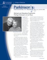
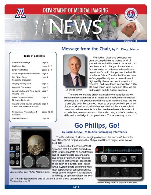
2DEPARTMENT OF MEDICAL IMAGING<strong>July</strong> <strong>2013</strong>Volume 1.2Message from the Chair, by Dr. Diego MartinTable <strong>of</strong> ContentsChairman’s Message page 1Go Philips, Go! pages 1 - 2Employee Pr<strong>of</strong>iles pages 2 - 3Graduating Residents & Fellows page 3Epic Team Status page 4Residents’ GraduationImaging Shining Stars page 5Awards & Graduation page 6Answers to Imaging Word Game page 7Imaging TriviaOther Department News page 8Birthdays, Births, etc.Imaging Grand Rounds Schedule page 9A Note from the Editor-In-Chief<strong>Public</strong>ations, Presentations & pages 10-24ResearchContact Information page 24Dr. Diego MartinWe had an awesome semester withgreat accomplishments thanks to all <strong>of</strong>your efforts and willingness to work with usdespite our rapid change. As a matter <strong>of</strong>fact, our external reviewers referred to ourdepartment’s rapid change over the last 18months as “vibrant” and noted that we havean “engaged faculty and a commitment tohigh quality clinical service, innovation inresearch, and excellence in education.” Westill have much to be done and I feel we areon the right path to further success.The next few months brings us much more transition as wewelcome new colleagues at all levels and introduce technologicaladvances that will position us with the other medical areas. As were-energize over the summer, I want to emphasize the importance<strong>of</strong> your work and input, which has resulted in all our accomplishmentsand advancements thus far. We have been able to recruittop scholars, researchers and others who bring a lot <strong>of</strong> experience,skills and knowledge to our great team. Thank you very much.Go Philips, Go!by Balazs Lengyel, M.D., Chief <strong>of</strong> Imaging InformaticsThe Department <strong>of</strong> Medical Imaging witnessed the successful completion<strong>of</strong> the PACS project when the Philips IntelliSpace project went live onApril 12th.The benefit <strong>of</strong> the Philips PACSsystem is that it enables our radiologiststo fully integrate all departmentsand all imaging data into one cohesivesingle system, thereby making“everything that’s image” accessibleat the push <strong>of</strong> a button. The PhilipsPACS System <strong>of</strong>fers enterprise wide,inter-departmental collaboration likeA screenshot from Philips PACS system. never before. Whether it is radiology,cardiology or ophthalmology, the systemlinks all departments and all divisions within those departments.(Continued on page 2)Another Philips PACS system screenshot..Page 1
Page 2Go Philips, Go! by Balazs Lengyel, M.D. (Continued from page 1)An added capability <strong>of</strong> the PACS system is that itenables us to reach out to external referring physicians.We can import studies directly into PACS, allowing thepatient’s images to arrive at the physician’s <strong>of</strong>fice longbefore the patient gets there.The PACS system gives a timeline for each imageand has the capacity to migrate images from the olderFUJI system; effectively accessing the images withinseconds. This gives us the capability to access virtuallyall images - x-rays, ultrasound, MRI, nuclear and IR imagesfor every patient, even the archived images. Userinitiated migration <strong>of</strong> images is conducted using an activetimeline query. The user needs to conduct this queryonce and only once since the image becomes residentwithin the Philips PACS system with the very first query.The PACS system installation was completed onsuch a tight schedule and in record-setting time. It was afirst-<strong>of</strong>-its-kind effort to implement the system within only6 months! This was only possible due to the amazingcontributions <strong>of</strong> our support team and collaborators.The success <strong>of</strong> this project was through the hard anddedicated work <strong>of</strong> a small contingent, which currentlyrepresents our core implementation team. I would liketo say thanks to many amazing and inspirational peoplefor their help: Lori Cavalli, Amy Brewer-Burton, VedranaDjurdjic, Mike Wolfe,Bob Borys, Dr. SanjaySuri, Mike Deeter, AmySchoenherr, KendraPotter, Robert Lovemore,CIO Shirley Gabriel, ourVice-Chariman Dr. BillErly, CMIO Dr. Peter Catinella,our Chairman Dr.Diego Martin and CEODr. Michael Waldrum.Based on a contractwith Philips, we are excitedand looking forward toour mutual collaborationover the next eight years.Dr. Balazs Lengyel, Chief <strong>of</strong>Imaging InformaticsA New Feature - Employee Pr<strong>of</strong>iles! by Mimi VillafaneA new feature that’s been added to the Department <strong>of</strong> Medical Imaging newsletter is the employee pr<strong>of</strong>ilesection. Each edition <strong>of</strong> the newsletter will contain a pr<strong>of</strong>ile on one or two department employees. This is a way torecognize the members <strong>of</strong> our team from all the different sections in our department. Thanks goes out to StephenAlden for suggesting this new feature!Bill Quirk - Radiology Technician, Clinical Instructor, Walking Imaging Archive!Quality control. Radiology Technologist. Clinical Instructor. Dark Room Technician. These are some <strong>of</strong> thetitles and jobs that Bill Quirk has taken on in his 40-year career here at the Department <strong>of</strong> Medical Imaging. Hiredin 1973 as a Dark Room Technician, Bill developed x-ray films back when the development process involvedmessy chemicals and the end result was part art, part chemistry, part miracle. Quality control soon became anotherone <strong>of</strong> his jobs and his mission.After receiving his Radiologic Technologist’s license through the Pima Community College (PCC) RadiologicTechnology Program, Bill became the x-ray trouble-shooting expert and the go-to man for processor problems.Next he took on the added responsibility <strong>of</strong> training and teaching the PCC radiology technologist students in <strong>University</strong>Medical Center’s (UMC) Radiology Department. Since that time back in 1976, he has trained generations <strong>of</strong>radiology technologists at UMC. On average, Bill trains seven to eight PCC radiology students a year. That’s wellover 400 radiology techs in his career!Back when the radiology department only <strong>of</strong>fered x-rays, Bill became the stabilizing force for learning andgrowing in our department with every new imaging service we added - Ultrasound, MRI, Nuclear Medicine, and IR.You could say Bill is a walking imaging archive with all his experience!Passionate about teaching and imaging, Bill’s proud <strong>of</strong> what he does. “We work with our students and staff toprovide the best image possible and available.” He strives to maintain the quest for excellent imaging. Bill says,“No other radiology department services the diversity <strong>of</strong> patients that we do.” In imaging everyone from Neonatalpatients, to Organ Transplant patients, to Trauma Level One patients, we excel. (Continued on page 3)
Page 3A New Feature - Employee Pr<strong>of</strong>iles (Continued from page 2)Bill has been working closely with the Neonatal ICU Unit since day one, working to make imaging easieron the smallest and most fragile patients we see, the premature babies. He’s also worked hard to createcharts and protocols to make imaging easier and less traumatic for all patients, including abused children.Bill’s compassion shines through in all his interactions with patients, with students and with staff. Bill is aninvaluable resource. He is an imaging shining star!Natalie McMillan -Chief Radiology Tech, Coordinator, Wunderkind!One look at Natalie and you might guess that she’s a student. Don’t let her youthfulappearance fool you. A natural born leader, she is the Chief Radiology Tech in theNuclear Medicine section.Natalie has been with the department for nine years. She came here from SouthDakota after finishing her nuclear medicine degree from Southeast Technical Instituteand started at <strong>University</strong> Medical Center as a staff technologist. And, just four yearsago, Natalie took over as the Chief Tech.The Nuclear Medicine department interacts and works very closely with every singledepartment in the hospital - Pediatrics, Orthopedics, OB-GYN, Neurology, Cardiology,Natalie McMillanetc. That’s just one <strong>of</strong> the reasons why Nuclear Medicine relies heavily on their technologistsand, <strong>of</strong> course, on Natalie. Natalie works well with everyone, in every department.Well known and respected, Natalie handles the clinical side equally well as the research side. “We do alot <strong>of</strong> clinical trials, more than I ever thought I’d do,” says Natalie. She enjoys the fast-paced, busy aspects <strong>of</strong>her job as well as the staff she works with. “We have a great team and support staff,” says Natalie.Her organizational and leadership skill level is equivalent to someone with decades <strong>of</strong> experience, somuch so that you could almost say she’s a leader-prodigy. Natalie’s natural leadership and ability to handlecomplex research protocols came into play when we began the huge challenge <strong>of</strong> orchestrating the PET CTProgram at UMI in November 2010. This wouldn’t have been possible without Natalie.Natalie is someone who takes on challenges in-the-blink-<strong>of</strong>-an-eye and easily shines. She admits thatsometimes it’s challenging to balance the administrative duties with the technical duties, but she still does itwith ease. Natalie scintillates, pure and simple.Congratulations to our Graduating Residents and Fellows!They’re on their way to the following Fellowships and positions:ResidentsMelanie Kuhlman, MD – <strong>University</strong> <strong>of</strong> <strong>Arizona</strong> – Interventional Radiology FellowAndrew Nash, MD – Brigham & Women’s Hospital, Boston, MA – Neuroradiology FellowMohammed Nawas, MD – Mallinckrodt Inst. <strong>of</strong> Radiology, St. Louis, MO - Neuroradiology FellowRavi Shastri, MD – <strong>University</strong> <strong>of</strong> Michigan, Ann Arbor, MI – Neuroradiology FellowSachin Shr<strong>of</strong>f, MD – <strong>University</strong> <strong>of</strong> <strong>Arizona</strong> – Neuroradiology FellowKaran Sundlass, MD – Medical College <strong>of</strong> Wisconsin, Milwaukee, WI - Interventional Radiology FellowFellowsSindhu Kumar, MD - Ab. Imag. Fellow (<strong>2013</strong>-14): UA Dept. <strong>of</strong> Med. Imaging - 2nd yr Body Imag.FellowStacey Black, MD - Vascular & Interventional - Faculty: UA Dept. <strong>of</strong> Med. Imaging, effec. 12/1/<strong>2013</strong>Monique Meyer, MD - Neuroradiology - Faculty: <strong>University</strong> <strong>of</strong> Louisville, Dept. <strong>of</strong> Rad., NeuroradiologyTamim Sultani, MD - Neuroradiology - MSK Fellow (<strong>2013</strong>-14) at UCLADanielle Carroll, MD - Breast Imaging - Radiologist: <strong>Arizona</strong> State Radiology, St. Mary’s Hospital, focuson Breast ImagingNeil Kaura, MD- Musculoskeletal - Position w/Lake Havasu Regional Med. Ctr. in Diagnostic Radiology
Page 4Epic Go-Live Delayed until Nov. 1UAHN President and CEO Dr. Michael Waldrum announced on Thursday, June27th, that “After very deliberate and thoughtful evaluation, and considering thoroughinternal and external multi-stakeholder assessments surrounding UAHN’s readinessto launch our new Electronic Health Records system, I have made the decision topush back our planned Epic Go-Live from Sept. 1 to Nov. 1, <strong>2013</strong>.” He also statedthat “All <strong>of</strong> us ‘own’ this project and we must renew our commitments and efforts toensure it is successful. Please take this extra time to assure your area is prepared.”The EPIC Team will work to strengthen the training process during this time.He suggested that you should know what your training requirements are. If youare unsure <strong>of</strong> your training needs or if you havequestions, please contact your direct supervisor.Please realize that your supervisor has beendirected to limit PTO requests that in any wayjeopardize the success <strong>of</strong> our Epic Go-Live.CEO Dr. Michael WaldrumResidents’ & Fellows’ Graduation Celebrationby Michele Dalmendray(Left to right, Residents Sachin Shr<strong>of</strong>f and Adam Baer, Dr. CharlesHennemeyer, who won the George Barnes Award, Dr. DorothyGilberson-Dahdal and Dr. Diego Martin)Annually, our department acknowledges the academicachievement <strong>of</strong> its graduating residents andfellows. Friends, family, faculty and staff were on hand atthe Skyline Country Club on Friday, June 14th to celebratetheir accomplishments.After a delightful buffet <strong>of</strong> appetizers, delicious maincourses and decadent desserts, the following fourth yearresidents received their Resident Certification from ResidencyProgram Director, Dr. Dorothy Gilbertson-Dahdal:Melanie S. Kuhlman, M.D.; Andrew K. Nash, M.D.;Mohammed T. Nawas, M.D.; Ravi K. Shastri, M.D.;Sachin H. Shr<strong>of</strong>f, M.D. and Karanjot Sundlass, M.D.Each <strong>of</strong> our graduating residents will move on to subspecialtyfellowships into which they matched with theirfirstchoice <strong>of</strong> programs. Congratulations to you all!Next in the program was the presentation <strong>of</strong> Fellowship Certifications.Monique A. Meyer, M.D. and Tamim Sultani, M.D.were awarded their certificates <strong>of</strong> completion for the NeuroradiologyFellowship by Program Director Dr. Wayne S. Kubal. Dr. LukeScalcione presented the Musculoskeletal Fellowship certificateto Neil V. Kaura, M.D. Stacey R. Black, M.D., Vascular andInterventional fellow, was awarded her certificate <strong>of</strong> completion byProgram Director Dr. Charles Hennemeyer. Dr. Kim Fitzpatrick,the Program Co-Director, presented the Breast Imaging Fellowshipcertificate <strong>of</strong> completion to Danielle M. Carroll, M.D. GraduatesHina Arif-Tiwari, M.D.; Samantha Matz, D.O. and SindhuKumar, M.D. received their Body Imaging Fellowship certificates<strong>of</strong> completion from Program Director Dr. James Costello. All <strong>of</strong>our graduating fellows have acquired job assignments with privateradiology organizations, academic institutions or have attained additionalfellowship positions around the country. (Continued on page 24)(Left to right, Dr. Dorothy Gilberson-Dahdal, Matthew Covingon,who won one <strong>of</strong> the Resident Teaching Awards, and Dr. DiegoMartin)
Page 5*Imaging Shining Stars*Each edition <strong>of</strong> the Department <strong>of</strong> Medical Imagingnewsletter will have a section called “Imaging ShiningStars.” This section recognizes employees who’ve goneabove and beyond the scope <strong>of</strong> their duties; peoplewho’ve been given a pat on the back from a patient,another department, or generally someone outside ourteam.***************The following report came in from Laurie McMahon,an RN in the Emergency Department:I wanted to share a very positive experience I hadtoday. I had patient in room 46 that was a 1:1, we hada very hard time getting her to sleep and a hard timekeeping her that way. The gentlemen that were at the CTscanner, named Greg and Julio, were without any doubtthe most pr<strong>of</strong>essional and caring CT peeps I have everdealt with. They helped me out so much by dimming thelights, whispering and using the slide board. They wentabove and beyond and were very helpful to help keep thispatient sleeping, thanks to them. Could you please givethem a pat on the back from us over here?***************Angela Castillo submitted these comments andrecognition:5E: Dennis Schrage jumped in to the PICC teamand prevented a patient from leaving AMA. He alsomade sure a patient on the same floor received theverification read necessary for discharge.Incredible collaboration <strong>of</strong> the PICC team and IR:The PICC team started Friday down a nurse and 8 patients.Several patients needed discharge PICCs. Carlquickly prioritized needs and requested assistancefrom Interventional Radiology. By noon, there were 12patients. Mike T. was able to come in to help out in theafternoon. With the help <strong>of</strong> all involved only 2 patientsrolled to the next day. This is incredible!D5: Phinney Ripley and Dulce Quiroga for arrivingso quickly to ensure a STAT x-ray was done forone <strong>of</strong> our precious patients and you were excellent.You both were friendly, fast and very pr<strong>of</strong>essional. Yourexcellent care is so appreciated.D3N: Mandy Vigil in the ED made me feel lessscared and really took steps to try and be gentle becauseI was in a lot <strong>of</strong> pain. I really feel like I receivedEXCELLENT care at UAMC. I would recommend thishospital to anyone. Everyone treated me very pr<strong>of</strong>essionallyand was wonderful!! Thank you so much.***************A patient called to inform hospital administration<strong>of</strong> what a wonderful experience she had at UAMC.She mentioned Carol Suida assisted with her CT.The patient was very scared and nervous for thescan; however, Carol explained everything in detail,answered all her questions, and made her feel verycomfortable and at ease. She stated she was coldand Carol provided her with a blanket.Prior to her CT, she also had an ultrasound andstated the staff was pleasant, courteous and helpful.When she had a biopsy, she stated that Mark Akersand Dr. Khan held her hand and stayed by her sideduring the whole procedure. She was very appreciative<strong>of</strong> their time and patience while she underwentthe procedure.***************Desi Spurr, MS CCC-SLP, Speech and LanguagePathologist, submitted this awesome report:I wanted to take a moment to recognize someamazing members <strong>of</strong> your staff in Radiology.Kimberly Guinter and Danielle Hobbs are trueassets to this hospital, and especially to our SpeechTherapy team. They both work very hard to accommodateour Speech Therapy pt’s, to meet ourschedules and communicate with us throughout theday about orders both new and pending. Their interactionswith patients are always very respectful,pr<strong>of</strong>essional, and supportive. They are excellent ptadvocates, always vigilant <strong>of</strong> orders that don’t quiteseem to make sense and page me about theseorders <strong>of</strong>ten for clarification. On multiple occasions,this vigilance has prevented an unsafe procedurefrom occurring (i.e., an NPO pt with severe dysphasiahaving an esophagram). They are also welleducated about the equipment in fluoro and helpthe doctors with this so very <strong>of</strong>ten. These womendo an excellent job every day they are here. Ihonestly do not know what I would do without them.Just wanted to acknowledge them for the amazingjob they do.***************Kudos goes out to Carl, Mike T., Julio Jimenezand Greg Rosas, Dennis Schrage, PhinneyRipley, Dulce Quiroga, Mandy Vigil, Carol Suida,Mark Akers, Dr. Rihan Khan, Kimberly Guinterand Danielle Hobbs for going above and beyondfor our patients! You ARE Imaging Shining Starsand truly embody the new department motto...We are here for you!
Page 7First Clinical X-ray in AmericaX-Ray event captured by photographer HenryH. Barrett on February 3, 1996, in Dartmouth’sReed Hall. (L to R - Pr<strong>of</strong>essor EdwinFrost, patient Eddie McCarthy, Gilman Frostand Margaret Mead Frost.)On January 19, 1896, young EddieMcCarthy <strong>of</strong> Hanover, New Hampshire, fellwhile skating on the Connecticut River andfractured his left wrist. His physician, GilmanD. Frost, contacted his brother, Edwin Frost,a Pr<strong>of</strong>essor <strong>of</strong> Astronomy at Dartmouth.On February 3, 1896 Eddie was broughtto the Physics Laboratory in Reed Hall atDartmouth where a battery powered Crookesvacuum tube was used to produce the firstclinical x-ray in America. This “Colles” fractureswas depicted with an exposure time <strong>of</strong>20 minutes on glass “plates.”For more information, go to the AmericanJournal <strong>of</strong> Roentgenology. January1995; 164:241-243 or click on this link:http://www.ajronline.org/doi/abs/10.2214/ajr.164.1.7998549Photograph <strong>of</strong> originalgelatin plate made with20-minute exposure. Ulnarfracture is visible but imagecertainly shows evidence <strong>of</strong>significant degradation bymotion.Imaging Word Search... Here are the answers to last month’s word search.
Page 8Other Department News and Updates...• Dr. Hina Arif-Tiwari won the 2012 Fresh Face Award at the 27th Annual Advances in Contrast Ultrasound- ICUS Bubble Conference. Sept 20 - 21, 2012 ,Chicago.• Dr. Ray Carmody received the “Distinguished Service Award” from the American Board <strong>of</strong> Radiology.• Dr. Rihan Khan won the ASHNR “Name That Neoplasm” gameshow contest at the American Society <strong>of</strong>Head & Neck Radiology (ASHNR), 46th Annual Meeting, Miami, FL.• Dr. Balazs Lengyel has been promoted to the Chief <strong>of</strong> Informatics, Informatics Division.• Dr. Kambiz Nael was promoted to Director <strong>of</strong> Neuroradiology MRI in April.• Dr. Isabel Oliva was appointed Director <strong>of</strong> Cardiothoracic Imaging, Director <strong>of</strong> Computed Tomography.• Dr. Russell Witte has been promoted to Associate Pr<strong>of</strong>essor with Tenure, effective <strong>July</strong> 1, <strong>2013</strong>.• The Radiology CT Department has a new addition - Debbie Beckett has joined our team as <strong>of</strong> Aprilthis year. She has been a CT/ MRI Technologist with 26 years <strong>of</strong> experience, with a total <strong>of</strong> 30 years inthe Radiology field. Debbie has experience in acute care and trauma faculties. She also comes from abackground in Siemens equipment. Debbie is certified in CT, MRI, and Mammography. Debbie originallycame from St. Louis, Missouri. She moved to Tucson <strong>Arizona</strong> in 2011, where she worked at St. Joseph’sHospital until she joined us at UAHN.• Midge Ochart retired after 35 years <strong>of</strong> service in the Medical Imaging Department, Nuclear Medicine section.Thank you for your years <strong>of</strong> dedicated service, Midge!Birthdays, Babies, Etc.Wishing a very Happy June, <strong>July</strong> and August Birthday to our team members!June:<strong>July</strong>:August:6/1 Balazs Lengyel6/2 Ryan Avery6/3 Sylvia Gomez6/5 Adam Bauer6/9 Evan Hall6/12 Natarajan Raghunand6/15 Hina Arif Tiwari6/17 Sterling HansenTerry MatsunagaJune Stavem6/19 Divya Pillai6/20 Zhonglin Liu6/22 Elizabeth HagueMir Shayegan Salek6/29 Lars Furenlid6/30 Tzu-Yu Wu7/1 Harrison BarrettMichael Capp7/9 Saveen Ahuja7/14 Danielle CarrollMichael MousaRussell Witte7/16 Hui Leung7/30 Matthew Risi7/31 Sean LewisArash Meshksar***If we’ve missed yourbirthday, please let us knowso we can help you celebrateand get you on our list fornext year!***Congratulations!8/1 Gail Stevenson8/3 Marisa Borders8/4 Margaret GoodmanSusan LeGendre-McGhee8/8 Xiang Fan8/10 Vevek Parikh8/11 Eugene Duke8/15 Tim Hunter8/17 Christy BarberZhitao Li8/18 Frank Morello8/19 Abhishek Pandey8/20 Mary Stuart8/22 Kurtis Tedesco8/25 Karanjot Sundlass8/26 Irwin Freundlich8/30 Alexander Barber*Dr. Rihan Khan and his wife, Mahjabeen, welcomed their baby boy, Dean, on May24th. He was 8 lbs, 3 oz. and 20 inches.*Dr. Stewart Rasmussen and his wife, Emily, welcomed their baby girl, NaomiFaith, on March 24th. She was 6 lbs,13 oz. and 18 1/2 inches.
Page 9Department <strong>of</strong> Medical ImagingGrand RoundsJune Lecture Topic DIV Faculty/Visiting Pr<strong>of</strong> . Location5 Lung Cancer Screening Chest Isabel Oliva, MD DuVal 260012 NO GRAND ROUNDS19 Imaging <strong>of</strong> Head and Neck Infections Neuro Wayne Kubal, MD DuVal 260026 Picturing the Future <strong>of</strong> Cancer Health Research Marty Pagel, PhD DuVal 2600Care with AcidoCEST MRI<strong>July</strong> TBA TBA TBA DuVal 2600August TBA TBA TBA DuVal 2600<strong>July</strong> and August GRAND ROUNDS Topics & Speakers TBAA Note from the Editor, by Dr. Rihan KhanOur end <strong>of</strong> the year newsletter edition is very encompassing and focuses on manyexciting accomplishments by both individuals and groups within Medical Imaging. Wehave a detailed list <strong>of</strong> research projects, publications, presentations, and awards won,in addition to sections spotlighting individuals and highlighting great jobs by shiningstars. We want to keep recognizing accomplishments no matter how large or small, soplease keep forwarding your news, ideas, and recommendations for future issues (seeemail addresses on the last page).This last year was a great one, and let’s continue to work hard together to makethe new academic year even better!RihanDr. Rihan Khan
Page 10<strong>Public</strong>ations, Presentations & ResearchThe following pages (pages 10 - 24) contain the publications, presentations, book chapters,invited lectures, poster and electronic exhibits and research that doctors from the Department<strong>of</strong> Medical Imaging are working on or have completed between <strong>July</strong> 2012 and June <strong>2013</strong>.<strong>Public</strong>ations• Huang C, Graff CG, Clarkson EW, Bilgin A, and Altbach MI. “MR Parameter Mapping from Highly Undersampleddata by REconstruction <strong>of</strong> Principal COmponent coefficient Maps (REPCOM) using CompressedSensing”, Magn Reson Med 67:1355–1366, 2012.• Altbach MI, Barr T, Singh J, Ainapurapu B, Kc D, Squire S, Galons JP, Huang C, Abidov A. T2 mapping<strong>of</strong> the heart with high temporal and spatial resolution using a radial double inversion fast spin-echo pulsesequence with view sharing, Journal <strong>of</strong> Cardiovascular Magnetic Resonance 2012, 14 (Suppl 1): 0112doi:10.1186/1532-429X-14-S1-O112• Huang C, Bilgin A, Barr T, and Altbach MI. “T2 Relaxometry with Indirect Echo Compensation from HighlyUndersampled Data”, Magn Reson Med : DOI: 10.1002/mrm.24540.• Anavy N, Avery R, Desai C and Kuo PH. Wandering Transplant Kidney Presents as a Pitfall in Interpretation<strong>of</strong> Renal Scintigraphy for Dual Kidney Transplant. Journal <strong>of</strong> Nuclear Medicine Technology, December 2012;40(4):275-7.• M. K. Kupinski, E. W. Clarkson, H. H. Barrett, Scanning linear estimation: improvements over region <strong>of</strong> interest(ROI) methods, Physics in Medicine and Biology, 58, 1283-1301, <strong>2013</strong>.• E. Clarkson, Asymptotic ideal observers and surrogate figures <strong>of</strong> merit for signal detection with list-modedata, Journal <strong>of</strong> the Optical Society <strong>of</strong> America A, 29, 2204-2216, 2012.• A. K. Jha, M. A. Kupinski, H. H. Barrett, E. Clarkson, J. H. Hartman, Three-dimensional Neumann-series approachto model light transport in nonuniform media, Journal <strong>of</strong> the Optical Society <strong>of</strong> America A, 29, 1885-1899, 2012.• A. K. Jha, M. A. Kupinski, T. Masumura, E. Clarkson, A. V. Maslov and H. H. Barrett, Simulating photontransportin unifrom media using the radiative transport equation: a study using the Neumann-series approach,Journal <strong>of</strong> the Optical Society <strong>of</strong> America A, 29, 1741-1757, 2012.• C. Huang, C. G. Graff, E. W. Clarkson, A. Bilgin, M. I. Altbach, T2 mapping from highly undersampled databy reconstruction <strong>of</strong> principal component coefficient maps using compressed sensing, Magnetic Resonancein Medicine, 67, 1355-1366, 2012.• Hunter TB. Nothing new under the Sun. Letter to the Editor. Radiology 2012; 262:370.• Kalb B, Sharma P, Tigges S, Raye G, Kitajima HD, Costello JR, Chen Z, Martin DR. Magnetic ResonanceImaging <strong>of</strong> Pulmonary Embolism: Diagnostic accuracy <strong>of</strong> contrast-enhanced 3D MRPA, contrast-enhancedlow flip angle 3D gradient echo and noncontrast steady-state free precession sequences. Radiology 2012;263: 271-278.• Khan R, Krupinski E, Graham JA, Benodin L, Lewis P Assessing First Year Radiology Resident CompetencePre-Call – Development and Implementation <strong>of</strong> a Computer-based Exam before and after the 12Month Training Requirement Acad Radiol. 2012 Jan 30. [Epub ahead <strong>of</strong> print]• Khan R, Nael K, Erly W. Acute Stroke Imaging: What Clinicians Need to Know. Am J Med. <strong>2013</strong>May;126(5):379-86.• Nguyen N, Ceizyk M, Vinh-Hung V, Sroka T, Jang S, Khan R, Locke A, Albala G, Truong C, Godinez J, VoR, Smith-Raymond L . Feasibility <strong>of</strong> Tomotherapy to Reduce Cochlea Radiation Dose in Patients with LocallyAdvanced Nasopharyngeal Cancer. Accepted for publication in Tumori. 2012 Nov;98(6):709-14.• Nguyen NP, Vock J, Chi A, Ewell L, Vos P, Mills M, Khan R, Almeida F, Davis R, Betz M, Jang S, GelumbauskasS, Vo RP, Vinh-Hung V Effectiveness <strong>of</strong> Intensity-Modulated and Image-Guided Radiotherapy toSpare the Mandible From Excessive Radiation. Oral Oncol. 2012 Feb 15. [Epub ahead <strong>of</strong> print]• Miller C, Khan R, Lemole GM, Jacob A. Osteoblastoma <strong>of</strong> the Lateral Skull Base: Work-Up, Surgical Management,and a Review <strong>of</strong> the Literature. Journal <strong>of</strong> Neurological Surgery Reports. e<strong>Public</strong>ation date: Jun13 <strong>2013</strong>.• Betts AM, Ritter JL, Kubal WS. Reversible Delayed Posthypoxic Leukoencephalopathy after Drug Overdose:MRI Findings in a Collection <strong>of</strong> Patients. Emergency Radiology 19:165-173, 2012.• Kubal WS. Updated Imaging <strong>of</strong> Traumatic Brain Injury. Radiologic Clinics <strong>of</strong> North America. 50:15-41, 2012• Collins JM, Krishnamoorthy AD, Kubal WS, Johnson MH, Poon CS. Multidetector CT <strong>of</strong> Temporal BoneFractures. Seminiars in Ultrasound, CT, and MRI. 33:418-431, 2012.(Continued on page 11)
Page 11<strong>Public</strong>ations, Presentations & Research (Continued from page 10)<strong>Public</strong>ations (Continued)• Kuhlman M, Meyer M, Krupinski E. Direct Reporting <strong>of</strong> Results to Patients: the Future <strong>of</strong> Radiology. AcademicRadiology 2012;19:646-650• Kuo PH, Avery R, Krupinski E, Lei H, Bauer A, Sherman S, McMillan N, Seibyl J, Zubal G: Receiver-Operator-CharacteristicAnalysis <strong>of</strong> an Automated Program for Analyzing Striatal Uptake <strong>of</strong> 123I-I<strong>of</strong>lupane SPECTimages: Calibration Using Visual Reads. Journal <strong>of</strong> Nuclear Medicine Technology; <strong>2013</strong>; 41:1-6.• Martin DR, Costello J, Kalb B, Sauer C, Goldschmid S. Magnetic Resonance Enterography in Crohn’s Disease:A Perspective on Methodology, Interpretation, and Utility. Imaging in Medicine 2012; 4(3): 329-342.• Martin DR. Impact <strong>of</strong> Magnetic Resonance Imaging (MRI) on Computed Tomography (CT)-based treatmentplanning and acute toxicity for prostate cancer patients treated with Intensity –Modulated radiotherapy(IMRT). Practical Radiation Oncology. PRACTICALRADONC-D-12-00005R2.• Nael K, Meshksar A, Liebeskind D.S, Wang D.J, Ellingson B.S, Salamon N, Villablanca JP. Peri-ProceduralArterial Spin Labeling and Dynamic Susceptibility contrast Perfusion in Detection <strong>of</strong> Cerebral Blood Flow inPatients with Acute Ischemic Syndrome. Stroke. <strong>2013</strong> Mar;44(3):664-70.• Nael K, Meshksar A, Liebeskind DS, Coull BM, Krupinski EA, Villablanca PJ. Quantitative Analysis <strong>of</strong> Hypoperfusionin Acute Stroke: Arterial Spin Labeling vs. Dynamic Susceptibility Contrast. Stroke. <strong>2013</strong> June;<strong>Public</strong>ation status: Accepted for publication.• Oliva I, Knox K. Medical image <strong>of</strong> the week: CREST plus ILD. Southwest J Pulm Crit Care. <strong>2013</strong>;6(6):275-6• Kuzmik GA, Detterbeck FC, Decker RH, B<strong>of</strong>fa DJ, Wang Z, Oliva IB, Kim AW. “Pulmonary resections followingprior definitive chemoradiation therapy are associated with acceptable survival”. Eur J Cardiothorac Surg.<strong>2013</strong> Apr 4. [Epub ahead <strong>of</strong> print]• Oliva IB, Davarpanah AH, Rybicki FJ, Desjardins B, Flamm SD, Francois CJ, Gerhard-Herman MD, KalvaSP, Ashraf Mansour M, Mohler ER 3rd, Schenker MP, Weiss C, Dill KE. “ACR appropriateness criteria(®)imaging <strong>of</strong> mesenteric ischemia”. Abdom Imaging. <strong>2013</strong> Jan 9. [Epub ahead <strong>of</strong> print]• Puchalski JT, Argento AC, Murphy, TE, Araujo KL, Oliva, IB, Rubinowitz AN, Pisani MA. “Etiologies <strong>of</strong> bilateralpleural effusions”. Respir Med. <strong>2013</strong> Feb;107(2):284-91.• Geetinder Goyal, Isabel OIiva, Pramod Bonde, Clemente Britto, Wassim Fares. “An unusual case <strong>of</strong> chestpain and dyspnea on exertion”. Case Reports in Clinical Medicine. Vol.2, No.2, 108-110 (<strong>2013</strong>).• Maxfield MW, Schuster KM, McGillicuddy EA, Young CJ, Ghita M, Bokhari SA, Oliva, IB, Brink JA, Davis KA.“Impact <strong>of</strong> adaptative statistical iterative reconstruction on radiation dose in evaluation <strong>of</strong> trauma patients”. JTrauma Acute Care Surg. 2012 Dec; 73(6):1404-9• Demehri S, Rybicki FJ, Desjardins B, Fan CM, Flamm SD, Francois CJ, Gerhard-Herman MD, Kalva SP, KimHS, Mansour MA, Mohler ER 3rd, Oliva IB, Schenker MP, Weiss C, Dill KE. ACR Appropriateness Criteria ®blunt chest trauma – suspected aortic injury. Emerg Radiol. 2012 Aug;19(4):287-92.• Lawrence DA, Oliva IB, Israel GM. “Detection <strong>of</strong> hepatic steatosis on contrast-enhanced Ct images: diagnosticaccuracy <strong>of</strong> identification <strong>of</strong> areas <strong>of</strong> presumed focal fatty sparing”. AJR Am J Roentgenol. 2012Jul;199(1):44-7• Tanpitukpongse TP, Klein MA, Scalcione LR, Katz DS. Large uterine fibroids with deep venous thrombosis,pulmonary embolus, and caval compression. ACR Case in point. <strong>July</strong> 1, <strong>2013</strong>.• Scalcione LR, Pathria MN, Chung C. The Athlete’s Hand: Ligament and Tendon Injury. Semin MusculoskeletRadiol. 2012; 16:338-350.• Lazzara B, Scalcione LR, Garnet DJ, Geller M, Katz DS. Radiology-Pathology Conference:Primary Perinephricand Extraosseous Ewing Sarcoma. Clin Imaging. 2012; 36:77-79.• Monu JU, Muyinda Z, Taljanovic M. ISS outreach sub-Saharan Africa insight: Uganda 2011. Skeletal Radiol.2012 Nov;41(11):1347-8. Epub 2012 Aug 17. No abstract available.• Monu JU, Kopakopa D, Taljanovic M. ISS outreach Sub-Saharan Africa: Zambia 2011. Skeletal Radiol. 2012Dec;41(12):1493-4. doi: 10.1007/s00256-012-1495-1. Epub 2012 Aug 15. No abstract available.• Nissim L, Krupinski E, Hunter T, Taljanovic M. Exposure to, understanding <strong>of</strong>, and interest in interventionalradiology in american medical students. Acad Radiol. <strong>2013</strong> Apr;20(4):493-9. doi: 10.1016/j.acra.2012.09.026.• Kraus VB, Feng S, Wang S, White S, Ainslie M, Le Graverand MP, Brett A, Eckstein F, Hunter DJ, Lane NE,Taljanovic MS, Schnitzer T, Charles HC. Subchondral bone trabecular integrity predicts and changes concurrentlywith radiographic and MRI determined knee osteoarthritis progression. Arthritis Rheum. <strong>2013</strong> Apr 10.doi: 10.1002/art.37970. [Epub ahead <strong>of</strong> print](Continued on page 12)
Page 12<strong>Public</strong>ations, Presentations & Research (Continued from page 11)<strong>Public</strong>ations (Continued)• Totenhagen J, Lope-Piedrafita S, Thome K, Borbon I, Erickson RP and Trouard TP. In Vivo Assessment <strong>of</strong>Myelination in Niemann-Pick Type C Mice across the lifespan via Quantitative T2 mapping and Diffusion TensorImaging. Journal <strong>of</strong> Magnetic Resonance Imaging. 2012.• Russell G, Harkins KD, Secomb TW, Galons JP and Trouard TP. () A Finite Difference Method for Large ScaleSimulations <strong>of</strong> Water Diffusion in Tissue. Physics in Medicine and Biology. 2012; 57: N35-N46.• Maue RA, Burgess RW, Wang B, Wooley CM, Seburn KL, Vanier MT, Rogers MA, Chang CC, Chang T-Y, HarrisBT, Graber DJ, Penatti CAA, Porter DM, Szwergold BS, Henderson LP, Totenhagen JW, Trouard TP, BorbonIA and Erickson RP. A novel mouse model <strong>of</strong> Niemann Pick type C Disease carrying a D1005G-Npc1 mutationcomparable to commonly observed human mutations. Human Molecular Genetics. 2012; 21:730-750.• Borbon IA, Totenhagen J, Fiorenza MT, Canterini S, Ke W, Trouard TP and Erickson RP. Niemann-Pick C1 mice,a model <strong>of</strong> “juvenile Alzheimer’s disease”, with normal gene expression in neurons and fibrillary astrocytes showlong term survival and delayed neurodegeneration. J. Alzheimer’s Disease. 2012; 30:875-887.• Culp WC, Woods SD, Skinner RD, Brown AT, Lowery JD, Johnson JL, Unger EC, MD; Borrelli MJ, Roberson PK.Dodecafluoropentane Emulsion Decreases Infarct Volume in a Rabbit Ischemic Stroke Model. Journal <strong>of</strong> VascularInterventional Radiology 2012;23:116-121.• Roos Sebastiaan T, Slikkerveer Jeroen, Unger Evan C, Porter Tom, Kamp Otto, Microbubble enhanced sonothrombolysisin patients: a literature review <strong>of</strong> clinical studies. submitted to Heart.• Unger E, Porter T, Lindner J, Grayburn P. Cardiovascular Drug Delivery with Ultrasound. Submitted to AdvancedDrug Delivery Review. June <strong>2013</strong>.• Unger E, Porter T, Lindner J, Grayburn P. Cardiovascular Drug Delivery with Ultrasound. Advanced Drug DeliveryReview. June <strong>2013</strong>.Invited Lectures/Oral Presentations• Barr T, Huang C, Bilgin A, Abidov A, and Altbach MI. Indirect Echo Corrected Fast T2 mapping <strong>of</strong> the Heart fromHighly Undersampled Radial FSE Data Using the CURLIE Reconstruction, Annual Meeting <strong>of</strong> the InternationalSociety for Magnetic Resonance in Medicine, Salt Lake City, Utah, April <strong>2013</strong>.• Huang C, Pandey A, Barr T, Bilgin and Altbach MI. An Indirect Echo Compensated Reconstruction Algorithm forT2 Mapping <strong>of</strong> The Liver from Highly Undersampled Radial FSE Data, Annual Meeting <strong>of</strong> the International Societyfor Magnetic Resonance in Medicine, Salt Lake City, Utah, April <strong>2013</strong>.• Berman BP, Li Z, Altbach MI, Galons JP, Martin DR, Dong B, Sharma P, Kalb BT, and Bilgin A. How to stack thestars: a variable center-dense k-space trajectory for 3D MRI, submitted to the Annual Meeting <strong>of</strong> the InternationalSociety for Magnetic Resonance in Medicine, Salt Lake City, Utah, April <strong>2013</strong>.• Li Z, Berman BP, Altbach MI, Galons JP, Martin DR, Dong B, Sharma P, Raghunand N, and Bilgin. A Highly Accelerated3D Dynamic Imaging with Variable Density Golden Angle Stack-<strong>of</strong>-Stars Sampling, Annual Meeting <strong>of</strong>the International Society for Magnetic Resonance in Medicine, Salt Lake City, Utah, April <strong>2013</strong>.• Pandey A, Bilgin A, Cumar S, Kalb B, Martin DR, and Altbach MI. Automated segmentation <strong>of</strong> liver , parenchymaand blood vessel with in-vivo radial Gradient and Spin-Echo (GRASE) datasets for characterization <strong>of</strong> diffuseliver disease, Annual Meeting <strong>of</strong> the International Society for Magnetic Resonance in Medicine, Salt Lake City,Utah, April <strong>2013</strong>.• Keerthivasan MB, Galons JP, Sharma P, Martin DR, Bilgin A, and Altbach MI. A Variable Bandwidth Radial Gradientand Spin-Echo (VB-radGRASE) Method for Improved T2 and Fat-Water Parameter Estimation, Annual Meeting<strong>of</strong> the International Society for Magnetic Resonance in Medicine, Salt Lake City, Utah, April <strong>2013</strong>.• Arif-Tiwari H. Magnetic Resonance Imaging <strong>of</strong> Infiltrative Hepatocellular Carcinoma – Characterization <strong>of</strong> ImagingFeatures and Tumor Conslpicuity. Abstract presentation at the American Roentgen Ray Society Annual Meeting,Washington D.C., April <strong>2013</strong>.• Arif-Tiwari H. Virtual MR Biopsy <strong>of</strong> Inflammatory Bowel Disease. Oral presentation at Society <strong>of</strong> Abdominal RadiologyMeeting, Maui, HI, February <strong>2013</strong>.• Arif-Tiwari H. Utility <strong>of</strong> Singe Shot T2W Images & Dynamic Contrast Enhanced MRI in Differentiating AtypicalAdrenal Adenomas and Malignant Adrenal Neoplasms. Oral presentation at the Radiological Society <strong>of</strong> NorthAmerica, Chicago, IL, November 2012.• Arif-Tiwari H. Defecography Phase in Dynamic Pelvic Floor MRI: Is there An Added Advantage? Oral presentationat the Radiological Society <strong>of</strong> North America, Chicago, IL, November 2012.(Continued on page 13)
Page 13<strong>Public</strong>ations, Presentations & Research (Continued from page 12)Invited Lectures/Oral Presentations (Continued)• Arif-Tiwari H. Spectrum <strong>of</strong> Magnetic Resonance Imaging Features <strong>of</strong> Autoimmune Pancreatitis: A Pictorial essay.Oral presentation t the Radiological Society <strong>of</strong> North America, Chicago, IL, November 2012.• Pierchecci C, Avery R, Kaplan S, Poston R. Evidence <strong>of</strong> pulmonary hypertension on postoperative CT scan predicts30-day unplanned readmission following robot-assisted coronary artery bypass graft surgery. Oral presentationat the American Roentgen Ray Society Annual Meeting, Washington D.C., April <strong>2013</strong>.• Kalb B. American College <strong>of</strong> Radiology, Body MRI course (Level II): Course faculty.• Kalb B. State <strong>of</strong> the Art Multidisciplinary Treatment <strong>of</strong> Gastrointestinal Cancers: Winship Cancer Institute, Emory<strong>University</strong> School <strong>of</strong> Medicine.• Kalb B. American College <strong>of</strong> Radiology, Body MRI course (Level II): Course faculty.• Khan R. Neurogenic Spread <strong>of</strong> Tumor. Presentation at the Western Neuroradiological Society (WNSR) 44th AnnualMeeting, Sedona, AZ, October 2012.• Khan R. Perineural Tumor Spread in the Head & Neck: The Essentials. Keynote talk at the Neuroradiology ScientificSession at the American Roentgen Ray Society (ARRS) <strong>2013</strong> Annual Meeting, Washington, D.C., April <strong>2013</strong>.• Krupinski EA. (2012). Current research base. Institute <strong>of</strong> Medicine Workshop on The Role <strong>of</strong> Telehealth in anEvolving Health Care Environment, Aug 8-9, Washington, DC.• Krupinski EA. (2012). Appreciating the role <strong>of</strong> visual perception & human factors in whole slide imaging evaluation.Pathology Informatics 2012 Histopathological Image Analysis (HIMA) Workshop, Oct 9-12, Chicago, IL.• Krupinski EA, Berbaum KS, Caldwell R, Schartz K, Madsen M. (2012). Impact <strong>of</strong> radiologists’ fatigue on visualsearch during the detection <strong>of</strong> bone Fractures. Radiological Society <strong>of</strong> North America 98th Scientific Assembly, Nov25 – Nov 30, Chicago, IL.• Schnall MD, Votaw JR, Krupinski EA, Knopp MV. (2012). Supporting radiology research: imaging cores, facultydevelopment, and finances. Radiological Society <strong>of</strong> North America 98th Scientific Assembly, Nov 25 – Nov 30,Chicago, IL.• Avanki A, Espig K, Marchessoux C, Krupinski EA, Bakic PR, Kimpe T, Maidment A. (<strong>2013</strong>). Integration <strong>of</strong> spatiotemporalcontrast sensitivity with a multislice channelized Hotelling observer. SPIE Medical Imaging Conference,Feb 9-14, Orlando, FL.• Barta D, Wibberly K, Krupinski EA. (<strong>2013</strong>). Telehealth Resource Centers – What can we do for you? National RuralHealth Association’s 36th Annual Rural Health Conference, May 7-10, Louisville, KY.• Hur S, Bauer A, Krupinski E, McMillan N, Kuo P. (<strong>2013</strong>). Optimizing the VQ scan for image quality and radiationexposure. Society <strong>of</strong> Nuclear Medicine and Molecular Imaging, Jun 8-12, Vancouver, BC, Canada.• Krupinski EA. (<strong>2013</strong>). How do you make sure WSI are safe to use? Molecular Med Tri-Con Digital Pathology Conference,February 11-15, San Francisco, CA.• Krupinski EA. (<strong>2013</strong>). Potential method for relieving fatigue in radiologists. SPIE Medical Imaging Conference, Feb9-14, Orlando, FL.• Krupinski EA. (<strong>2013</strong>). Do long radiology work days impact diagnostic accuracy? Dept Medical Imaging GrandRounds <strong>University</strong> <strong>of</strong> AZ, 27 March, Tucson, AZ.• Krupinski EA. (<strong>2013</strong>). The role <strong>of</strong> color in telemedicine applications. International Color Consortium Summit onColor in Medical Imaging. May 8-9, Silver Spring, MD.• Krupinski EA, Turvey C, Budhrani S, Davis TM. (<strong>2013</strong>). ATA Practice Guidelines shaping service delivery. AmericanTelemedicine Association Annual Meeting, May 5-7, Austin, TX.• Kuo P, Lei H, Avery R, Sherman S, Bauer A, Krupinski EA, Seibyl J, Zubal G. (<strong>2013</strong>). Evaluation <strong>of</strong> an objectivestriatal analysis program for determining laterality in uptake <strong>of</strong> DaTscan SPECT images: comparison to clinicalsymptoms and to visual reads. <strong>2013</strong> American Roentgen Ray Society Meeting, Apr 14-19, Washington, DC.• Taljanovic MS, Holden DA, Krupinski EA, Sheppard JE. (<strong>2013</strong>). High-resolution ultrasonography <strong>of</strong> the dorsal andpalmar extrinsic wrist ligaments in correlation with 3T magnetic resonance imaging in 40 normal volunteers and 10cadaveric specimens with surgical correlation. Society <strong>of</strong> Skeletal Radiology Annual Meeting, March 17 – 20, SanAntonio, TX.• Kubal WS. Stroke Imaging for the Emergency Radiologist. RSNA, November 2012. Based on his high score(4.79/5.00, Dr. Kubal has been invited to reprise his presentation at RSNA <strong>2013</strong>.• Kubal WS. Gave three invited presentations, National Diagnostic Imaging Symposium, Orlando, Florida. Dr. Kubalwas also invited to speak at the National Diagnostic Imaging Symposium in <strong>2013</strong>.• Kubal WS. MRI in the Evaluation <strong>of</strong> Cervical Spine Trauma. Annual Meeting <strong>of</strong> American Roentgen Ray Society(ARRS), April 14-19, <strong>2013</strong>, Washington D.C.(Continued on page 14)
Page 14<strong>Public</strong>ations, Presentations & Research (Continued from page 13)Invited Lectures/Oral Presentations (Continued)• Kuo PH, Nauman RW, Symanowski J, Nguyen B, Harb W and Edelman MJ: “99mTc-etarfolatide (EC20) SPECTimaging for the identification <strong>of</strong> ovarian and non-small cell lung cancer (NSCLC) patients who are most likely tobenefit from folate-receptor targeted agent vintafolide (EC145), ” Plenary session talk and scientific poster at theAACR-SNMMI State-<strong>of</strong>-the-Art Molecular Imaging in Cancer Biology and Therapy Joint Conference, San Diego,CA (February <strong>2013</strong>).• Kuo PH, Avery R, Lei H, Sherman S, Krupinski E, Bauer A, Seibyl J, Zubal G: “Evaluation <strong>of</strong> an Objective StriatalAnalysis program for determining laterality in uptake <strong>of</strong> DaTscan SPECT images: Comparison to clinical symptomsand to visual reads,” Oral presentation at the American Roentgen Ray Society Annual Meeting, Washington D.C.,(April <strong>2013</strong>).• Ho A, Avery R, Krupinski E and Kuo PH: “Importance <strong>of</strong> Sampling Nodes with Lower Radioactive Counts in SelectiveSentinel Lymph Node Dissection for Melanoma and the Role <strong>of</strong> Lymphoscintigraphy,” Oral presentation at theAmerican Roentgen Ray Society Annual Meeting, Washington D.C., (April <strong>2013</strong>).• Cheeney G, Guerrero M and Kuo PH: “The Advantages <strong>of</strong> SPECT in Parathyroid Adenoma Detection and OperatingRoom Cost Reduction,” Scientific exhibit at the American Roentgen Ray Society Annual Meeting, WashingtonD.C., (April <strong>2013</strong>)• Kuo PH. Instructor for the American College <strong>of</strong> Radiology PET/CT Course (Reston, VA) in March <strong>2013</strong>.• Kuo PH. Course Instructor for Molecular Neuroimaging at the Annual Meeting <strong>of</strong> the American Roentgen RaySociety in April <strong>2013</strong>.• Kuo PH. Member <strong>of</strong> expert panel for white paper on “Efficient and Effective Diagnosis <strong>of</strong> Parkinson’s Disease:Guidance for Managed Care” (Miami, FL) in January <strong>2013</strong>.• Woodhead G, Avery R and Kuo PH: “Extraosseous Findings and Mechanism <strong>of</strong> Uptake in Sodium Fluoride NaF-18 PET/CT,” Education exhibit at the American Roentgen Ray Society Annual Meeting, Washington D.C., (April<strong>2013</strong>).• Martin D. Keynote Speaker, International Society <strong>of</strong> Magnetic Resnonance In Imaging – Functional Renal Imaging,2012.• Martin D. ACR Annual Body MRI Review Course, 2012.• Martin D. Body MRI Case-Review Practicum, American College <strong>of</strong> Radiology Learning Center, Reston, VA. 2012.• Martin D. New Techniques and Applications: Liver and HCC, Pancreas, Enterography. 29th Annual MRI 2012National Symposium. Las Vegas, Nevada, 2012.• Martin D. Magnetic Resonance Enterography. International Society <strong>of</strong> Magnetic Resonance Technologist. Melbourne,Australia, 2012.• Meshksar A, Khan R, Carmody R, Nael K. The Role <strong>of</strong> Echo-planar Fluid-Attenuated Inversion Recovery (EPI-FLAIR) in Acute Stroke Setting: A Feasibility Study. 44th annual WNRS meeting, Sedona, AZ, Oct 18-20, 2012 andASNR 51st Annual Meeting, San Diego, CA. May 22, <strong>2013</strong>, Paper # O-394.• Javan R, Durham NC, Choudhri AF, Meshksar A, Petrella JR. The Fourth Dimension in Diffusion Tensor FiberTractography: Color 3D Printing. Poster Presentations at the RSNA 2012, Nov; Chicago, IL.• Meshksar A, Villablanca PJ, Pirastehfar M, Ellingson B, Salamon N, Nael K. Addition <strong>of</strong> a Low-Dose ContrastEnhanced MRA at 3.0T in the Assessment <strong>of</strong> Acute Stroke: A More Efficient and Accurate Stroke Protocol. Oralpresentation at the RSNA 2012, Nov; Chicago, IL.• Nael K, Meshksar A, Khan R, Salamon N. Pseudo-Continuous Arterial Spin Labeling Perfusion in Acute IschemicSyndrome: A Feasibility Study. ASNR 51st Annual Meeting, San Diego, CA. May 22, <strong>2013</strong>, Paper # O-390.• Carmody R, Khan R, Shastri R, Nael K. CT and MR Imaging Findings in Orbital Lymphoma. ASNR 51st AnnualMeeting, San Diego, CA. May 20, <strong>2013</strong>. Education Exhibit #: 55.• Nael K, Meshksar A, Ellingson B, Villablanca JP, Salamon N. Quantitative Evaluation <strong>of</strong> Mismatch in RecanalizedAcute Stroke Patients: Comparison Between Arterial Spin Labeling and Dynamic Susceptibility Contrast Perfusion.21ST annual ISMRM meeting, Salt lake City, Utah, Apr 22 <strong>2013</strong> page: ?.• Nael K, Meshksar A, Liebeskind DS, Ellingson B, Salamon N, Villablanca JP. Comparison <strong>of</strong> Arterial Spin Labelingand Dynamic Susceptibility Contrast Perfusion for Quantitative Evaluation <strong>of</strong> Mismatch in Successfully RecanalizedAcute Stroke Patients. International Stroke Conference (ISC), Honolulu, Hawaii, Feb 6-8, <strong>2013</strong>.• Nael K, Meshksar A, Salamon N, Ellingson B, Villablanca JP. Peri-Procedural ASL and DSC Perfusion in Detection<strong>of</strong> Cerebral Blood Flow in Patients with Acute Ischemic Syndrome. International Stroke Conference (ISC), Honolulu,Hawaii, Feb 6-8, <strong>2013</strong>.(Continued on page 15)
Page 15<strong>Public</strong>ations, Presentations & Research (Continued from page 14)Invited Lectures/Oral Presentations (Continued)• Nael K, Meshksar A Linetsky M, Pirastehfar M, Salamon N, Villablanca JP. Addition <strong>of</strong> a Low-dose ContrastEnhanced MRA at 3.0T in the Assessment <strong>of</strong> Acute Stroke: A More Efficient and Accurate Stroke Protocol. RSNA98th Scientific Assembly and Annual Meeting, Chicago, November 26, 2012.• Nael K, Meshksar A, Ellingson B, Salamon N, Villablanca JP. Reducing the Contrast Dose for Dynamic SusceptibilityContrast Perfusion at 3.0 Tesla for Stroke Evaluation: A Feasibility and Comparative Analysis. RSNA 98thScientific Assembly and Annual Meeting, Chicago, November 27, 2012.• Scalcione LR. Wrist and Hand: Occult fractures, osteonecrosis, and tendon injuries. Invited speaker, ACR MRIcourse, San Francisco, CA, June 21, <strong>2013</strong>.• Taljanovic MS. Normal Anatomy <strong>of</strong> the Extrinsic Capsular Wrist Ligaments by 3T MRI and High-ResolutionUltrasonography”, presented at the 19th Annual Congress <strong>of</strong> the European Society <strong>of</strong> MusculoskeletalRadiology(Refresher Course on the Hand and Wrist), Innsbruck, Austria June 28-30, 2012, Moderated Session6- Tendons- Muscles.• Taljanovic MS. “Ultrasound <strong>of</strong> the Foot and Ankle- Rheumatologic Applications”, presented at the 39th Annualmeeting <strong>of</strong> The International Skeletal Society (ISS), 9/11/2012, Rome, Italy• Taljanovic MS. “Fingers: So Small, What Could Go Wrong?”, presented at the 39th Annual meeting <strong>of</strong> TheInternational Skeletal Society (ISS), Musculoskeletal Imaging Course: Basic Principles and Advanced Concepts,Sports Medicine Imaging, Musculoskeletal diseases and Tumours, 9/12-15/2012, Rome, Italy• Taljanovic MS. “Rheumatological Applications: Foot and Ankle Ultrasound”, presented in the Ankle and Foot UltrasoundWorkshop, 2/15/<strong>2013</strong>, 2nd Joint Meeting <strong>of</strong> the Asian Musculoskeletal Society (AMS) and The ArabianGulf Society <strong>of</strong> Skeletal Radiology (AGSSR), Doha, Qatar.• Taljanovic MS. “Imaging and Treatment <strong>of</strong> Peroneal Tendon Injuries”, presented in the Refresher Course, at the2nd Joint Meeting <strong>of</strong> the Asian Musculoskeletal Society (AMS) and The Arabian Gulf Society <strong>of</strong> Skeletal Radiology(AGSSR), 2/16-2/17/<strong>2013</strong>, Doha, Qatar.• Taljanovic MS. “Imaging <strong>of</strong> Benign Bone Tumors” presented to participants <strong>of</strong> the International Skeletal SocietyRegional Outreach Program during the 7th PACORI (Pan African Congress <strong>of</strong> Radiology and Imaging) meeting,Kigali, Rwanda, April 24-26, <strong>2013</strong>, also moderated 2hrs session on 4/24/<strong>2013</strong>.• Taljanovic MS. “US Imaging <strong>of</strong> the Wrist and Hand”, presented to participants <strong>of</strong> the International Skeletal SocietyRegional Outreach Program during the 7th PACORI (Pan African Congress <strong>of</strong> Radiology and Imaging) meeting,Kigali, Rwanda, April 24-26, <strong>2013</strong>.• Taljanovic MS. “US Imaging <strong>of</strong> the Shoulder”, presented to participants <strong>of</strong> the International Skeletal SocietyRegional Outreach Program during the 7th PACORI (Pan African Congress <strong>of</strong> Radiology and Imaging) meeting,Kigali, Rwanda, April 24-26, <strong>2013</strong>.• Taljanovic MS. “US Imaging <strong>of</strong> the Ankle and Foot”, presented to participants <strong>of</strong> the International Skeletal SocietyRegional Outreach Program during the 7th PACORI (Pan African Congress <strong>of</strong> Radiology and Imaging) meeting,Kigali, Rwanda, April 24-26, <strong>2013</strong>.• Taljanovic MS. “Extremity Fractures- Imaging and Management”, presented to participants <strong>of</strong> the InternationalSkeletal Society Regional Outreach Program during the 7th PACORI (Pan African Congress <strong>of</strong> Radiology andImaging) meeting, Kigali, Rwanda, April 24-26, <strong>2013</strong>.• Taljanovic MS. “Glenoid Labrum and Shoulder Instability”, presented in the Department <strong>of</strong> Radiology, <strong>University</strong><strong>of</strong> Rochester, Rochester, NY, May 10th, <strong>2013</strong>.• Taljanovic MS. “Imaging and Management <strong>of</strong> Carpal Fractures and Fracture Dislocations”, presented at RochesterRoentgen Ray Society monthly dinner meeting, May 9th, <strong>2013</strong>.• Taljanovic MS. “MRI and Ultrasound <strong>of</strong> the Wrist Ligaments”, presented in the Department <strong>of</strong> Radiology, <strong>University</strong><strong>of</strong> Rochester, Rochester, NY, May 10th, <strong>2013</strong>.• Taljanovic MS. “MR Arthrography <strong>of</strong> the Knee”, presented at the 20th Annual Congress <strong>of</strong> the European Society<strong>of</strong> Musculoskeletal Radiology (Refresher Course on Sports Injuries), Marbella, Spain, June13-15, <strong>2013</strong>.• Taljanovic MS. “Labral Tear and Shoulder Instability”, presented at ACR MRI course “MRI from Head to Toe”,San Francisco, CA, June 20-22 <strong>2013</strong>.• Taljanovic MS. “Wrist and Hand Ligaments: Normal Anatomy and Injuries”, presented at ACR MRI course “MRIfrom Head to Toe”, San Francisco, CA, June 20-22 <strong>2013</strong>.• Taljanovic MS. “Tendons <strong>of</strong> the Ankle and Foot”, presented at ACR MRI course “MRI from Head to Toe”, SanFrancisco, CA, June 20-22 <strong>2013</strong>.(Continued on page 16)
<strong>Public</strong>ations, Presentations & Research (Continued from page 15)Invited Lectures/Oral Presentations (Continued)• Yoshimaru E, Cardenas-Rodriguez J, Pagel M, Erickson RP and Trouard TP (2012). In Vivo 1H/19F UTE Imaging<strong>of</strong> Lung Disease, Proceedings <strong>of</strong> the International Society for Magnetic Resonance in Medicine annual ScientificMeeting, Melbourne, Australia, 2012.• Valdez M, Yoshimaru E, Ingram P, Totenhagen J, Forbes A, Moore S, Helquist P, Matsunaga T, Witte R, FurenlidL, Liu Z, Erickson R and Trouard TP (2012) Trans-Blood-Brain Barrier Drug Delivery via Ultrasound and Microbubblesfor Neurodegenerative Diseaseses, Proceedings <strong>of</strong> the International Society for Magnetic Resonance inMedicine annual Scientific Meeting, Melbourne, Australia.• Plange K, Burke SN, Thome A, Engle JR, Trouard TP Gothard KM Barnes. CA (2012) Grey matter volume in theorbital prefrontal cortex correlates with reinforcer devaluation but not reversal learning performance in bonnet macaques.Annual Meeting for the Society for Neuroscience, Washington DC.• Fitzugh MC, Totenhagen JW, Toshimaru ES, Richards A, Hoang LT, Allen AN, Turk M, Krate J, Biwer LA, Hale TM,Chen K, Moeller JR, Mitchell KD, Huentelman MJ, Barnes CA Trouard TP and Alexander GE. (2012) Regionalbrain network <strong>of</strong> MRI gray matter with gradual induction <strong>of</strong> hypertension in the Cyp1a1-Ren2 transgenic rat. AnnualMeeting for the Society for Neuroscience, Washington D.C.• Yoshimaru E, Cardenas-Rodriguez J, Pagel M, Erickson RP and Trouard TP. (2012) In Vivo 1H/19F UTE Imaging<strong>of</strong> Lung Disease, <strong>Arizona</strong> Alzheimer’s Consortium Annual Scientific Meeting, Glendale AZ.• Unger EC, Marinelli E, El Mehdi D, Williams S, Touroo J, Kaplan H, Johnson JLH, E-selectin Targeted Nanocompositesfor Imaging Uveitis. The 27th Annual Advances in Contrast Ultrasound September 21st, 2012, Chicago,Illinois.• Unger E. Invited lecture at the 16th annual meeting <strong>of</strong> the American Society <strong>of</strong> Gene and Cell Therapy, entitled“Gene Delivery with Ultrasound and Microbubbles,” in Salt Lake City, Utah, May 18, <strong>2013</strong>.Poster & Electronic Exhibits/Case Reports• Arif-Tiwari H. Spectrum <strong>of</strong> Magnetic Resonance Imaging Features <strong>of</strong> Autoimmune Pacreatitis: A Pictorial Essaypresented at the Radiological Society <strong>of</strong> North America Annual Meeting, November 2012.• Arif-Tiwari, H. Autoimmune Pancreatitis. Electronic poster at Society <strong>of</strong> Abdominal Radiology Meeting, Maui, HI,February <strong>2013</strong>.• Arif-Tiwari H. Adrenal Adenoma. Exhibit at International Society for Magnetic Resonance in Medicine Meeting,Salt Lake City, UT, April <strong>2013</strong>.• Chundru, S. Novel MR Fine Texture Spectroscopy Technique Enabling Visualization <strong>of</strong> Fine Structures: Fibrosisdetection in chronic Liver Disease. Exhibit at the International Society for Magnetic Resonance in Medicine, 2012.• Fisher JG, Kalb B, Dhere T, Martin DR, Galloway DR, Srinivasan J. Magnetic Resonance Imaging AccuratelyDifferentiates Bowel Fibrosis from Acute Inflammation and Identifies Extraluminal Complications (with or withoutGIcontrast) in Patients with Inflammatory Bowel Disease. American College <strong>of</strong> Surgeons’ 98th Annual ClinicalCongress. Chicago; October 2012.• Bauer A, Khan R. Normal Cerebral Myelination: Birth and Beyond. Educational Exhibit presented at the RadiologicalSociety <strong>of</strong> North America Annual Meeting, Chicago, IL, November 2012. Won RSNA Cum Laude Award.• Bauer A, Khan R. Revisiting Neurovascular MR Angiography (MRA): Techniques, Clinical Applications, andPitfalls. Educational Exhibit presented at the Radiological Society <strong>of</strong> North America Annual Meeting, Chicago, IL,November 2012. Won RSNA Certificate <strong>of</strong> Merit Award.• Lamb T, Shr<strong>of</strong>f SH, Shastri R, Nawas MT, Khan R. Holes in the Skull: What Should I Worry About? EducationalExhibit presented at Radiological Society <strong>of</strong> North America, 98th Scientific Assembly and Scientific Meeting, Chicago,IL, November 2012.• Shastri R, Nawas MT, Shr<strong>of</strong>f SH, Lamb T, Rihan Khan. Clinical Implementation <strong>of</strong> 3D Volumetric MRI for StereotacticSurgical Planning. Educational Poster Presentation at the Radiological Society <strong>of</strong> North America, 98th ScientificAssembly and Scientific Meeting, Chicago, IL, November 2012.• Carmody R, Shastri R, Khan R, Nael K, Bauer A. “CT and MRI <strong>of</strong> Orbital Lymphoproliferative Disorders” exhibitat the ASNR 51st Annual Meeting, San Diego, CA, May <strong>2013</strong>. Meyer M, Shastri R, Khan R. Clinical CorrelationsBetween Imaging Findings in Spinal Hemorrhage and Clinical Outcomes in Trauma Patients. American Society <strong>of</strong>Neuroradiology, 51st Annual Meeting, San Diego, CA, May <strong>2013</strong>.(Continued on page 17)
Page 17<strong>Public</strong>ations, Presentations & Research (Continued from page 16)Poster & Electronic Exhibits/Case Reports (Continued)• Meyer M, Shastri R, Shr<strong>of</strong>f S, Khan R. An Illustrative Review <strong>of</strong> Cavernous Angiomas and Their Variants. AmericanSociety <strong>of</strong> Neuroradiology, 51st Annual Meeting, San Diego, CA, May <strong>2013</strong>.• Kubal WS. Collaborated with radiologists from Yale and the <strong>University</strong> <strong>of</strong> Chicago on exhibit. Head and Neck Cancer:Pitfalls in Image Interpretation and How to Avoid Them. Annual Meeting <strong>of</strong> American Roentgen Ray Society(ARRS), April 14-19, <strong>2013</strong>, Washington D.C.• Kuo PH, Avery R, Sherman S, McMillan N, Seibyl J, Zubal G. ROC analysis <strong>of</strong> an automated program for analyzingstriatal uptake <strong>of</strong> DaTscan SPECT images: Comparison to visual reads. Society <strong>of</strong> Nuclear Medicine AnnualMeeting, Miami, FL, June 2012.• Nawas M, Kuo P, Kubal WS. Decreased Cerebellar Activity on FDG-PET/CT Secondary to Diffuse IdiopathicCerebellar Calcification. Clinical Nuclear Medicine (in press).• Alan Goldstein, Isabel Oliva, Hedieh Honarpisheh, Ami Rubinowitz. “A tour <strong>of</strong> the Thymus: Thymic Lesions withRadiologic, Pathologic, and Clinical Correlation.” Education Exhibit, ARRS 113th Annual Meeting, April 14-19,<strong>2013</strong>. Washington, DC.• Mohammad M Samim, Amir Davarpanahfakhr, Isabel Oliva. “The Role <strong>of</strong> Cardiac MR in the Evaluation <strong>of</strong> Tetralogy<strong>of</strong> Fallot Status Postsurgical Repair: Imaging Appearance <strong>of</strong> Postsurgical Anatomy and Common Complications”.Education Exhibit, RSNA 98th Scientific Assembly and Annual Meeting, November 25-30, 2012. Chicago,IL.• Mohammad M Samim, Amir Davarpanahfakhr, Isabel Oliva. “MR Imaging <strong>of</strong> Congenital Heart Disease StatusPost-Repair: Understanding the New Anatomy and Common Complications”. Education Exhibit, RSNA 98th ScientificAssembly and Annual Meeting, November 25-30, 2012. Chicago, IL.• Gimber LH, Scalcione LR, Rowan A, Hardy JC, Taljanovic MS. Diagnosis and management <strong>of</strong> acute knee dislocations.ARRS <strong>2013</strong>.• Scalcione LR, Gimber LH, Ho AM, Johnston S, Sheppard JE, Taljanovic MS. Spectrum <strong>of</strong> carpal fractures/dislocations:imaging and management. ARRS <strong>2013</strong>.• Scalcione LR, Gimber LH, Latt DL, Chilvers M, Taljanovic MS. Hallux valgus:spectrum <strong>of</strong> imaging, surgical procedures,and complications. SSR <strong>2013</strong>. Received award <strong>of</strong> excellence.• Garnet DJ, Scalcione LR, Jlayer Z, Ortiz O, Haas J, Brooks M, Katz D. Radiation-induced changes in bone: pictorialand literature review. RSNA 2012.• Scalcione LR, Flug J, Katz DS, Chen KC, Dwek JR, Chung C. Osteochondritis dissecans: varied appearancesand misleading mimickers. RSNA 2012, AUR 2012, ARRS 2012.• Lee R, Flug JA, Giordano M, Cohen S, Scalcione LR, Irwin GAL, Katz DS, Rackson M, Mindelzun RE. Gone ButNot Completely Forgotten: A Pictorial Review <strong>of</strong> ‘Antiquated’ Radiology Procedures. RSNA 2012, ARRS <strong>2013</strong>(received a certificate <strong>of</strong> merit).• Garnet DJ, Scalcione LR, Barkan A, Katz DS. Enterolith Ileus: Liberated Large Jejunal Diverticulum EnterolithCausing Small Bowel Obstruction in the Setting <strong>of</strong> Jejunal Diverticulitis. ASER 2012 Case <strong>of</strong> the day.• Taljanovic MS, Karantanas A, Griffith JF, Desilva GL, Rieke JD, Sheppard JE. “Imaging and Treatment <strong>of</strong> ScaphoidFractures and their Complications”, presented at the 19th Annual Congress <strong>of</strong> the European Society <strong>of</strong> MusculoskeletalRadiology (Refresher Course on the Hand and Wrist), Innsbruck, Austria, June 28-30, 2012.• Walsh J, Avery R. Semantic Dementia diagnosed by FDG-PET/CT. ACR Case in Point.Book Chapters• Arif H, Kalb B, Semelka R, Martin DR. Imaging the Kidneys. In: NFK’s Primer on Kidney Diseases, 6th ed.• Costello J, Saindane A, Kalb B, Martin D. MRA (Brain, Neck, Chest, Abdomen, and Pelvis), in Review <strong>of</strong> CardiothoracicSurgery, 1st edition (2012).• Carmody RF. Craniocerebral Trauma: Part I: Fracture and Hemorrhage. In: Imaging <strong>of</strong> the Brain and Spine. NaidichTP, Castillo M, Cha SM. Raybaud C, Smirniotopoulis J. (Eds). (2012)• Carmody RF. Craniocerebral Trauma: Part II: Vascular Injury and Parenchymal Changes. In: Imaging <strong>of</strong> theBrain and Spine. Naidich TP, Castillo M, Cha SM. Raybaud C, Smirniotopoulis J. (Eds). (2012)• Khan R. Neuroimaging.Book chapter in Oxford American Handbook <strong>of</strong> Radiology. <strong>2013</strong>• Khan R. Radiology Essentials. Book chapter in Oxford American Handbook <strong>of</strong> Radiology. <strong>2013</strong>.(Continued on page 18)
<strong>Public</strong>ations, Presentations & Research (Continued from page 17)Research ActivitiesDr. Borders said they are currently evaluating BI-RADS Category 3 findings and follow-up rates <strong>of</strong> patients. Amedical student or resident could certainly help with retrospective data collection for this project if interested in breastimaging.Dr. Buadu has three research projects in progress where she is the Principal Investigator:1. Cost-Benefit Analysis <strong>of</strong> lung nodule follow up on Computed Tomography; Principal Investigator;2. Evaluation <strong>of</strong> Liver surface Nodularity with the High Frequency Transducer; Systematic Review <strong>of</strong> the literature;Principal Investigator3. Renal transplant ultrasound complications-Principal InvestigatorDr. Carmody has several ongoing Alzheimer research projects with Ge<strong>of</strong>f Ahearn. He also started a new projectwith Elizabeth Krupinski last <strong>July</strong>, to study the effect <strong>of</strong> the iPad on the practice <strong>of</strong> neurosurgery.Dr. Carmody also began a Phase III Trial: Safety and Efficacy Evaluation <strong>of</strong> Dotarem in Magnetic Resonance Imagingin Patients with Central Nervous System Lesions (SENTIO Study). Phase III Trial. Competitive enrollment grant,paying approximately $7000/enrollee. Carmody R (PI), Khan R, Kubal W, Smith G (Sub Is), May 2010.Dr. Devis is working on a phase III research project with oncology.Dr. Erly is researching force vectors in mixed martial arts knock-out punches. He also has interest in mappingvascular grooves in the cervical spine.Kim Fitzpatrick has four ongoing research studies. She is the Principal Investigator for the first one and Co-Investigatorfor the last three:BI-RADS 3 Study, Principal Investigator- Evaluating current ACR recommendations for follow-up <strong>of</strong> BI-RADS 3 lesions, as well asreasons for or against compliance with follow-up, 2011-presentI-SPY2 Clinical Trial, Co-Investigator- Interpreting breast MRIs for monitoring <strong>of</strong> tumor volume and performing serial breastbiopsies for monitoring tumor histology pr<strong>of</strong>ile in the setting <strong>of</strong> a novel neoadjuvantchemotherapy regimen, 2009-presentBreast Density and Chemoprevention Study, Co-Investigator- MRI evaluation <strong>of</strong> breast tissue changes in women with mammographically dense breastsreceiving breast cancer chemoprevention therapy, 2009-presentLetrozole Study, Co-Investigator- Identifying women who meet criteria for appropriate referral to the study, which isevaluating the efficacy <strong>of</strong> Letrozole therapy for prevention <strong>of</strong> breast cancer in high-riskwomen, 2010-presentDr. Gilbertson has several ongoing projects:“Is the Greulich and Pyle Bone Age Atlas Accurate for the Diverse Pediatric Population <strong>of</strong> Today? A RetrospectiveReview” D Gilbertson-Dahdal, A Peterson, M Taljanovic. IRB Approved Oct, 2010.“Comparison <strong>of</strong> Ultrasound and Computer Tomography in the Evaluation <strong>of</strong> Pediatric Acute Appendicitis” D Gilbertson-Dahdal, A Bauer. IRB approved April 2011.“Evaluation <strong>of</strong> Central Venous Catheter-Related Thrombosis in Pediatric Patients with Traumatic Brain Injury” KTyppo, R Friese, D Gilbertson-Dahdal, B Hall, A Theodorou. IRB in progress.(Continued on page 19)
Page 19<strong>Public</strong>ations, Presentations & Research (Continued from page 18)Research Activities (Continued)Dr. GilbertsonCo-Investigator, 5% salary support:Title: “Gene-by-Gene Interactions and Lung Fluid Balance in Patients with Heart Failure”Source: National Institutes <strong>of</strong> Health (R01) $2,230,000 (PI : Eric Snyder)Dr. Hunter is developing a website to display the wide range <strong>of</strong> medical devices and foreign bodies found oneveryday imaging studies. This website will be the electronic outgrowth and update on the textbook Radiologic Guide toMedical Devices and Foreign Bodies by Hunter and Bragg and the series <strong>of</strong> RadioGraphics articles on medical devicesand foreign bodies by Hunter and Taljanovic.Dr. Khan has two ongoing studies:Visualase, Inc. Protocol Number: VIS-10-001“Pilot Study to Evaluate MR-guided Laser Ablation <strong>of</strong> Epileptic Foci.”Role: Rihan Khan, M.D. Sub-I, study Neuroradiologist. PI: David Labiner, M.D.2011- Janssen Alzheimer Immunotherapy. Protocol # AAB-001-SC-ALZ-2003:“A Phase 2, Randomized, Double-Blind, Placebo-Controlled, Parallel Group, Multi-Center, Biomarker, Safety, and PharmacokineticStudy <strong>of</strong> Bapineuzumab (AAB-001) Administered Subcutaneously at Monthly Intervals in Subjects with Mildto Moderate Alzheimer’s Disease”Role: Rihan Khan, M.D. Sub-I, one <strong>of</strong> two local study Neuroradiologists. PI: Ge<strong>of</strong>frey Ahean, M.D.Dr. Elizabeth Krupinski:• Eyestrain in Radiologists. NIH/NIBIB. Elizabeth Krupinski, PhD PI.• New Methods for Analysis <strong>of</strong> Eye-Tracking Data for Medical Image Perception Research. NIH/NIBIB. Elizabeth Krupinski,PhD PI <strong>of</strong> sub-award from <strong>University</strong> <strong>of</strong> Pittsburgh• Satisfaction <strong>of</strong> Search in Diagnostic Radiology. NIH/NIBIB. Elizabeth Krupinski, PhD PI <strong>of</strong> sub-award from <strong>University</strong><strong>of</strong> Iowa• Southwest Regional Telehealth Resource Center. HRSA. Elizabeth Krupinski, PhD PI.• MIPS XV Conference. NIH. Elizabeth Krupinski, PhD PI. Human Observer Study Design for Quantitative CT Image-Quality Assessment. GE Corp. Elizabeth Krupinski, PhD PI.• Understanding Visual Search Patterns <strong>of</strong> Dermatologists Assessing Pigmented Lesions. <strong>University</strong> <strong>of</strong> <strong>Arizona</strong> CancerCenter Skin Cancer Institute. Elizabeth Krupinski, PhD PI.• Compromised Diagnostic Radiology Interpretation from Observer Fatigue. NIH/NIBIB. Elizabeth Krupinski, PhD PI.Dr. Kuo is the Principal Investigator for two <strong>University</strong> <strong>of</strong> <strong>Arizona</strong> studies (the first two listed below) and the Subinvestigatorfor three more studies (numbers four through six):1. Principal investigator for <strong>University</strong> <strong>of</strong> <strong>Arizona</strong> for “Multicenter, open-label study to evaluate the safety and efficacy(by blinded reading) <strong>of</strong> contrast-enhanced magnetic resonance angiography (MRA) after a single intravenousinjection <strong>of</strong> 0.1 mmol/kg gadobutrol in subjects with known or suspected vascular disease <strong>of</strong> the supra-aortic vessels”(GEMSAV). 2011- present.2. Principal investigator for <strong>University</strong> <strong>of</strong> <strong>Arizona</strong> for “Efficacy and safety <strong>of</strong> 1.0 molar gadobutrol (Gadovist) forbreast MRI” (GEMMA).3. Sub-investigator for “Diagnostic and prognostic value <strong>of</strong> the stress-induced right ventricular uptake on Lexiscanstress MPI in patients with known and suspected CAD.” Investigator initiated grant from Astellas. (A. Abidov PI)4. Sub-investigator for S1001 “A Phase II Trial <strong>of</strong> PET-Directed Therapy for Limited Stage Diffuse Large B-Cell Lymphoma(DLBCL).” (D. Persky PI)5. Sub-investigator for “Investigation <strong>of</strong> Serial Studies to Predict Your Therapeutic Response with Imaging And mo-Lecular Analysis 2” (I-SPY 2).6. Sub-investigator for “Prospective Multi-center Imaging Study for the Evaluation <strong>of</strong> Chest Pain” (PROMISE).7. Most recently, Adam Bernstein, a recent UA graduate with a bachelor <strong>of</strong> science degree in physiology and biomedicalengineering with a minor in mathematics, worked with Dr. Kuo. The goal <strong>of</strong> Adam’s project with Dr. Kuo isto demonstrate that significant reductions in radiation dose for bone PET (positron emission tomography) scans areachievable with minimal detriment to image quality. (Continued on page 20)
Page 20<strong>Public</strong>ations, Presentations & Research (Continued from page 19)Research Activities (Continued)Dr. Martin has several research grants:Grant Title: The Early Collaborative Clinical Studies in PKD: The Consortium for Radiologic Imaging Studies <strong>of</strong>PKD ((CRISP) Extended Cohort Institute)Funding Agency: NIH/NIDDK U01 DK62408Role: Co-Investigator (0.60 calendar months) (PI: Arlene Chapman)Dates: August 2002 – January 2014otal Costs: $768,048Grant Title: Core A Imaging Core - Emory Molecular and Translational Imaging Research CenterFunding Agency: NIH P 50 CA 128301-01A1Role: Co-investigator (0.60 calendar months) (PI: Carolyn Meltzer)Dates: September 2008 – <strong>2013</strong>Total Costs: $967,742Grant Title: Virtual MRI Biopsy <strong>of</strong> Diffuse Liver Disease (Bayer Pharmaceuticals)Role: Principal Investigator (0.12 calendar months)Dates: 11/4/09 – 11/3/13Total Costs: $23,077Grant Title: Emory-Bracco Educational MRI Fellowship FundRole: Principal InvestigatorDates: <strong>July</strong> 2007-2012Total Costs: $325,000Dr. Nael has several projects and grants:IRB: Principal Investigator for:Multimodal MR imaging in evaluation <strong>of</strong> patients with acute stroke and TIA (UOA IRB #2012-464-01)Multimodal MR imaging in evaluation <strong>of</strong> patients with mild traumatic brain Injury (UOA IRB #2012-820-01)Grant proposal: Multi-modality MRI in Patients with TIA: A Paradigm for Assessment <strong>of</strong> Outcome and Risk <strong>of</strong> FutureStroke. Application will be submitted for National Institute <strong>of</strong> Health (NIH), Stroke Section. Role: PIGrant proposal: “Neural correlates <strong>of</strong> recovery from aphasia after acute stroke”. Application has been submitted toNIH. Role: Co-PI (PI: Stephen Wilson, PhD).Grant Proposal: “Detection and Quantification <strong>of</strong> Mild TBI using Biomarkers Derived from MR Phase”. NIH CareerDevelopment Award (K). Role: Co-MentorDr. Stern is currently working with Ravi and Veronica on a retrospective PE analysis study.Dr. Taljanovic is working with Medical Students and Residents on many <strong>of</strong> her projects (currently working withtwo Radiology residents, two Orthopaedic residents, a Medical Student and a Rheumatology fellow).Co-investigator in “Ultrasound elasticity imaging for the diagnosis <strong>of</strong> human PTTD” (PI- Daniel Latt MD- Faculty seedgrant- $75,000):- <strong>Arizona</strong> Department <strong>of</strong> Health and Human Services <strong>Public</strong> Health Services. <strong>University</strong> <strong>of</strong> <strong>Arizona</strong>BIO5 Institute Pilot GrantDr. Trouard has these grants:Agency: NIH-NIAGrant #: R03Title: 3D UTE Imaging in Alzheimer’s MiceRole, % effort: Principal Investigator, 5%Dates: 07/01/2010 – 06/30/2012DC, current project year: $50,000DC, total project period: $100,000IC rate: 51.5% (Continued on Page 21)
<strong>Public</strong>ations, Presentations & Research (Continued from page 20)Research Activities (Continued)Page 21Dr. Trouard - grants:Agency: NIHGrant #: SBIRTitle: Whole-brain fluorescence and brightfield imaging at single-cell levelRole, % effort: Co-PI 5%Dates: 012/01/2009 – 11/30/2014DC, total project period: $3,652,829IC rate: 51.5%Dr. Unger has research work on several fronts:Member NIH Gene and Drug Delivery Study Section 2008 to 2012Member NCI Site Visit, <strong>University</strong> <strong>of</strong> Chicago Comprehensive Cancer Center, Cancer Center Review, September2012.Member <strong>of</strong> the study section for the National Cancer Institute to review UCLA’s Cancer Center Support Grant,May 21, <strong>2013</strong>.Member NCI Nanotechnology Platforms in Cancer Study Section, <strong>July</strong> 11th-12th <strong>2013</strong>Grants:Program Co-Leader, <strong>Arizona</strong> Cancer Center Imaging Program, NCI P50, January 2010 to present provides about10% <strong>of</strong> salary support from NCI.PI <strong>of</strong> subcontract, NIH 1 R01 EB009050-01, Ultrasound and Targeted Microbubbles to Treat Myocardial Infarction,PI Xie, $201,600 in funding to my lab in sub-contract 2008 to <strong>2013</strong>. His lab makes non-targeted bubbles andbubbles targeted to GPIIBIIIA and to fibrin for the project.Projects:Clinical translation <strong>of</strong> a new oxygen therapeutic with broad ranging medical applications. Filed pre-IND for dodecafluoropentane(DDFPe), met with FDA on April 4th, 2012. Received guidance and now proposing plan to FDA tocomplete additional pre-clinical studies to enable entry into clinical trials for DDFPe as oxygen therapeutic. Conductingpreclinical studies in stroke, radiation sensitization <strong>of</strong> hypoxic tumors and as fluid for organ preservation intransplantation. A number <strong>of</strong> publications related to this project are presently in progress.Development <strong>of</strong> new contrast agents for ultrasound and combination imaging and therapy. Novel new nanoparticletechnology targeted to inflammation. We have synthesized peptides targeted to E-selectin and incorporatedthese into novel nanoparticles. These nanomaterials are active in murine and human cells lines. Nanoparticles notonly bind to inflamed endothelial cells but also are accumulated intracellularly by target cells. Applications includeultrasound and fluorescence imaging and gene and drug delivery.Dr. Witte is working on a DARPA Project “Defense Award Helps <strong>University</strong> <strong>of</strong> <strong>Arizona</strong> Advance Detection <strong>of</strong>Bombs and Breast Cancer”, http://www.engr.arizona.edu/news/story.php?id=593(PI Hao Xin [ECE], co-PI Russell Witte [Medical Imaging], Raytheon Missile Systems, and NIST)Patents:US Patent (Application no. 61/687,788). System and Method for Image Processing with Highly UndersampledImaging. Altbach MI, Bilgin A, Huang C, Patent application filed on February 2, <strong>2013</strong>.UA-13-073. Variable Density Stack-<strong>of</strong>-Stars Sampling for 3D CS MRI, Bilgin A, Altbach MI, Galons, JP, Martin D,Berman BP, Li Z. US provisional patent filed on January 14, <strong>2013</strong>.US Provisional Patent (Application No. 61/656,442). Fluid for Suspended Animation. Unger E, Patent applicationfiled on June 6, 2012.U.S. Patent 8427906, Acoustic Sensor Utilizing the Acoustoelectric Effect, Witte RS, Olafsson R, Huang S, HallTL, O’Donnell M, Patent issued April <strong>2013</strong>. (Continued on page 22)
Page 22<strong>Public</strong>ations, Presentations & Research (Continued from page 21)Society Leadership PositionsMaria Altbach:Charter Member <strong>of</strong> the Medical Imaging (MEDI) NIH Study Section from <strong>July</strong> 2012Special Emphasis Review Panel Member for NIBIB (March <strong>2013</strong>)M. Paul Capp: Despite being semiretired, Dr. Capp still serves on one or two committees <strong>of</strong> the ARRS and RSNAand still helps examine for the ABR.Bill Erly: Program committee for Western Neuroradiological Society.Bobby Kalb: RSNA Education Exhibits CommitteeRihan Khan: American Society <strong>of</strong> Neuroradiology (ASNR), Education Exhibits CommitteeWestern Neuroradiological Society (WNRS), Membership CommitteeAmerican Society <strong>of</strong> Neuroradiology (ASNR), Abstract Reviewer for Annual MeetingWestern Neuroradiological Society (WNSR), Gabriel H. Wilson Award CommitteeModerator for Scientific Session (Oral presentations) at 2012 WNSR Annual Meeting, Sedona, AZAmerican Roentgen Ray Society (ARRS), Scientific Program SubcommitteeWayne Kubal:Vice President <strong>of</strong> the American Society <strong>of</strong> Emergency RadiologyScientific Program Committee <strong>of</strong> the American Society <strong>of</strong> Emergency RadiologyNominating Committee <strong>of</strong> the American Society <strong>of</strong> Emergency RadiologyAmerican Society <strong>of</strong> Neuroradiology (ASNR) Fellowship Directors CommitteeAmerican College <strong>of</strong> Radiology (ACR) Committee on Emergency RadiologyRSNA <strong>2013</strong> Education Exhibit Subcommittee, reviewer <strong>of</strong> exhibit abstracts at <strong>2013</strong> RSNA meetingDiego Martin:RSNA Research and Education Foundation Corporate Giving SubcommitteeDirector, American College <strong>of</strong> Radiology, Annual Body MRI Review CourseAuthor and Director, American College <strong>of</strong> Radiology, Level II Practical Body MRI Case Review Course and CredentialingChair, American College <strong>of</strong> Radiology, Continued Pr<strong>of</strong>essional Improvement (CPI) Continued Medical Education(CME) on Body MRIRobert Stern:Member, Editorial Board <strong>of</strong> the American Journal <strong>of</strong> MedicineSpecialty Editor, Imaging – AJMMihra Taljanovic:Pan African Congress <strong>of</strong> Radiology and Imaging (PACORI)- scientific advisorEditorial Board <strong>of</strong> Skeletal Radiology journalTed Trouard:Appointed Assistant Director <strong>of</strong> BIO5 Institute (2012)Reviewers/Advisory Board Members:Dr. AltbachJournal <strong>of</strong> Magnetic Resonance ImagingMagnetic Resonance in MedicineIEEE Transactions in Medical Imaging(Continued on page 23)
Page 23<strong>Public</strong>ations, Presentations & Research (Continued from page 22)Reviewers/Advisory Board Members: (Continued)Dr. KalbReviewer for RadiographicsJournal <strong>of</strong> Computer Assisted TomographyJournal <strong>of</strong> Magnetic Resonance ImagingJournal <strong>of</strong> the PancreasInternational Journal <strong>of</strong> Cardiovascular ImagingEuropean RadiologyDr. KhanAcademic Radiology, ReviewerInternational Geriatric Radiotherapy Group, Scientific AdvisorAmerican Journal <strong>of</strong> Neuroradiology, ReviewerOxford <strong>University</strong> Press Radiology Advisory BoardOxford <strong>University</strong> Press, ReviewerDr. KuoReviewer for American Journal <strong>of</strong> Roentgenology, Reviewer for European RadiologyDr. KubalReviewer for the Journal RadioGraphicsReviewer for Emergency RadiologyReviewer for NeuroradiologyDr. MartinAmerican Journal <strong>of</strong> RadiologyJournal <strong>of</strong> Magnetic Resonance ImagingMagnetic Resonance in MedicineRadioGraphicsDr. NaelReviewer for Investigative Radiology, American Journal <strong>of</strong> Roentgenology, Journal <strong>of</strong> Magnetic Resonance Imaging,Magnetic Resonance in Medicine, European RadiologyIsabel Oliva:RadiographicsJournal <strong>of</strong> Thoracic ImagingTerry OvittState ACR Councilor, Member ACR Communications CommitteeEuropean Journal <strong>of</strong> RadiologyEuropean RadiologyAmerican Journal <strong>of</strong> Kidney DiseaseHepatologyActa RadiologicaEditorial Board Journal <strong>of</strong> Magnetic Resonance Imaging, Associate Editor for Book Reviews (Continued on page 24)Dr. UngerConsultant to the Editor, RadiologyIndustry - Chairman, Board <strong>of</strong> Directors, NuvOx Pharma, LLC, Tucson, AZ.Scientific Advisory Board - Focused Ultrasound Surgery Foundation, 2007-present.Resident Committee Memberships:Monique Meyer - RSNA Resident and Fellow Committee (last meeting 6/6/12)Monique Meyer - ABR Initial Certification Advisory Group (last meeting 5/30/12)(Continued on page 24)
Page 24<strong>Public</strong>ations, Presentations & Research (Continued from page 23)Resident Committee Memberships: (Continued)Melanie Kuhlman and Greg Horsley - <strong>University</strong> <strong>of</strong> <strong>Arizona</strong> College <strong>of</strong> Medicine ACGME Committee(last meeting 6/15/12)Ravi Shastri - <strong>University</strong> <strong>of</strong> <strong>Arizona</strong> Medial Center Quality CommitteeResidents’ & Fellows’ Graduation Celebration(Continued from page 4)Dr. Gilbertson and Department Chair Dr. Diego Martin presented multiple awards during the evening as well.During the course <strong>of</strong> an academic year, <strong>July</strong> through June, medicalstudents vote on the resident they feel provided the bestinstruction during their rotation in our department. This year therewas a three way tie between Drs. Matthew Covington, EugeneDuke and Amy Janicek. Each <strong>of</strong> these residents was presentedthe Resident Teaching Award. Medical students also vote on thebest teacher among our Attendings. Dr. Veronica Arteaga wonthat honor, The Saguaro Award, for the third consecutive year.We wish to extend a hearty THANK YOU to all who spend time inthe reading rooms and presenting lectures to our MD candidatesevery week.The Outstanding Exam Award was designated this year toDr. Michael Davis for achieving the highest score on the residentexam.The Roentgen Resident/Fellow Research Award is presentedby the RSNA to a program nominated resident or fellow. Thisaward seeks to recognize and encourage outstanding residents andfellows in radiologic research as evidenced by presentations <strong>of</strong> scientificpapers at regional or national meetings, publication <strong>of</strong> scientificpapers in peer-reviewed journals, receipt <strong>of</strong> a research grant or(Left to right, Dr. Dorothy Gilberson-Dahdal, Neuro FellowMonique Meyer, winner <strong>of</strong> the Roentgen Resident/Fellow Research Award, and Dr. Diego Martin)contributions to the success <strong>of</strong> a research program within the department and other research activities. Our <strong>2013</strong>-14recipient <strong>of</strong> the this prestigious award was Neuroradiology fellow Dr. Monique A. Meyer. Look for her achievementto be acknowledged nationally in upcoming RSNA publications.Each year, our resident house staff chooses a faculty member who stands out as the one who most contributesto resident education. Originally this award was called the Outstanding Teacher in Radiology. Over a period <strong>of</strong> twentyyears, Dr. George Barnes was a tireless member <strong>of</strong> the academic staff in the Department <strong>of</strong> Radiology who had apassion for teaching. He wore a bow-tie every day and instilled his love <strong>of</strong> radiology to residents and fellows. He wonthe Outstanding Teacher award so many times that it was subsequently named in his honor. This year the GeorgeBarnes Award was presented by fourth year residents Dr. Sachin Shr<strong>of</strong>f and Dr. Adam Bauer to Vascular/IR SectionChief Charles Hennemeyer, M.D.CONGRATULATIONS to all our graduates and award winners. We wish you continued success wherever yourcareer may take you.KUDOS also goes out to Norma Leon and Dr. Dorothy Gilbertson-Dahdal for coordinating this special event!Newsletter Editorial StaffRihan Khan, MD Mimi Villafañe Michele DalmendrayEditor-in-Chief Associate Editor Associate Editorrkhan@radiology.arizona.edu rmv@radiology.arizona.edu kmd@radiology.arizona.edu