
Create successful ePaper yourself
Turn your PDF publications into a flip-book with our unique Google optimized e-Paper software.
EXAMION ® <strong>AQS</strong> <strong>Software</strong> <strong>and</strong> <strong>PACS</strong><br />
The EXAMION ® <strong>AQS</strong> <strong>Software</strong> is ‘the’ universal image acquisition software of the future, with superior<br />
connectivity to a widest range of different CR digitizers or DR detectors. The innovative graphical user interface<br />
easily <strong>and</strong> quickly guides the user through the complete imaging process <strong>and</strong> allows integrated X-ray generator<br />
control setting. As this full range of X-ray modalities are all managed by this intuitive single graphical user<br />
interface, training requirements for the software remain uniquely low.<br />
The EXAMION ® <strong>AQS</strong> <strong>Software</strong> works with the most advanced, latest generation image processing algorithms<br />
(MTFA / de-noising, etc.).<br />
The EXAMION ® <strong>AQS</strong> <strong>Software</strong> is focused around its user-friendly interface with touch screen compatibility.<br />
The large, clear <strong>and</strong> self-explanatory buttons allow the user to instantly obtain perfect image results with just<br />
fewer clicks. The EXAMION ® <strong>AQS</strong> <strong>Software</strong> controls all imaging processes, regardless whether a CR digitizer<br />
or DR detector is connected.<br />
After image acquisition, the integrated image viewer opens up automatically. The fully integrated EXAMION ®<br />
<strong>AQS</strong> <strong>Software</strong> solution h<strong>and</strong>les all digital X-ray processes with optimum ease.<br />
Integrated medical measurement functions provide comprehensive tools, with professional medical precision.<br />
The EXAMION ® <strong>AQS</strong> <strong>Software</strong> provides all relevant human measurement functions with built-in step-by-step<br />
guiding functions. benefit from the extended <strong>and</strong> integrated guided measurement functions for Cobb Angle,<br />
ullman-Sharp Angle, Wiberg Angle <strong>and</strong> more. For maximum diagnostic flexibility, all measurement lines <strong>and</strong><br />
position markers can be freely adjusted, temporarily hidden or even deleted.<br />
The EXAMION ® <strong>AQS</strong> <strong>Software</strong> saves your precious clinic time from start to finish.<br />
This thanks to its automated, fast <strong>and</strong> fewer clicks step through workflow, its automated processing, through<br />
to its <strong>PACS</strong> archiving, all from within a single user screen<br />
f Enjoy better diagnostic radiographs <strong>and</strong> benefit from the following features:<br />
f Pre-diagnostic viewer with full communication <strong>and</strong> selection tools<br />
f Complete multiple <strong>and</strong> free user-adaptable worklists<br />
f Complete specific measurements with guided tools<br />
f Editable organ list<br />
f Multi-lingual interface<br />
Follow the QR Code <strong>and</strong> experience the EXAMION ® <strong>AQS</strong> 2.0 live<br />
www.examion.com/software.html<br />
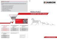
DR detector unit<br />
CR<br />
examion® aqs<br />
Acquisition software<br />
X-ray unit (motorized)<br />
raw images � � position protocol<br />
dr detector control � � collimator/motor control<br />
f control of X-ray generator<br />
<strong>and</strong> DR panel<br />
f advanced image processing<br />
f image management<br />
f local image database<br />
X-ray generator<br />
f integrated diagnostic<br />
scanned raw images �<br />
viewer<br />
� exposure protocol<br />
film scanner control �<br />
DICOM Worklist � � DICOM Store<br />
PaCs<br />
� kV, mAs, body part, etc.<br />
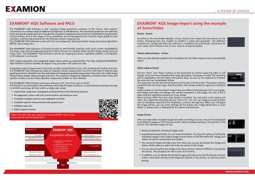
EXAMION ® <strong>AQS</strong> Image-Import using the example<br />
of Sono/Video<br />
Device - Screen<br />
To switch to the Sono/Video Module, all you need to do is select the sono device on the<br />
screen. Changing from one module to another is quick <strong>and</strong> automatic. The different<br />
modules are configured when the software is installed <strong>and</strong> individually customized for<br />
your needs. Each module is has its own, easy-to-recognize symbol.<br />
Patient adminsitration - Screen<br />
After you have selected a patient from the patient list, the Video Capture screen automatically<br />
opens.<br />
Video-Capture Screen<br />
use the “Start” <strong>and</strong> “Stop” buttons or the footswitch to control capturing video or still<br />
images. Once you have completed the study description, the system is ready. The recording<br />
time is displayed in the bar above the patient data. As soon as the exposure is finished,<br />
the next one can immediately follow.<br />
The exposures can be mirrored along the vertical axis in the live view. The button remains<br />
pressed for the mirrored image. Press the button with camera symbol to capture an individual<br />
image.<br />
Small symbols on the thumbnail images help you differentiate between the X-ray images,<br />
still images <strong>and</strong> video recordings: the camera represents a still image, the roll of film a<br />
video <strong>and</strong> the radioactive symbol an X-ray image.<br />
Other settings differ from one video device to another. You can select a new device <strong>and</strong><br />
select the supported recording devices from a list. You can use image processing filters<br />
such as histogram operations for brightness, contrast <strong>and</strong> gamma. When you configure<br />
the image format, you can enter settings for the frame rate, image dimensions or color<br />
depth. A dialog menu is displayed by the device manufacturer.<br />
Image Viewer<br />
After you have taken multiple image <strong>and</strong> video recordings in a row, they are first displayed<br />
as thumbnail images on the Overview screen. before postprocessing or saving them in the<br />
archive, the selected images can be:<br />
f directly printed (for individual images only)<br />
f exported <strong>and</strong> saved locally or to an external medium. You have the option of saving the<br />
individual images in the original image format <strong>and</strong>/or as DICOM <strong>and</strong>/or JPG. Images <strong>and</strong><br />
videos can also be anonymized <strong>and</strong> zipped.<br />
f You can send images <strong>and</strong> video via e-mail. Here, too, you can anonymize the images <strong>and</strong><br />
videos. All the data you select <strong>and</strong> mark are noted on the image.<br />
f You can also anonymize the images <strong>and</strong> videos <strong>and</strong> burn them to CD/DVD in the desired<br />
file format. The procedure for this is quick <strong>and</strong> intuitive.<br />
f In addition, you can delete the selected images <strong>and</strong> videos from the thumbnail overview<br />
screen, move them directly to the diagnosis worklist or the archive, or start the viewer<br />
directly.<br />
... A SYSTEM OF CHOICES<br />
www.examion.com