European Journal of Radiology Radiofrequency ablation in the ...
European Journal of Radiology Radiofrequency ablation in the ...
European Journal of Radiology Radiofrequency ablation in the ...

Create successful ePaper yourself
Turn your PDF publications into a flip-book with our unique Google optimized e-Paper software.
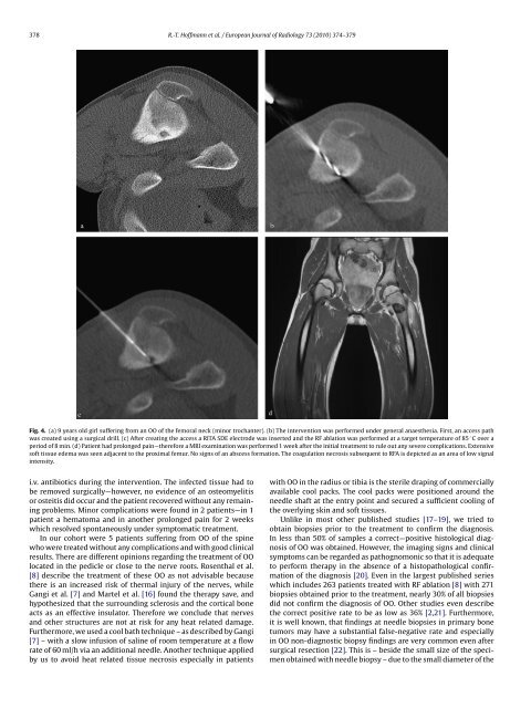
378 R.-T. H<strong>of</strong>fmann et al. / <strong>European</strong> <strong>Journal</strong> <strong>of</strong> <strong>Radiology</strong> 73 (2010) 374–379Fig. 4. (a) 9 years old girl suffer<strong>in</strong>g from an OO <strong>of</strong> <strong>the</strong> femoral neck (m<strong>in</strong>or trochanter). (b) The <strong>in</strong>tervention was performed under general anaes<strong>the</strong>sia. First, an access pathwas created us<strong>in</strong>g a surgical drill. (c) After creat<strong>in</strong>g <strong>the</strong> access a RITA SDE electrode was <strong>in</strong>serted and <strong>the</strong> RF <strong>ablation</strong> was performed at a target temperature<strong>of</strong>85 ◦ Coveraperiod <strong>of</strong> 8 m<strong>in</strong>. (d) Patient had prolonged pa<strong>in</strong>—<strong>the</strong>refore a MRI exam<strong>in</strong>ation was performed 1 week after <strong>the</strong> <strong>in</strong>itial treatment to rule out any severe complications. Extensives<strong>of</strong>t tissue edema was seen adjacent to <strong>the</strong> proximal femur. No signs <strong>of</strong> an abscess formation. The coagulation necrosis subsequent to RFA is depicted as an area <strong>of</strong> low signal<strong>in</strong>tensity.i.v. antibiotics dur<strong>in</strong>g <strong>the</strong> <strong>in</strong>tervention. The <strong>in</strong>fected tissue had tobe removed surgically—however, no evidence <strong>of</strong> an osteomyelitisor osteitis did occur and <strong>the</strong> patient recovered without any rema<strong>in</strong><strong>in</strong>gproblems. M<strong>in</strong>or complications were found <strong>in</strong> 2 patients—<strong>in</strong> 1patient a hematoma and <strong>in</strong> ano<strong>the</strong>r prolonged pa<strong>in</strong> for 2 weekswhich resolved spontaneously under symptomatic treatment.In our cohort were 5 patients suffer<strong>in</strong>g from OO <strong>of</strong> <strong>the</strong> sp<strong>in</strong>ewho were treated without any complications and with good cl<strong>in</strong>icalresults. There are different op<strong>in</strong>ions regard<strong>in</strong>g <strong>the</strong> treatment <strong>of</strong> OOlocated <strong>in</strong> <strong>the</strong> pedicle or close to <strong>the</strong> nerve roots. Rosenthal et al.[8] describe <strong>the</strong> treatment <strong>of</strong> <strong>the</strong>se OO as not advisable because<strong>the</strong>re is an <strong>in</strong>creased risk <strong>of</strong> <strong>the</strong>rmal <strong>in</strong>jury <strong>of</strong> <strong>the</strong> nerves, whileGangi et al. [7] and Martel et al. [16] found <strong>the</strong> <strong>the</strong>rapy save, andhypo<strong>the</strong>sized that <strong>the</strong> surround<strong>in</strong>g sclerosis and <strong>the</strong> cortical boneacts as an effective <strong>in</strong>sulator. Therefore we conclude that nervesand o<strong>the</strong>r structures are not at risk for any heat related damage.Fur<strong>the</strong>rmore, we used a cool bath technique – as described by Gangi[7] – with a slow <strong>in</strong>fusion <strong>of</strong> sal<strong>in</strong>e <strong>of</strong> room temperature at a flowrate <strong>of</strong> 60 ml/h via an additional needle. Ano<strong>the</strong>r technique appliedby us to avoid heat related tissue necrosis especially <strong>in</strong> patientswith OO <strong>in</strong> <strong>the</strong> radius or tibia is <strong>the</strong> sterile drap<strong>in</strong>g <strong>of</strong> commerciallyavailable cool packs. The cool packs were positioned around <strong>the</strong>needle shaft at <strong>the</strong> entry po<strong>in</strong>t and secured a sufficient cool<strong>in</strong>g <strong>of</strong><strong>the</strong> overly<strong>in</strong>g sk<strong>in</strong> and s<strong>of</strong>t tissues.Unlike <strong>in</strong> most o<strong>the</strong>r published studies [17–19], we tried toobta<strong>in</strong> biopsies prior to <strong>the</strong> treatment to confirm <strong>the</strong> diagnosis.In less than 50% <strong>of</strong> samples a correct—positive histological diagnosis<strong>of</strong> OO was obta<strong>in</strong>ed. However, <strong>the</strong> imag<strong>in</strong>g signs and cl<strong>in</strong>icalsymptoms can be regarded as pathognomonic so that it is adequateto perform <strong>the</strong>rapy <strong>in</strong> <strong>the</strong> absence <strong>of</strong> a histopathological confirmation<strong>of</strong> <strong>the</strong> diagnosis [20]. Even <strong>in</strong> <strong>the</strong> largest published serieswhich <strong>in</strong>cludes 263 patients treated with RF <strong>ablation</strong> [8] with 271biopsies obta<strong>in</strong>ed prior to <strong>the</strong> treatment, nearly 30% <strong>of</strong> all biopsiesdid not confirm <strong>the</strong> diagnosis <strong>of</strong> OO. O<strong>the</strong>r studies even describe<strong>the</strong> correct positive rate to be as low as 36% [2,21]. Fur<strong>the</strong>rmore,it is well known, that f<strong>in</strong>d<strong>in</strong>gs at needle biopsies <strong>in</strong> primary bonetumors may have a substantial false-negative rate and especially<strong>in</strong> OO non-diagnostic biopsy f<strong>in</strong>d<strong>in</strong>gs are very common even aftersurgical resection [22]. This is – beside <strong>the</strong> small size <strong>of</strong> <strong>the</strong> specimenobta<strong>in</strong>ed with needle biopsy – due to <strong>the</strong> small diameter <strong>of</strong> <strong>the</strong>