- Page 2 and 3:
Local Organizing CommitteeThomas Ba
- Page 4 and 5:
Women in Physics, why so few?Petra
- Page 6 and 7:
Carbon nanotubes in materials scien
- Page 8 and 9:
Temperature Study of Surface Plasmo
- Page 10 and 11:
Transmission Electron Microscopy Te
- Page 12 and 13:
Transmission Electron Microscopy St
- Page 14 and 15:
Effects of pressure on the Boson pe
- Page 16 and 17:
Session MON-3Physics of surfaces, t
- Page 18 and 19:
Transmission Electron Microscopy of
- Page 20 and 21:
Magnetic and Fluorescent ZnO/Iron O
- Page 22 and 23:
Slip/no-slip existence at the nanos
- Page 24 and 25:
Session MON-4Strongly correlated el
- Page 26 and 27:
The multilayer growth was studied b
- Page 28 and 29:
Fig.1b: orthorhombic, Pmmn, a=4.78
- Page 30 and 31:
Next we present our preliminary res
- Page 32 and 33:
Dielectric Properties of La 2 CuO 4
- Page 34 and 35:
Photonic interfacing by laser light
- Page 36 and 37:
Figure 2. Phononic band structure a
- Page 38 and 39:
can support, in addition, phononic
- Page 40 and 41:
WO 3 films electrodeposited on a pi
- Page 42 and 43:
Compared to electrochromics, the ph
- Page 44 and 45:
All-angle Homogenization of Anisotr
- Page 46 and 47:
Implantation and Diffusion of Galli
- Page 48 and 49:
Session TUE-1Physics of surfaces, t
- Page 50 and 51:
Nano-crystalline Mg 2 Si Prepared b
- Page 52 and 53:
Strain State and Internal Polarizat
- Page 54 and 55:
From Quantum Dynamics to Supramolec
- Page 56 and 57:
A neutron diffraction and magnetiza
- Page 58 and 59:
(a)0.12(b)0.050TRM0.110.100.09t w=2
- Page 60 and 61:
systems. It is also well known that
- Page 62 and 63:
we flip the cluster and n B is the
- Page 64 and 65:
the rare earth cation towards 3+ [4
- Page 66 and 67:
Development of Smart Polymer-functi
- Page 68 and 69:
Silicon Oxy Carbide Nanorings From
- Page 70 and 71:
Ordered Mesoporous Silicas as Addit
- Page 72 and 73:
Investigation of the Nanomechanical
- Page 74 and 75:
Nonlinearity-induced Anomalous Char
- Page 76 and 77:
Growth and Properties of Porous Ano
- Page 78 and 79:
Session TUE-4Ceramics, composites,m
- Page 80 and 81:
the layered materials was examined
- Page 82 and 83:
Normalised TL (a.u.)Normalised TL (
- Page 84 and 85:
Figure 1: Summary of reactions betw
- Page 86 and 87:
without the intermediate drying ste
- Page 88 and 89:
For the evaluation of the insertion
- Page 90 and 91:
One of another great value is that
- Page 92 and 93:
Modeling Nanostructured Materials S
- Page 94 and 95:
conductance at the conductance thre
- Page 96 and 97:
Pb 5d3/2 peak height (a.u.)0.180.16
- Page 98 and 99:
slope might be the same for all cas
- Page 100 and 101:
Session WED-2Physics of surfaces, t
- Page 102 and 103:
Load, μN500400300200Load, μΝ5000
- Page 104 and 105:
In Figs. 2 and 3 the load vs. displ
- Page 106 and 107:
Thermally Poled Glasses with Non-li
- Page 108 and 109:
Figure 1. HRTEM image of a SiO x fi
- Page 110 and 111:
A compared to film B and C, by one
- Page 112 and 113:
Surface Enhanced Raman Spectroscopy
- Page 114 and 115:
equirements for a variety of bias v
- Page 116 and 117:
Phase Solitons in the Spin Ground S
- Page 118 and 119:
Poster Session 1Photonics and optoe
- Page 120 and 121:
Diffraction Grating Thin Film Senso
- Page 122 and 123:
Using a polarised laser source at 6
- Page 124 and 125:
EPR and Conductivity Study of the M
- Page 126 and 127:
Electronic transport in nanocrystal
- Page 128 and 129:
Carrier Scattering and Hybrid Phono
- Page 130 and 131:
One-phonon processes versus Multi-p
- Page 132 and 133:
Far Infrared Spectra of the AsS 2 a
- Page 134 and 135:
The Effect of Tin Impurities in the
- Page 136 and 137:
Poster Session 1Structural-dynamica
- Page 138 and 139:
Figure 1(b) displays the Raman spec
- Page 140 and 141:
Figure 2. Selected area diffraction
- Page 142 and 143:
Figure 1: Bright-field XTEM images
- Page 144 and 145:
Figure 2: Cross-sectional HRTEM ima
- Page 146 and 147:
1 GPa -1 ) [6]. Moreover, the press
- Page 148 and 149:
Poster Session 1Strongly correlated
- Page 150 and 151:
Effect Of [Fe(CN) 6 ] 4- Substituti
- Page 152 and 153:
Investigations of Orbital and Magne
- Page 154 and 155:
Fig.1 Hysteresis loops of [Ni/Pt] 6
- Page 156 and 157:
the other hand at sample Fe4 the ch
- Page 158 and 159:
the absolute magnetization per site
- Page 160 and 161:
1J1+J2p( Jij ) = ⎡δ( Jij J1) δ(
- Page 162 and 163:
Spin order and lattice frustration
- Page 164 and 165:
CCIntensity (Counts)120010008006004
- Page 166 and 167:
Figure 2. CTEM and HRTEM of Fe 3 O
- Page 168 and 169:
the combined processes of grain gro
- Page 170 and 171:
Assemblies of Magnetic Nanoparticle
- Page 172 and 173:
Poster Session 1Physics of surfaces
- Page 174 and 175:
Figure 1. The magnetization, M, as
- Page 176 and 177:
2). Furthermore, the number of As a
- Page 178 and 179:
Development of a model Ziegler-Natt
- Page 180 and 181:
Ab initio study of Semipolar AlN Su
- Page 182 and 183:
Adsorption, diffusion and incorpora
- Page 184 and 185:
Self-Assembled InGaN Quantum Dot Su
- Page 186 and 187:
Formation of Hexagonal Ni in Ru/Ni
- Page 188 and 189:
Deposition of Superparamagnetic Col
- Page 190 and 191:
Electron Energy Loss Near Edge Stru
- Page 192 and 193:
Controllable synthesis of iron oxy-
- Page 194 and 195: Growth and Characterization of Al 2
- Page 196 and 197: The interaction of metallic Cr with
- Page 198 and 199: Optical Properties of Metallic Nano
- Page 200 and 201: Load, μN600500400300200100Load, μ
- Page 202 and 203: Figure 2a: AFM image showing thetop
- Page 204 and 205: Structural and electronic propertie
- Page 206 and 207: [2] Wildgoose G. G., Banks C. E., C
- Page 208 and 209: Poster Session 2Physics of surfaces
- Page 210 and 211: Ab-Initio Calculations of Cu Nanowi
- Page 212 and 213: Surface Functionalization by means
- Page 214 and 215: Formation of micro-cones by oxygen
- Page 216 and 217: this temperature there is no remark
- Page 218 and 219: Results and DiscussionNanocomposite
- Page 220 and 221: At 300 K, a significant hysteresis
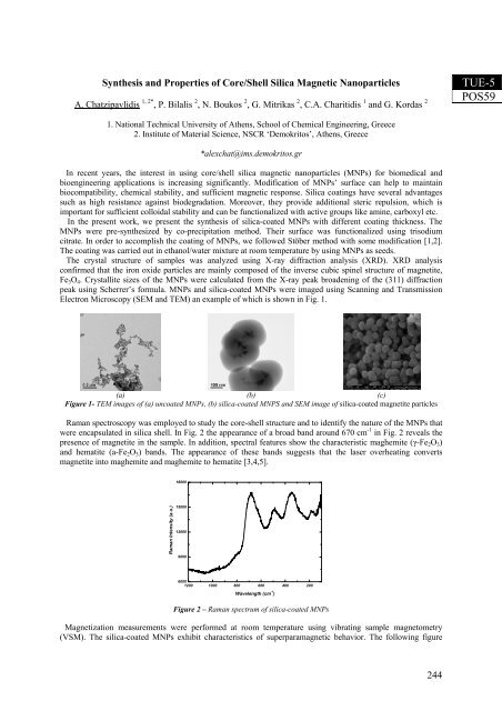
- Page 222 and 223: Figure 1: TEM micrograph of a film
- Page 224 and 225: 12,6A oSi_GOIntensity (a.u.)10,4 A
- Page 226 and 227: In Fig. I we show two representativ
- Page 228 and 229: working area of the periodic table
- Page 230 and 231: Thermal Treatment of PEDOT:PSS Film
- Page 232 and 233: A Novel Approach for Preparation of
- Page 234 and 235: Detection of Residual Solvent in Th
- Page 236 and 237: Brownian dynamics simulations on se
- Page 238 and 239: 120016010010001401201200100080E' (
- Page 240 and 241: Metathesis Polymerization of Phenyl
- Page 242 and 243: Microphase Separation in (AB)-Multi
- Page 246 and 247: Poster Session 2Ceramics, composite
- Page 248 and 249: Fig. 1: Influence of temperature an
- Page 250 and 251: Sample 5-TiO 2 P25 U-5-TiO 2 U-P25
- Page 252 and 253: transient state in the relaxation o
- Page 254 and 255: σ (A . m 2 /kg)0.650.600.550.500.4
- Page 256 and 257: (a)(b)Fig.2. a) Mean values of flex
- Page 258 and 259: transformation to Si-II and subsequ
- Page 260 and 261: ER/MWCNTs nanocomposites [6]. Moreo
- Page 262 and 263: a H (Å) Me-O (Å)1.9651.9551.9451.
- Page 264 and 265: suspension on glass slide. XRD anal
- Page 266 and 267: Table 1. Rietveld refinement struct
- Page 268 and 269: the real effect of a native oxide a
- Page 270 and 271: where it was shown that further pro
- Page 272 and 273: Calculation of the Penetration Dept
- Page 274 and 275: Oxidation of zinc hot-dip galvanize
- Page 276 and 277: An XRD study of galvanized coatings
- Page 278 and 279: Influence of Wollastonite on the Qu
- Page 280 and 281: Study of the Bioactive Hydroxyapati
- Page 282 and 283: Study of the Bioactive Hydroxyapati
- Page 284 and 285: An EPR-Based Method to Probe the Lo
- Page 286 and 287: Surprisingly, the spectra indicate
- Page 288 and 289: with O 2 . In the XRD pattern only
- Page 290 and 291: characteristic strong reflection at
- Page 292 and 293: indicates abundant volatile compone
- Page 294 and 295:
The layers are alternating in a sta
- Page 296 and 297:
(111)(220)MS200MS202Mg2Siintensity
- Page 298 and 299:
MEMS charging the FTIR spectra and
- Page 300 and 301:
possible this particular route for
- Page 302 and 303:
produces significantly more accurat
- Page 304 and 305:
Hyperthermia Response of Iron Oxide
- Page 306 and 307:
probably used, however, either this
- Page 308 and 309:
1110 cm-1 attributed to the ν 1 vi
- Page 310 and 311:
Choulis S.A. ......................
- Page 312 and 313:
Ho Rong-Ming ......................
- Page 314 and 315:
Malliakas Christos.................
- Page 316 and 317:
Simserides C.......................
- Page 318 and 319:
318
- Page 320 and 321:
23. All-angle Homogenization of Ani
- Page 322 and 323:
K. Karkas and N. Guskos ...........
- Page 324 and 325:
C. A. Charitidis,A. Skarmoutsou, A.
- Page 326 and 327:
G. Litsardakis and S. Nikolaki.....