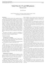
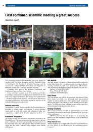
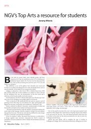
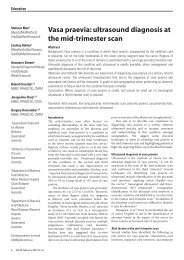
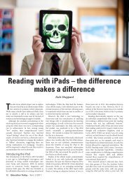
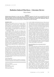
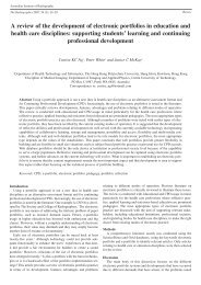
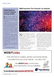
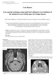
<strong>3D</strong> <strong>ultrasound</strong> <strong>in</strong> <strong>first</strong> <strong>and</strong> <strong>second</strong> <strong>trimester</strong> – hype or helpful?Fig. 3: <strong>3D</strong> surface image at 12wks demonstrates large omphalocoele<strong>and</strong> shortish limbs. Trisomy 13.Fig. 4: <strong>3D</strong> surface image at anatomy scan show<strong>in</strong>g persistent subtleoverlapp<strong>in</strong>g f<strong>in</strong>gers 2 <strong>and</strong> 3 right h<strong>and</strong>. Left h<strong>and</strong> <strong>and</strong> feet appearednormal. Trisomy 18.Fig. 5: <strong>3D</strong> skeletal image sp<strong>in</strong>e shows lower thoracic hemivertebrawith rib disparity.Anatomy scanFacial features, especially cleft lip <strong>and</strong> hard palate are wellsuited for <strong>3D</strong> imag<strong>in</strong>g, particularly when try<strong>in</strong>g to depictthese for the parents or cl<strong>in</strong>icians. A well performed 2Danatomy scan should detect virtually all cleft lips, but sometimescleft of the hard palate can only be found us<strong>in</strong>g volumeimag<strong>in</strong>g with either a reverse face <strong>3D</strong> 2 or an extended midl<strong>in</strong>esagittal image acquisition <strong>3D</strong> 1 .LimbsH<strong>and</strong>, wrist <strong>and</strong> foot anomalies, <strong>in</strong>clud<strong>in</strong>g clubfoot, polydactyly<strong>and</strong> clench<strong>in</strong>g, may be very difficult to depict <strong>in</strong> 2Dimag<strong>in</strong>g but are often well seen us<strong>in</strong>g <strong>3D</strong> surface (Fig. 4).Skeletal assessmentIn particular, where sp<strong>in</strong>al anomalies such as hemivertebraare detected, <strong>3D</strong> imag<strong>in</strong>g of the sp<strong>in</strong>e helps confirm the f<strong>in</strong>d<strong>in</strong>gby depict<strong>in</strong>g the hemivertebra, <strong>and</strong> also allows accuratecount<strong>in</strong>g of ribs on either side (Fig. 5). Sp<strong>in</strong>a bifida, skull<strong>and</strong> long bones can also be further demonstrated with <strong>3D</strong>.HeartThe Spatio-Temporal Image Correlation (STIC) volumeacquisition, can allow rapid depiction of the cardiac anatomy,<strong>and</strong> with an enthusiastic sonographer can give a quickFig. 6: <strong>3D</strong> surface image face at anatomy scan show<strong>in</strong>g descriptivefacial <strong>and</strong> h<strong>and</strong> expressions.<strong>and</strong> accurate depiction of the chambers <strong>and</strong> major vessels3,4,5,6 . STIC has many benefits both for screen<strong>in</strong>g <strong>in</strong> therout<strong>in</strong>e anatomy scan, <strong>and</strong> for further evaluat<strong>in</strong>g the heartwhere an abnormality is suspected. It improves resolution,offers unlimited images <strong>in</strong> any plane, allows simultaneouscorrelation between image planes that are perpendicular tothe acquisition plane <strong>and</strong> <strong>3D</strong> rendered images can be reconstructed3 . With an experienced sonologist, evaluation timemay be shortened, especially with complex defects. It alsoallows all images <strong>in</strong> all planes to be reviewed <strong>in</strong> c<strong>in</strong>e-loop,<strong>and</strong> the complete data volume can be stored, reviewed <strong>and</strong>manipulated later. This means the data can be assessed at aremote site by a more expert cl<strong>in</strong>ician who is not limited bythe f<strong>in</strong>d<strong>in</strong>gs detected by the <strong>first</strong> exam<strong>in</strong>er 3 . In conventional2D <strong>ultrasound</strong> review of the exam<strong>in</strong>ation is generally limitedto the f<strong>in</strong>d<strong>in</strong>gs detected <strong>and</strong> documented by the sonologist.Some <strong>3D</strong>/4D imag<strong>in</strong>g can be <strong>in</strong>corporated <strong>in</strong>to the rout<strong>in</strong>escreen<strong>in</strong>g anatomy scan with not much effort or <strong>in</strong>crease<strong>in</strong> scann<strong>in</strong>g time <strong>in</strong> the majority of patients. Certa<strong>in</strong>ly, tolook at the face <strong>and</strong> limbs only takes a few moments, <strong>and</strong>with enthusiasm <strong>and</strong> some practice, the heart <strong>and</strong> sp<strong>in</strong>e canoften be shown <strong>in</strong> a more useful way. Just as the developmentof reasonable technique for th<strong>in</strong>gs such as NT scann<strong>in</strong>g,nasal bone, tricuspid <strong>and</strong> ductus, requires 50–100 orAustralasian Journal of Ultrasound <strong>in</strong> Medic<strong>in</strong>e August 2009; 12 (3)29
Richard Davisso cases, similarly basic expertise <strong>in</strong> <strong>3D</strong> scann<strong>in</strong>g can beacquired <strong>and</strong> used dur<strong>in</strong>g anatomy assessment 6 .So far, we are only really scratch<strong>in</strong>g at the surface with<strong>3D</strong>. It has opened up the possibilities of what we can dowith <strong>ultrasound</strong>, <strong>and</strong> its beneficial uses will <strong>in</strong>crease withpeople’s imag<strong>in</strong>ation.Indirect benefitsThere are numerous <strong>in</strong>direct ways <strong>in</strong> which <strong>3D</strong> <strong>ultrasound</strong>helps <strong>in</strong> <strong>first</strong> <strong>and</strong> <strong>second</strong> <strong>trimester</strong> <strong>ultrasound</strong>. Althoughsome of these are debatable, <strong>and</strong> also unscientific, they stillare real, <strong>and</strong> for me they have been a giant leap forward <strong>in</strong>the use of <strong>ultrasound</strong>.<strong>3D</strong> <strong>ultrasound</strong> further “humanises” the fetus <strong>in</strong> the eyesof the parents <strong>and</strong> sonographers. Perhaps this will helpclarify the rights of the fetus/unborn child, <strong>and</strong> may also helpwith ethical considerations. <strong>3D</strong> will enable a better underst<strong>and</strong><strong>in</strong>gof fetal behaviour. At the moment our focus on fetalwellbe<strong>in</strong>g (“happ<strong>in</strong>ess”) is largely conf<strong>in</strong>ed to third <strong>trimester</strong>biophysical profile <strong>and</strong> Doppler. With 4D <strong>ultrasound</strong> we cansee the “happy” 12-week fetus, not only jump<strong>in</strong>g around, butbreak-danc<strong>in</strong>g, kickbox<strong>in</strong>g, wav<strong>in</strong>g <strong>and</strong> explor<strong>in</strong>g its world.The happy <strong>second</strong> <strong>trimester</strong> fetus can be seen mak<strong>in</strong>g facialexpressions, tongue-pok<strong>in</strong>g, explor<strong>in</strong>g its body with h<strong>and</strong>s,<strong>and</strong> even giv<strong>in</strong>g the hippy peace sign or other “gangsta”h<strong>and</strong> signals (Fig. 6). The reaction of parents <strong>and</strong> sonographersupon see<strong>in</strong>g this is special.Parental <strong>and</strong> family bond<strong>in</strong>g is enhanced. Is this justsome psycho mumbo-jumbo? No, I th<strong>in</strong>k it’s real, <strong>and</strong> comesat a time when sometimes there may be extra stra<strong>in</strong>s with<strong>in</strong> arelationship, or with<strong>in</strong> a family. Mum knows she is pregnant<strong>and</strong> feels the babe, but now Dad can really see the cutie caus<strong>in</strong>gthe havoc, <strong>and</strong> big brother or sister can better underst<strong>and</strong>Mum’s distraction 7,8 .What about the enjoyment <strong>and</strong> attentiveness of thesonographer? With<strong>in</strong> my practice I have seen a newfoundstaff enthusiasm with the use of 4D. It has added a spr<strong>in</strong>g <strong>in</strong>their step <strong>and</strong> put a smile on their face, to the extent that mypolicy now is that the brief use of 4D is encouraged as partof the rout<strong>in</strong>e obstetric scan. Why? Well I believe this makesus better sonographers. Why is it that even now, there aresome cases of sp<strong>in</strong>a bifida, cleft lip, club-foot, h<strong>and</strong> anomaliesetc. that are not detected at the rout<strong>in</strong>e anatomy scan?Why are we not better at detect<strong>in</strong>g major heart abnormalities.We all know we should do better. My belief is that someof the “missed” diagnoses arise because of complacency,or boredom. Most anatomy scans are “normal”. For thoseof us do<strong>in</strong>g rout<strong>in</strong>e scans, the <strong>in</strong>cidence of most anomaliesmeans we can do hundreds of anatomy scans without see<strong>in</strong>ga major abnormality. To f<strong>in</strong>d the “wolf <strong>in</strong> sheep’s cloth<strong>in</strong>g”requires a constant vigilance <strong>in</strong> the face of endless normals.It is harder to stay “switched on” when there is no variation,so the cerebellum is overlooked, the top lip not viewed, orboth h<strong>and</strong>s not checked. 4D provides the variation, the sparkor stimulus to keep us alert for every scan. The vitality of<strong>3D</strong> is also seen add<strong>in</strong>g enthusiasm to non-cl<strong>in</strong>ical reception<strong>and</strong> office staff.Another <strong>in</strong>direct benefit of 4D is it encourages thesonographer to obta<strong>in</strong> a nice midl<strong>in</strong>e sagital view of the face<strong>in</strong> 2D, this be<strong>in</strong>g the position to obta<strong>in</strong> a frontal face <strong>3D</strong>. Italso happens to be an important position to assess the nasalbone, the hard palate, the m<strong>and</strong>ible for micrognathia, theBabe with attitude.frontal bone <strong>and</strong> the premaxillary soft tissue, amongst otherth<strong>in</strong>gs. That’s useful.The advent of 4D is forc<strong>in</strong>g private practices <strong>and</strong> hospitaldepartments to upgrade, so we have the latest hardware <strong>and</strong>software benefits these mach<strong>in</strong>es br<strong>in</strong>g, rather than mak<strong>in</strong>gdo with an older mach<strong>in</strong>e <strong>and</strong> a fuzzier image. Furtherto this, 4D is generally only available on the higher endmach<strong>in</strong>es, which tend to have a clearer image <strong>and</strong> betteruser characteristics. Aga<strong>in</strong> this helps by, not only the clearerimage assist<strong>in</strong>g diagnosis, but it also enthuses the operator<strong>and</strong> enhances performance.ConclusionsEven sett<strong>in</strong>g narrow parameters on how we judge the helpfulnessof <strong>3D</strong> <strong>in</strong> <strong>first</strong> <strong>and</strong> <strong>second</strong> <strong>trimester</strong> <strong>ultrasound</strong> (versusconventional 2D), we can see it clearly makes a difference<strong>in</strong> areas such as anatomic anomalies (cleft lip, clubfoot etc.),<strong>in</strong> better assessment of the heart, <strong>and</strong> by giv<strong>in</strong>g a reassur<strong>in</strong>goverview of limbs, face <strong>and</strong> body surface.With wider parameters <strong>and</strong> by assess<strong>in</strong>g <strong>in</strong>direct benefitsof <strong>3D</strong>, the helpfulness of this imag<strong>in</strong>g modality is even morecompell<strong>in</strong>g. So much so that, at the current po<strong>in</strong>t <strong>in</strong> time, itwould be very hard to argue aga<strong>in</strong>st replac<strong>in</strong>g any outdatedUS mach<strong>in</strong>e with anyth<strong>in</strong>g but a 4D capable unit.Then there’s the future. I have never been great at crystalball gaz<strong>in</strong>g, but we can look at how history treats significanttechnological advances. In 15 years we have gone frombe<strong>in</strong>g amazed at a brick we could use as a mobile phone,to now be<strong>in</strong>g complacent about a th<strong>in</strong>g the size of a pack ofcards which is not only a phone, but a great camera, computer,GPS navigation system <strong>and</strong> portable stereo all <strong>in</strong> one!To me the future uses of 4D <strong>ultrasound</strong> will exp<strong>and</strong> greatly,mak<strong>in</strong>g our assessment of the fetus <strong>in</strong> the <strong>first</strong> <strong>and</strong> <strong>second</strong><strong>trimester</strong>s, not only a more complete, but also a more enjoyableexperience.Yes there are problems, <strong>and</strong> probably the biggest problemis to actually recognise what is just hype, <strong>and</strong> what isreally useful with 4D. While the real uses, the th<strong>in</strong>gs we canreally use 4D for <strong>in</strong> our everyday scann<strong>in</strong>g, are plentiful <strong>and</strong>compell<strong>in</strong>g enough that we all need to utilise it, the hype willbe even more plentiful. The reality of academia, the bus<strong>in</strong>essworld, <strong>and</strong> human <strong>in</strong>quisitiveness means that alreadythere are huge amounts of hype about obscure <strong>and</strong> difficultuses for 4D. Articles need publish<strong>in</strong>g, <strong>and</strong> the “volumes”30 Australasian Journal of Ultrasound <strong>in</strong> Medic<strong>in</strong>e August 2009; 12 (3)