Skull Series Radiographic Views
Skull Series Radiographic Views
Skull Series Radiographic Views
- No tags were found...

You also want an ePaper? Increase the reach of your titles
YUMPU automatically turns print PDFs into web optimized ePapers that Google loves.
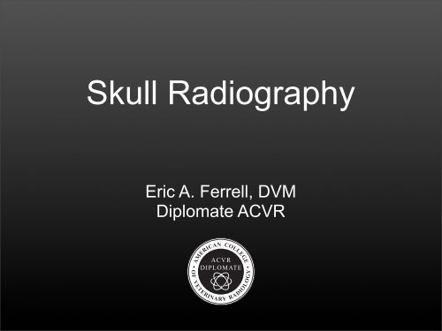
<strong>Skull</strong> RadiographyEric A. Ferrell, DVMDiplomate ACVRACVRDIPLOMATE• AMERICAN COLLEGE•OF VETERINARY RADIOLOGY
Reference materials- Veterinary Diagnostic Radiology -Thrall- Diagnostic Radiology and Ultrasonography of theDog and the Cat - Kealy, McAllister & Graham- Techniques of Veterinary Radiography - Morgan- Miller's Anatomy of the Dog- Other
Objectives- Technical considerations / QC- Normal anatomy- <strong>Radiographic</strong> views- Disease processes- Additional modalities
image transition slide
General overview- Review for normal skull radiography• nasal cavities, sinuses, maxilla, mandible, ...• also pharynx& larynx
General overview- Complex anatomy
General overview- Multiple species
General overview- Breed differences- Infrequent procedure w/ lack of familiarityCN, MesaticephalicCN, Brachycephalicbrachy vs meta vsdolio....imagesFE, rostrocaudal bullae
General overview- Reference material & anatomic specimen
General overview- Systematic assessment- Röentgen signs• Size• Shape• Margination• Opacity• Number• Position / locationWilhelm Conrad Röentgen
Technical considerations- General anesthesia- Positioning aids
Technical considerations- Positioning markers• R, L, VD, DV• important for obliques- Collimation- SF radiography• exposure factors• grid if greater than 10 cm
Technical considerations- Digital (CR/DR)• exposure factors• intraoral projections, DR
Radiation safety- NOT acceptabletechnique forskull radiography...
Completeness- Projections needed dependent uponquestion to be answered- “ Do you need a rostrocaudal frontal sinuswhen assessing the mandible?”or“ Do you need a rostrocaudal of the frontalsinus on a brachycephalic?”
Completeness<strong>Skull</strong> <strong>Series</strong>Routine surveyFrontal sinusNasal cavityMaxillaMandibleMaxillary teethMandibular teethMaxillary incisorsMandibular incisorsTympanic bullaeTMJZygomatic and orbital<strong>Radiographic</strong> <strong>Views</strong>DV/VD, lateralDV/VD, lateral, lateral oblique, open mouth frontalDV/VD, lateral, open mouth VDDV/VD, lateral, open mouth lateral obliqueDV/VD, lateral, open mouth lateral obliqueDV/VD, open mouth lateral oblique, intraoral DVDV/VD, open mouth lateral oblique, intraoral VDlateral, intraoral DVlateral, intraoral VDDV/VD, lateral oblique, open mouth rostrocaudalDV/VD, lateral oblique, sagittal obliqueDV/VD, lateral oblique, open mouth VD, rostrocaudal
The Normal <strong>Skull</strong>- Normal positions & images- Select abnormalities
<strong>Radiographic</strong> views- Lateral<strong>Radiographic</strong> Techniques: The Dog. Morgan JP, et al.
<strong>Radiographic</strong> views• Lateral
<strong>Radiographic</strong> views- DV<strong>Radiographic</strong> Techniques: The Dog. Morgan JP, et al.
<strong>Radiographic</strong> views• DV
<strong>Radiographic</strong> views- VD<strong>Radiographic</strong> Techniques: The Dog. Morgan JP, et al.
<strong>Radiographic</strong> views• DV
<strong>Radiographic</strong> views- DV, intraoral<strong>Radiographic</strong> Techniques: The Dog. Morgan JP, et al.
<strong>Radiographic</strong> views• DV, intraoral
<strong>Radiographic</strong> views- VD, intraoral<strong>Radiographic</strong> Techniques: The Dog. Morgan JP, et al.
<strong>Radiographic</strong> views• VD, intraoral
<strong>Radiographic</strong> views- Lateral oblique, mandible<strong>Radiographic</strong> Techniques: The Dog. Morgan JP, et al.
<strong>Radiographic</strong> views• Lateral oblique,mandible
<strong>Radiographic</strong> views- Lateral oblique, maxilla<strong>Radiographic</strong> Techniques: The Dog. Morgan JP, et al.
<strong>Radiographic</strong> views• Lateral oblique,maxilla
<strong>Radiographic</strong> views- Ventrodorsal, open-mouth<strong>Radiographic</strong> Techniques: The Dog. Morgan JP, et al.
<strong>Radiographic</strong> views• Ventrodorsal,open-mouth
<strong>Radiographic</strong> views- Rostrocaudal,open-mouth bulla<strong>Radiographic</strong> Techniques: The Dog. Morgan JP, et al.
<strong>Radiographic</strong> views- Rostrocaudal,open-mouth bulla
<strong>Radiographic</strong> views- Rostrocaudal, bulla• rostro 10° ventral caudodorsal obliqueVet Radiol Ultrasound 1995;36:14–15
<strong>Radiographic</strong> views• Rostrocaudal, bulla
<strong>Radiographic</strong> views- Lateral oblique, bulla• same lateral oblique as for maxilla<strong>Radiographic</strong> Techniques: The Dog. Morgan JP, et al.
<strong>Radiographic</strong> views• Lateral oblique, bulla
<strong>Radiographic</strong> views- Lateral oblique, TMJ• same lateral oblique as for bulla & maxilla<strong>Radiographic</strong> Techniques: The Dog. Morgan JP, et al.
<strong>Radiographic</strong> views• Lateral oblique, bulla
<strong>Radiographic</strong> views- Lateral oblique, TMJ (aka sagittal oblique)- fig 6-4<strong>Radiographic</strong> Techniques: The Dog. Morgan JP, et al.
<strong>Radiographic</strong> views• Lateral oblique,TMJ• fig 6-4
<strong>Radiographic</strong> views- Rostrocaudal, frontal sinus<strong>Radiographic</strong> Techniques: The Dog. Morgan JP, et al.
<strong>Radiographic</strong> views• Rostrocaudal,frontal sinus
<strong>Radiographic</strong> views- Pharynx & larynx
<strong>Radiographic</strong> views- Pharynx & larynx
Tooth root abscess
Nasopharyngeal polyp & otitis
Nasopharyngeal polyp & otitis
Nasal adenocarcinoma
Periodontal epulis
Rhinitis
Fungal rhinitis
Fibrosarcoma
Cross sectional imaging- Consider CT/MR compared to radiographs• Anesthesia, cost,availability & moredetailed information
Cross sectional imaging
<strong>Skull</strong> radiography- Questions- Thank you• FVMA• My wonderful wife,Jen & our beautifulgirls, Ella and Anna