Analysis of mRNA Levels by RT-PCR 433. 10X RT buffer: 200 mM Tris–HCl, pH 8.4 (room temperature), 500 mM KCl, 30mM MgCl 2 , 1 mg/mL BSA <strong>in</strong> DEPC–H 2 O, store at –20°C.4. 100 µM RT-primer, deprotected, desalted, and redissolved <strong>in</strong> DEPC–H 2 O: 5'-NNNNNC -3' (N = G, A, T, or C), store at –20°C.5. 40 U/µL RNAs<strong>in</strong> (Promega, Madison, WI), store at –20°C.6. 200 U/µL M-MLV reverse transcriptase, (Gibco BRL), store <strong>in</strong> aliquots at –70°C,sensitive to repeated freeze–thaw cycles).2.4. PCR Reaction1. 5 U/µL AmpliTaq DNA polymerase (Perk<strong>in</strong> Elmer, Branchburg, NJ), store at –20°C.2. 370 MBq/mL (10 mCi/mL) [α- 32 P]-deoxycytid<strong>in</strong>triphosphate , 110 TBq/mmol(3000 Ci/mmol) (Caution: radioactive material must be handled with great careand accord<strong>in</strong>g to local radiation safety regulations!).3. Light m<strong>in</strong>eral oil.4. PCR–dNTP mix (10 mM each: dCTP, dGTP, dATP, dTTP diluted <strong>in</strong> dH 2 O),store at –20°C.5. 10X PCR buffer: 100 mM Tris–HCl, pH 8.4 (room temperature), 500 mM KCl,15 mM MgCl 2 , 0.01 % (w/v) gelat<strong>in</strong>e <strong>in</strong> dH 2 O, made up from autoclaved stocksolutions, store at –20°C.6. 25 µM PCR primers, deprotected, desalted, and redissolved <strong>in</strong> dH 2 O, store at –20°C.7. Standard radiation safety devices.2.5. Electrophoresis and Detection1. Acrylamide/bisacrylamide solution (29:1) <strong>in</strong> dH 2 O.2. Blott<strong>in</strong>g paper.3. 5X DNA-load<strong>in</strong>g dye: 0.25 % bromphenol blue, 0.25% xylene cyanol, 15% Ficoll400 <strong>in</strong> dH 2 O.4. 10X TBE electrophoresis buffer: 1 M Tris–HCl, pH 8.6 (room temperature), 0.83M boric acid, 10 mM EDTA.5. Vertical polyacrylamide gel electrophoresis system with DC power supply.6. Vacuum gel dryer (caution: use cool<strong>in</strong>g trap or an activated charcoal filter toscavenge radioactive aerosols).7. Phosphor imager system or, as m<strong>in</strong>imal equipment, X-ray film and autoradiographycassettes with <strong>in</strong>tensify<strong>in</strong>g screens.3. <strong>Methods</strong>3.1. RNA PurificationBecause of its speed, high sample throughput, and reliability, we favor asimplified version of the s<strong>in</strong>gle-step extraction method by Chomczynski andSacchi (4). Stabilized, monophasic guanid<strong>in</strong>ium thiocyanate–phenol extractionsolutions are available from several manufacturers. Although RNA preparedby this method is contam<strong>in</strong>ated with variable amounts of genomic DNA,this is usually not sufficient to generate false-positive signals (see Note 3).
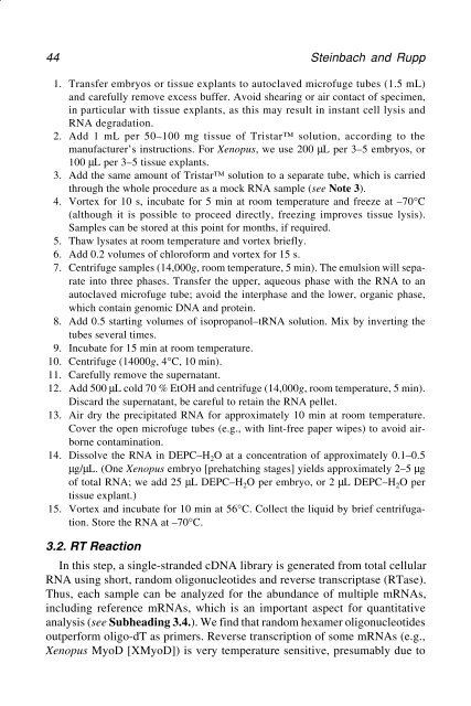
44 Ste<strong>in</strong>bach and Rupp1. Transfer embryos or tissue explants to autoclaved microfuge tubes (1.5 mL)and carefully remove excess buffer. Avoid shear<strong>in</strong>g or air contact of specimen,<strong>in</strong> particular with tissue explants, as this may result <strong>in</strong> <strong>in</strong>stant cell lysis andRNA degradation.2. Add 1 mL per 50–100 mg tissue of Tristar solution, accord<strong>in</strong>g to themanufacturer’s <strong>in</strong>structions. For Xenopus, we use 200 µL per 3–5 embryos, or100 µL per 3–5 tissue explants.3. Add the same amount of Tristar solution to a separate tube, which is carriedthrough the whole procedure as a mock RNA sample (see Note 3).4. Vortex for 10 s, <strong>in</strong>cubate for 5 m<strong>in</strong> at room temperature and freeze at –70°C(although it is possible to proceed directly, freez<strong>in</strong>g improves tissue lysis).Samples can be stored at this po<strong>in</strong>t for months, if required.5. Thaw lysates at room temperature and vortex briefly.6. Add 0.2 volumes of chloroform and vortex for 15 s.7. Centrifuge samples (14,000g, room temperature, 5 m<strong>in</strong>). The emulsion will separate<strong>in</strong>to three phases. Transfer the upper, aqueous phase with the RNA to anautoclaved microfuge tube; avoid the <strong>in</strong>terphase and the lower, organic phase,which conta<strong>in</strong> genomic DNA and prote<strong>in</strong>.8. Add 0.5 start<strong>in</strong>g volumes of isopropanol–tRNA solution. Mix by <strong>in</strong>vert<strong>in</strong>g thetubes several times.9. Incubate for 15 m<strong>in</strong> at room temperature.10. Centrifuge (14000g, 4°C, 10 m<strong>in</strong>).11. Carefully remove the supernatant.12. Add 500 µL cold 70 % EtOH and centrifuge (14,000g, room temperature, 5 m<strong>in</strong>).Discard the supernatant, be careful to reta<strong>in</strong> the RNA pellet.13. Air dry the precipitated RNA for approximately 10 m<strong>in</strong> at room temperature.Cover the open microfuge tubes (e.g., with l<strong>in</strong>t-free paper wipes) to avoid airbornecontam<strong>in</strong>ation.14. Dissolve the RNA <strong>in</strong> DEPC–H 2 O at a concentration of approximately 0.1–0.5µg/µL. (One Xenopus embryo [prehatch<strong>in</strong>g stages] yields approximately 2–5 µgof total RNA; we add 25 µL DEPC–H 2 O per embryo, or 2 µL DEPC–H 2 O pertissue explant.)15. Vortex and <strong>in</strong>cubate for 10 m<strong>in</strong> at 56°C. Collect the liquid by brief centrifugation.Store the RNA at –70°C.3.2. RT ReactionIn this step, a s<strong>in</strong>gle-stranded cDNA library is generated from total cellularRNA us<strong>in</strong>g short, random oligonucleotides and reverse transcriptase (RTase).Thus, each sample can be analyzed for the abundance of multiple mRNAs,<strong>in</strong>clud<strong>in</strong>g reference mRNAs, which is an important aspect for quantitativeanalysis (see Subhead<strong>in</strong>g 3.4.). We f<strong>in</strong>d that random hexamer oligonucleotidesoutperform oligo-dT as primers. Reverse transcription of some mRNAs (e.g.,Xenopus MyoD [XMyoD]) is very temperature sensitive, presumably due to
- Page 1 and 2: Methods in Molecular BiologyTMVOLUM
- Page 3 and 4: 2 Greentally active genes. Importan
- Page 5 and 6: 4 Greentwo or three gentle strokes
- Page 7 and 8: 6 Green
- Page 9 and 10: 8 GreenStage Assay Purpose10.5 RNA
- Page 11 and 12: 10 Greentein factors are to be used
- Page 13 and 14: 12 Greennature of much gene express
- Page 16 and 17: Cell/Tissue Transplantation in Zebr
- Page 18 and 19: Cell/Tissue Transplantation in Zebr
- Page 20 and 21: Cell/Tissue Transplantation in Zebr
- Page 22 and 23: Cell/Tissue Transplantation in Zebr
- Page 24 and 25: Cell/Tissue Transplantation in Zebr
- Page 26 and 27: Cell/Tissue Transplantation in Zebr
- Page 28 and 29: Cell/Tissue Transplantation in Zebr
- Page 30 and 31: Ribonuclease Protection Analysis 29
- Page 32 and 33: Ribonuclease Protection Analysis 31
- Page 34 and 35: Ribonuclease Protection Analysis 33
- Page 36 and 37: Ribonuclease Protection Analysis 35
- Page 38 and 39: Ribonuclease Protection Analysis 37
- Page 40 and 41: Ribonuclease Protection Analysis 39
- Page 42 and 43: Analysis of mRNA Levels by RT-PCR 4
- Page 46 and 47: Analysis of mRNA Levels by RT-PCR 4
- Page 48 and 49: Analysis of mRNA Levels by RT-PCR 4
- Page 50 and 51: Analysis of mRNA Levels by RT-PCR 4
- Page 52 and 53: Analysis of mRNA Levels by RT-PCR 5
- Page 54 and 55: Analysis of mRNA Levels by RT-PCR 5
- Page 56 and 57: Analysis of mRNA Levels by RT-PCR 5
- Page 58 and 59: WISH of Xenopus and Zebrafish Embry
- Page 60 and 61: WISH of Xenopus and Zebrafish Embry
- Page 62 and 63: WISH of Xenopus and Zebrafish Embry
- Page 64 and 65: WISH of Xenopus and Zebrafish Embry
- Page 66 and 67: WISH of Xenopus and Zebrafish Embry
- Page 68: WISH of Xenopus and Zebrafish Embry
- Page 71 and 72: 70 Bertwistleparative techniques. F
- Page 73 and 74: 72 Bertwistle3. Methods3.1. In Vitr
- Page 75 and 76: 74 Bertwistle7. Soak in 0.2 M HCl f
- Page 77 and 78: 76 Bertwistlegenes along the dorsov
- Page 79 and 80: 78 MacdonaldFig. 1. Immunohistochem
- Page 81 and 82: 80 Macdonaldlarger experiments, use
- Page 83 and 84: 82 Macdonaldblocked. This requires
- Page 85 and 86: 84 Macdonald9. The detection step i
- Page 87 and 88: 86 Macdonaldattached to the seconda
- Page 89 and 90: 88 Macdonaldparticular, I would lik
- Page 91 and 92: 90 Robinson and GuilleFig. 1. Visua
- Page 93 and 94: 92 Robinson and Guille6. BM Purple
- Page 95 and 96:
94 Robinson and Guille7. Wash off e
- Page 97 and 98:
96 Robinson and Guille12. Remove th
- Page 100 and 101:
Synthetic mRNA for Microinjection 9
- Page 102 and 103:
Synthetic mRNA for Microinjection 1
- Page 104 and 105:
Synthetic mRNA for Microinjection 1
- Page 106 and 107:
Synthetic mRNA for Microinjection 1
- Page 108 and 109:
Synthetic mRNA for Microinjection 1
- Page 110:
Synthetic mRNA for Microinjection 1
- Page 113 and 114:
112 Guilleantibodies (16), and anti
- Page 115 and 116:
114 GuilleFig. 1. A typical microin
- Page 117 and 118:
116 Guille3.1.1. Removal of Total O
- Page 119 and 120:
118 Guille4. The next morning, the
- Page 121 and 122:
120 Guille10. Incubate the oocytes
- Page 123 and 124:
122 Guilleencoding a protein (4F2hc
- Page 126 and 127:
Microinjection into Zebrafish Embry
- Page 128 and 129:
Microinjection into Zebrafish Embry
- Page 130 and 131:
Microinjection into Zebrafish Embry
- Page 132 and 133:
Microinjection into Zebrafish Embry
- Page 134 and 135:
Expression from DNA Injected into X
- Page 136 and 137:
Expression from DNA Injected into X
- Page 138 and 139:
Expression from DNA Injected into X
- Page 140 and 141:
Expression from DNA Injected into X
- Page 142 and 143:
Expression from DNA Injected into X
- Page 144 and 145:
Expression from DNA Injected into X
- Page 146 and 147:
Expression from DNA Injected into X
- Page 148 and 149:
Expression from DNA Injected into X
- Page 150 and 151:
Expression from DNA Injected into X
- Page 152 and 153:
Expression from DNA Injected into X
- Page 154:
Expression from DNA Injected into X
- Page 157 and 158:
156 JooreFig. 1. Typical example of
- Page 159 and 160:
158 JooreFig. 2. (A) A plastic mold
- Page 161 and 162:
160 Joorements). Be sure to adjust
- Page 163 and 164:
162 Joorethe yolk cell. Second, for
- Page 165 and 166:
164 Joore2. Microinjection in zebra
- Page 167 and 168:
166 Joore5. Kroll, K. L. and Amaya,
- Page 169 and 170:
168 Fu, Kan, and Evansconstruction
- Page 171 and 172:
170 Fu, Kan, and Evans2. MaterialsA
- Page 173 and 174:
172 Fu, Kan, and EvansITRs, we use
- Page 176 and 177:
Band-Shift Analysis of Oocyte and E
- Page 178 and 179:
Band-Shift Analysis of Oocyte and E
- Page 180 and 181:
Band-Shift Analysis of Oocyte and E
- Page 182 and 183:
Band-Shift Analysis of Oocyte and E
- Page 184 and 185:
Band-Shift Analysis of Oocyte and E
- Page 186 and 187:
Band-Shift Analysis of Oocyte and E
- Page 188 and 189:
DNA Footprinting Using Embryonic Ex
- Page 190 and 191:
DNA Footprinting Using Embryonic Ex
- Page 192 and 193:
DNA Footprinting Using Embryonic Ex
- Page 194 and 195:
DNA Footprinting Using Embryonic Ex
- Page 196 and 197:
DNA Footprinting Using Embryonic Ex
- Page 198 and 199:
DNA Footprinting Using Embryonic Ex
- Page 200 and 201:
Mapping Protein-DNA Interactions 19
- Page 202 and 203:
Mapping Protein-DNA Interactions 20
- Page 204 and 205:
Mapping Protein-DNA Interactions 20
- Page 206 and 207:
Mapping Protein-DNA Interactions 20
- Page 208 and 209:
Mapping Protein-DNA Interactions 20
- Page 210 and 211:
Mapping Protein-DNA Interactions 20
- Page 212 and 213:
Mapping Protein-DNA Interactions 21