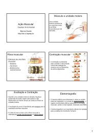
Biomechanics and Analysis of Running Gait - De Motu
Biomechanics and Analysis of Running Gait - De Motu
Biomechanics and Analysis of Running Gait - De Motu

Create successful ePaper yourself
Turn your PDF publications into a flip-book with our unique Google optimized e-Paper software.
608 DUGAN & BHAT<br />
The anatomic uniqueness <strong>of</strong> the plantar fascia also helps to create the<br />
solid structural platform that is needed for propulsion. It originates from<br />
the medial tubercle <strong>of</strong> the calcaneus <strong>and</strong> inserts around the metatarsal heads<br />
to the base <strong>of</strong> the proximal phalanges. It crosses the transverse tarsal<br />
<strong>and</strong> metatarsophalangeal joints <strong>and</strong> serves as a passive restraint. At the<br />
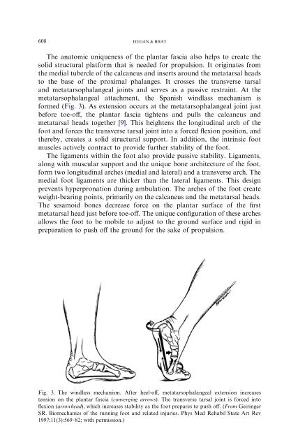
metatarsophalangeal attachment, the Spanish windlass mechanism is<br />
formed (Fig. 3). As extension occurs at the metatarsophalangeal joint just<br />
before toe-<strong>of</strong>f, the plantar fascia tightens <strong>and</strong> pulls the calcaneus <strong>and</strong><br />
metatarsal heads together [9]. This heightens the longitudinal arch <strong>of</strong> the<br />
foot <strong>and</strong> forces the transverse tarsal joint into a forced flexion position, <strong>and</strong><br />
thereby, creates a solid structural support. In addition, the intrinsic foot<br />
muscles actively contract to provide further stability <strong>of</strong> the foot.<br />
The ligaments within the foot also provide passive stability. Ligaments,<br />
along with muscular support <strong>and</strong> the unique bone architecture <strong>of</strong> the foot,<br />
form two longitudinal arches (medial <strong>and</strong> lateral) <strong>and</strong> a transverse arch. The<br />
medial foot ligaments are thicker than the lateral ligaments. This design<br />
prevents hyperpronation during ambulation. The arches <strong>of</strong> the foot create<br />
weight-bearing points, primarily on the calcaneus <strong>and</strong> the metatarsal heads.<br />
The sesamoid bones decrease force on the plantar surface <strong>of</strong> the first<br />
metatarsal head just before toe-<strong>of</strong>f. The unique configuration <strong>of</strong> these arches<br />
allows the foot to be mobile to adjust to the ground surface <strong>and</strong> rigid in<br />
preparation to push <strong>of</strong>f the ground for the sake <strong>of</strong> propulsion.<br />
Fig. 3. The windlass mechanism. After heel-<strong>of</strong>f, metatarsophalangeal extension increases<br />
tension on the plantar fascia (converging arrows). The transverse tarsal joint is forced into<br />
flexion (arrowhead), which increases stability as the foot prepares to push <strong>of</strong>f. (From Geiringer<br />
SR. <strong>Biomechanics</strong> <strong>of</strong> the running foot <strong>and</strong> related injuries. Phys Med Rehabil State Art Rev<br />
1997;11(3):569–82; with permission.)