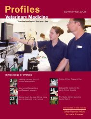
veterinary medical center veterinary medical centerState-<strong>of</strong>-the-art MRInow available at VMCThe <strong>University</strong> <strong>of</strong> <strong>Minnesota</strong><strong>Veterinary</strong> Medical Center(VMC) now has a valuable newtool for veterinarians who want to providethe best possible diagnostic services:a 3T magnetic resonance imaging (MRI)machine, the most powerful MR systemin a veterinary hospital in the world.What is MRI?As the name implies, MRI uses a strongmagnetic field to image parts <strong>of</strong> the body.It takes advantage <strong>of</strong> the natural magneticproperties <strong>of</strong> hydrogen atoms in thebody to create a detailed image that candifferentiate between myriad s<strong>of</strong>t tissues,bone structures, and body fluids.Aren’t all MRI systemsthe same?No! There are “high-field” and “lowfield”MR systems, and high-field magnets(systems with field strengths <strong>of</strong> 1.0T,1.5T, and 3.0T) are far superior to lowfieldmagnets in all aspects <strong>of</strong> image qualityand speed <strong>of</strong> acquisition. The VMC’snew 3T MRI unit allows radiologists toperform imaging exams that are not possiblewith lower field scanners.Why is MRI important?MRI can give information that cannotbe obtained by other imaging modalities.Its image clarity and detail areunparalleled. Compared to radiographyand CT, it also has the advantage <strong>of</strong>not using potentially harmful ionizingradiation. MRI allows doctors to makediagnoses that were previously difficultor impossible to confirm, enhancingtheir ability to recommend appropriatetreatment options for patients.What kinds <strong>of</strong> cases maybenefit from MRI?There are many kinds <strong>of</strong> cases for whichMRI is an excellent diagnostic tool. MRIis the best modality for imaging <strong>of</strong> neurologicdisease, musculoskeletal disease, andoncologic disease. For musculoskeletaldisease in dogs and horses, MRI is invaluablefor evaluating injuries to joint structures,tendons, and ligaments. Chroniclameness that has been localized to ageneral area, but for which an underlyingcause cannot be determined using traditionalmethods, can <strong>of</strong>ten be diagnosedusing MRI. MRI is also very versatilefor imaging <strong>of</strong> the structures <strong>of</strong> the headoutside <strong>of</strong> the brain, abdominal diseases,vascular diseases, certain thoracic diseases,and some cardiovascular disorders.How long does an MRIexam take?It depends on the body part beingexamined. The total procedure, includingpreparation, imaging, and recovery,generally lasts one to two hours. Equinepatients and some small animal patientsare hospitalized overnight so they canbe monitored closely for several hoursafter recovering from anesthesia.Who reviews the MR images?The exams are monitored by the<strong>Veterinary</strong> Medical Center’s board-certifiedradiologists. The images are interpretedon-site within hours. If a lesionis found, board-certified surgeons andother specialists are available to providethe necessary treatment.<strong>Veterinary</strong> radiologist Travis Saveraid and frienddisplay the <strong>Veterinary</strong> Medical Center’s new 3TMRI machine, the most powerful MR system ina veterinary hospital in the world. Photo by SueKirch<strong>of</strong>fAre there any risks in performingan MR exam?There are no known side effects <strong>of</strong> MRI,but patients are placed under generalanesthesia for the procedure. As withanything requiring general anesthesia,there are risks associated with the drugsand the recovery period. But the VMC'sboard-certified anesthesiologists workclosely with the imaging group to minimizethose risks.When will MRI exams be availableat the VMC?MRI exams are available now for VMCpatients. Starting this spring, veterinariansin the region will be able torefer cases directly to a new outpatientimaging service. Advanced <strong>Veterinary</strong>Imaging Direct (AVID) will provideimaging services, including MR, CT,fluoroscopy, and ultrasound (includingbiopsies) on an outpatient basis.“We want to make these tremendousresources accessible to all veterinarians inthe area,” says VMC director David Lee.12
veterinary medical center veterinary medical centerLinear accelerator <strong>of</strong>fers newoptions for animals with cancerIn August 2007, the <strong>Veterinary</strong>Medical Center unveiled a new linearaccelerator facility, becoming one<strong>of</strong> the only veterinary hospitals in theUpper Midwest to <strong>of</strong>fer state-<strong>of</strong>-the-artradiation therapy to animals with cancer.“Each year, thousands <strong>of</strong> dogs and cats in<strong>Minnesota</strong> and our neighboring states arediagnosed with cancer, and their ownersare faced with very difficult decisions,”says David Lee, <strong>Veterinary</strong> MedicalCenter director. “With the linear accelerator,our veterinarians are able to provideleading-edge cancer treatment.”The linear accelerator replaced cobaltradiation equipment used to treat cancerpatients for nearly 25 years. Identical tothe linear accelerators used in humanradiation treatment, it allows veterinarycancer specialists to map tumors inthree dimensions and focus radiationon cancerous lesions, minimizing theimpact to surrounding healthy tissue.The linear accelerator is also a key elementin the <strong>College</strong> <strong>of</strong> <strong>Veterinary</strong> <strong>Medicine</strong>’scomparative cancer research program.“Comparative cancer research involvesthe study <strong>of</strong> cancer in one species withthe goal <strong>of</strong> applying the lessons learnedto other species,” says Robert Washabau,chair <strong>of</strong> the <strong>College</strong>’s <strong>Veterinary</strong> ClinicalSciences Department. “Cancer in animalsis very similar to cancer in humans,both in cause and in response to therapy.Our research will benefit both animalsand humans.”Key features <strong>of</strong> the linear acceleratorinclude:■ Ability to adjust the strength <strong>of</strong> theproton beam to customize the treatment<strong>of</strong> deep masses<strong>Veterinary</strong> technician Kim Janisch, veterinary radiologist Dan Feeney, and Mason, an oncology patient,display the <strong>Veterinary</strong> Medical Center’s new linear accelerator. Photo by Sue Kirch<strong>of</strong>f.■ A computerized tool called a multileafcollimator, which uses informationfrom treatment planning s<strong>of</strong>tware toautomatically shield normal adjacenttissue from radiation, thereby lesseningthe treatment impact to surroundinghealthy tissues■ Electron capability, which will allowtreatment <strong>of</strong> skin masses while protectingdeeper tissues from exposureA fund-raising campaign, “AcceleratingHope,” is underway to <strong>of</strong>fset the cost<strong>of</strong> this new technology so it can benefitas many animals as possible. Corporateand private donations are being acceptedby the <strong>College</strong> <strong>of</strong> <strong>Veterinary</strong> <strong>Medicine</strong>and the <strong>University</strong> <strong>of</strong> <strong>Minnesota</strong>Foundation. For more information, contactKatharine Anderson, development<strong>of</strong>ficer, at 612-626-2343 or ksander@umn.edu.13