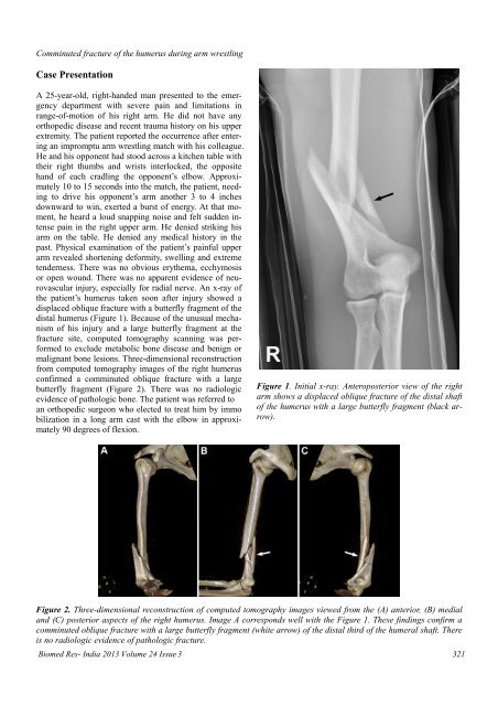
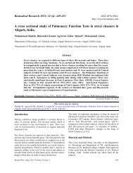
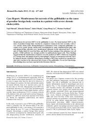
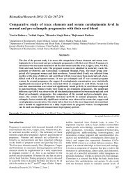
<strong>Comminuted</strong> <strong>fracture</strong> <strong>of</strong> <strong>the</strong> <strong>humerus</strong> during arm wrestlingCase PresentationA 25-year-old, right-handed man presented to <strong>the</strong> emergencydepartment <strong>with</strong> severe pain and limitations inrange-<strong>of</strong>-motion <strong>of</strong> his right arm. He did not have anyorthopedic disease and recent trauma history on his upperextremity. The patient reported <strong>the</strong> occurrence after enteringan impromptu arm wrestling match <strong>with</strong> his colleague.He and his opponent had stood across a kitchen table <strong>with</strong><strong>the</strong>ir right thumbs and wrists interlocked, <strong>the</strong> oppositehand <strong>of</strong> each cradling <strong>the</strong> opponent’s elbow. Approximately10 to 15 seconds into <strong>the</strong> match, <strong>the</strong> patient, needingto drive his opponent’s arm ano<strong>the</strong>r 3 to 4 inchesdownward to win, exerted a burst <strong>of</strong> energy. At that moment,he heard a loud snapping noise and felt sudden intensepain in <strong>the</strong> right upper arm. He denied striking hisarm on <strong>the</strong> table. He denied any medical history in <strong>the</strong>past. Physical examination <strong>of</strong> <strong>the</strong> patient’s painful upperarm revealed shortening deformity, swelling and extremetenderness. There was no obvious ery<strong>the</strong>ma, ecchymosisor open wound. There was no apparent evidence <strong>of</strong> neurovascularinjury, especially for radial nerve. An x-ray <strong>of</strong><strong>the</strong> patient’s <strong>humerus</strong> taken soon after injury showed adisplaced oblique <strong>fracture</strong> <strong>with</strong> a <strong>butterfly</strong> <strong>fragment</strong> <strong>of</strong> <strong>the</strong>distal <strong>humerus</strong> (Figure 1). Because <strong>of</strong> <strong>the</strong> unusual mechanism<strong>of</strong> his injury and a large <strong>butterfly</strong> <strong>fragment</strong> at <strong>the</strong><strong>fracture</strong> site, computed tomography scanning was performedto exclude metabolic bone disease and benign ormalignant bone lesions. Three-dimensional reconstructionfrom computed tomography images <strong>of</strong> <strong>the</strong> right <strong>humerus</strong>confirmed a comminuted oblique <strong>fracture</strong> <strong>with</strong> a large<strong>butterfly</strong> <strong>fragment</strong> (Figure 2). There was no radiologicevidence <strong>of</strong> pathologic bone. The patient was referred toan orthopedic surgeon who elected to treat him by immobilization in a long arm cast <strong>with</strong> <strong>the</strong> elbow in approximately90 degrees <strong>of</strong> flexion.Figure 1. Initial x-ray. Anteroposterior view <strong>of</strong> <strong>the</strong> rightarm shows a displaced oblique <strong>fracture</strong> <strong>of</strong> <strong>the</strong> distal shaft<strong>of</strong> <strong>the</strong> <strong>humerus</strong> <strong>with</strong> a large <strong>butterfly</strong> <strong>fragment</strong> (black arrow).Figure 2. Three-dimensional reconstruction <strong>of</strong> computed tomography images viewed from <strong>the</strong> (A) anterior, (B) medialand (C) posterior aspects <strong>of</strong> <strong>the</strong> right <strong>humerus</strong>. Image A corresponds well <strong>with</strong> <strong>the</strong> Figure 1. These findings confirm acomminuted oblique <strong>fracture</strong> <strong>with</strong> a large <strong>butterfly</strong> <strong>fragment</strong> (white arrow) <strong>of</strong> <strong>the</strong> distal third <strong>of</strong> <strong>the</strong> humeral shaft. Thereis no radiologic evidence <strong>of</strong> pathologic <strong>fracture</strong>.Biomed Res- India 2013 Volume 24 Issue 3 321
<strong>Comminuted</strong> <strong>fracture</strong> <strong>of</strong> <strong>the</strong> <strong>humerus</strong> during arm wrestlingThis article must be cited as:Hyun-Soo Kim, Young Ho Shin, Youn Wha Kim. <strong>Comminuted</strong> <strong>fracture</strong> <strong>with</strong> <strong>butterfly</strong> <strong>fragment</strong> <strong>of</strong> <strong>the</strong> <strong>humerus</strong> sustainedduring arm wrestling. Biomed Res- India; 2013; 24 (3): 320-323.DiscussionIntense muscular contractions can be a mechanism <strong>of</strong> injuryin various sports and can affect various bones. Thehumeral shaft <strong>fracture</strong> during arm wrestling occurs whenmoments <strong>of</strong> torsional forces induced by intense muscularcontractions are transmitted onto it. The moments <strong>of</strong> <strong>the</strong>seforces are resultant <strong>of</strong> forces acting in <strong>the</strong> direction <strong>of</strong>internal rotation by <strong>the</strong> pectoralis major, latissimus dorsi,teres major and subscapularis, and resultant <strong>of</strong> externalrotational forces applied by <strong>the</strong> opponent and transmittedby <strong>the</strong> long forearm lever through <strong>the</strong> elbow. In both cases<strong>the</strong> results <strong>of</strong> opposite directional forces occur thanksto <strong>the</strong> work <strong>of</strong> <strong>the</strong> internal rotator shoulder muscles.When <strong>the</strong> dominant competitor exerts force, <strong>the</strong> internalrotator muscles <strong>of</strong> <strong>the</strong> opponent undergo rapid stress from<strong>the</strong>ir maximum concentric contraction to eccentric passivecompensatory relaxation, causing an increase in <strong>the</strong> humeralrotational force. This results in <strong>the</strong> transmission <strong>of</strong><strong>the</strong> stress through <strong>the</strong> distal part <strong>of</strong> <strong>the</strong> arm and elbow,causing a shifting <strong>of</strong> <strong>the</strong> maximum force to <strong>the</strong> <strong>humerus</strong>and its <strong>fracture</strong>. Nyska et al. [15] note that <strong>the</strong> <strong>fracture</strong> isusually in <strong>the</strong> distal third <strong>of</strong> <strong>the</strong> <strong>humerus</strong>, and suggest that<strong>the</strong> forces acting on <strong>the</strong> <strong>humerus</strong> are bending, axial compressionand torsional forces, while <strong>the</strong> flexor muscles <strong>of</strong><strong>the</strong> forearm are forcefully contracted.In our case, we could easily consider <strong>the</strong> possibility <strong>of</strong>humeral <strong>fracture</strong> <strong>with</strong> <strong>the</strong> patients’ clinical presentations,so we performed imaging studies <strong>with</strong>out any hesitations.Some <strong>fracture</strong>s, however, which are not related to director indirect contact forces, can be missed if <strong>the</strong>y do notcause severe symptoms. Intense muscular contractionsrelated<strong>fracture</strong>s <strong>of</strong> protruded bone, such as those <strong>of</strong> <strong>the</strong>olecranon, anterior superior ischial spine <strong>of</strong> <strong>the</strong> pelvis andfifth metatarsal base <strong>of</strong> <strong>the</strong> foot, are good examples. We<strong>the</strong>refore suggest that a diagnosis <strong>of</strong> <strong>fracture</strong> should beconsidered in cases <strong>of</strong> intense muscular contractions. Acareful history taking and radiologic examination willenable to prevent misdiagnosis and mismanagement.Every long bone <strong>fracture</strong>s do not require computed tomographyscanning routinely, but in our case, we performedit due to <strong>the</strong> presence <strong>of</strong> a large <strong>butterfly</strong> <strong>fragment</strong>. With<strong>the</strong> understanding <strong>of</strong> three-dimensional morphology <strong>of</strong><strong>fracture</strong> site as well as its location, we could decide properoperative methods. Also, <strong>fracture</strong> <strong>of</strong> <strong>the</strong> distal one third<strong>of</strong> <strong>humerus</strong> is deeply associated <strong>with</strong> <strong>the</strong> occurrence <strong>of</strong>radial nerve palsy. Of <strong>the</strong> few complications <strong>with</strong> <strong>humerus</strong><strong>fracture</strong> sustained during arm wrestling that have beenreported, <strong>the</strong> most common is radial nerve palsy, whichusually manifests as wrist drop. It can be caused by direct322damage due to <strong>the</strong> <strong>butterfly</strong> <strong>fragment</strong>, so it is very importantto understand <strong>the</strong> morphology <strong>of</strong> <strong>the</strong> <strong>fracture</strong> site.For <strong>the</strong> treatment <strong>of</strong> such injury, conservative managementwas mainly recommended at past. However, because<strong>of</strong> developed instruments and increased patients’ requirements,operative treatments have been recently performedby many orthopedic surgeons.In summary, <strong>the</strong> present case illustrates that <strong>the</strong> extremelyhigh indirect forces generated by sudden intense muscularcontractions during arm wrestling can cause a comminuted<strong>fracture</strong> <strong>of</strong> <strong>the</strong> <strong>humerus</strong>. Severe pain in arm wrestlersshould not be attributed solely to musculoskeletal sprainsand ruptures. All patients who present <strong>with</strong> histories <strong>of</strong>sudden arm pain after arm wrestling should receive appropriateradiologic studies and a careful neurologic examinationto exclude <strong>fracture</strong> and prevent misdiagnosisand mismanagement. This type <strong>of</strong> <strong>fracture</strong> may occur inanyone <strong>of</strong> any age engaging in this type <strong>of</strong> sport. Especially,amateurs are injury-prone because <strong>the</strong>y use badwrestling techniques. Because <strong>of</strong> <strong>the</strong> possibility <strong>of</strong> participantssustaining such an injury, arm wrestling shouldnot be considered a totally benign injury.AcknowledgementsThe views and opinions expressed in this article are those<strong>of</strong> <strong>the</strong> author and do not reflect <strong>the</strong> <strong>of</strong>ficial policy or position<strong>of</strong> <strong>the</strong> Republic <strong>of</strong> Korea Air Force or Republic <strong>of</strong>Korea Ministry <strong>of</strong> National Defense. The author thanksmedical librarian Ja Ok Kim (Aerospace Medical Library,Aerospace Medical Center, Republic <strong>of</strong> Korea Air Force)for her assistance in searching for literature.References1. Brinker MR, O'Conner DP. The incidence <strong>of</strong> <strong>fracture</strong>sand dislocations referred for orthopaedic services in acapitated population. J Bone Joint Surg Am 2004; 86:290-297.2. Fenyo G. On <strong>fracture</strong>s <strong>of</strong> <strong>the</strong> shaft <strong>of</strong> <strong>the</strong> <strong>humerus</strong>: areview covering a 12-year period <strong>with</strong> special consideration<strong>of</strong> <strong>the</strong> surgically treated cases. Acta Chir Scand1971; 137: 221-226.3. Baker DM. Fractures <strong>of</strong> <strong>the</strong> humeral shaft associated<strong>with</strong> ipsilateral <strong>fracture</strong> dislocation <strong>of</strong> <strong>the</strong> shoulder: report<strong>of</strong> a case. J Trauma 1971; 11: 532-534.4. Chao SL, Miller M, Teng SW. A mechanism <strong>of</strong> spiral<strong>fracture</strong> <strong>of</strong> <strong>the</strong> <strong>humerus</strong>: a report <strong>of</strong> 129 cases following<strong>the</strong> throwing <strong>of</strong> hand grenades. J Trauma 1971; 11:602-605.5. Reed WJ, Mueller RW. Spiral <strong>fracture</strong> <strong>of</strong> <strong>the</strong> <strong>humerus</strong>in a ball thrower. Am J Emerg Med 1998; 16: 306-308.Biomed Res- India 2013 Volume 24 Issue 3