Western Blot Analysis on the Odyssey Infrared Imaging System
Western Blot Analysis on the Odyssey Infrared Imaging System
Western Blot Analysis on the Odyssey Infrared Imaging System
You also want an ePaper? Increase the reach of your titles
YUMPU automatically turns print PDFs into web optimized ePapers that Google loves.
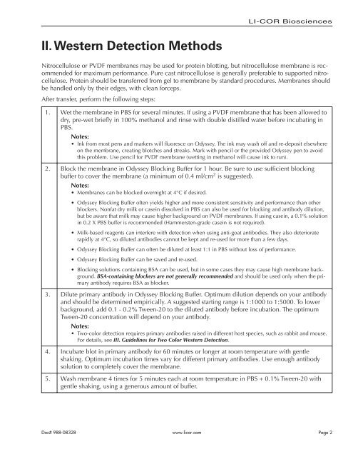
II. <str<strong>on</strong>g>Western</str<strong>on</strong>g> Detecti<strong>on</strong> Methods<br />
LI-COR Biosciences<br />
Nitrocellulose or PVDF membranes may be used for protein blotting, but nitrocellulose membrane is recommended<br />
for maximum performance. Pure cast nitrocellulose is generally preferable to supported nitrocellulose.<br />
Protein should be transferred from gel to membrane by standard procedures. Membranes should<br />
be handled <strong>on</strong>ly by <strong>the</strong>ir edges, with clean forceps.<br />
After transfer, perform <strong>the</strong> following steps:<br />
1. Wet <strong>the</strong> membrane in PBS for several minutes. If using a PVDF membrane that has been allowed to<br />
dry, pre-wet briefly in 100% methanol and rinse with double distilled water before incubating in<br />
PBS.<br />
Notes:<br />
• Ink from most pens and markers will fluoresce <strong>on</strong> <strong>Odyssey</strong>. The ink may wash off and re-deposit elsewhere<br />
<strong>on</strong> <strong>the</strong> membrane, creating blotches and streaks. Mark with pencil or <strong>the</strong> provided <strong>Odyssey</strong> pen to avoid<br />
this problem. Use pencil for PVDF membrane (wetting in methanol will cause ink to run).<br />
2. Block <strong>the</strong> membrane in <strong>Odyssey</strong> Blocking Buffer for 1 hour. Be sure to use sufficient blocking<br />
buffer to cover <strong>the</strong> membrane (a minimum of 0.4 ml/cm2<br />
is suggested).<br />
Notes:<br />
• Membranes can be blocked overnight at 4°C if desired.<br />
• <strong>Odyssey</strong> Blocking Buffer often yields higher and more c<strong>on</strong>sistent sensitivity and performance than o<strong>the</strong>r<br />
blockers. N<strong>on</strong>fat dry milk or casein dissolved in PBS can also be used for blocking and antibody diluti<strong>on</strong>,<br />
but be aware that milk may cause higher background <strong>on</strong> PVDF membranes. If using casein, a 0.1% soluti<strong>on</strong><br />
in 0.2 X PBS buffer is recommended (Hammersten-grade casein is not required).<br />
• Milk-based reagents can interfere with detecti<strong>on</strong> when using anti-goat antibodies. They also deteriorate<br />
rapidly at 4°C, so diluted antibodies cannot be kept and re-used for more than a few days.<br />
• <strong>Odyssey</strong> Blocking Buffer can often be diluted at least 1:1 in PBS without loss of performance.<br />
• <strong>Odyssey</strong> Blocking Buffer can be saved and re-used.<br />
• Blocking soluti<strong>on</strong>s c<strong>on</strong>taining BSA can be used, but in some cases <strong>the</strong>y may cause high membrane background.<br />
BSA-c<strong>on</strong>taining blockers are not generally recommended and should be used <strong>on</strong>ly when <strong>the</strong> primary<br />
antibody requires BSA as blocker.<br />
3. Dilute primary antibody in <strong>Odyssey</strong> Blocking Buffer. Optimum diluti<strong>on</strong> depends <strong>on</strong> your antibody<br />
and should be determined empirically. A suggested starting range is 1:1000 to 1:5000. To lower<br />
background, add 0.1 - 0.2% Tween-20 to <strong>the</strong> diluted antibody before incubati<strong>on</strong>. The optimum<br />
Tween-20 c<strong>on</strong>centrati<strong>on</strong> will depend <strong>on</strong> your antibody.<br />
Notes:<br />
• Two-color detecti<strong>on</strong> requires primary antibodies raised in different host species, such as rabbit and mouse.<br />
For details, see III. Guidelines for Two Color <str<strong>on</strong>g>Western</str<strong>on</strong>g> Detecti<strong>on</strong>.<br />
4. Incubate blot in primary antibody for 60 minutes or l<strong>on</strong>ger at room temperature with gentle<br />
shaking. Optimum incubati<strong>on</strong> times vary for different primary antibodies. Use enough antibody<br />
soluti<strong>on</strong> to completely cover <strong>the</strong> membrane.<br />
5. Wash membrane 4 times for 5 minutes each at room temperature in PBS + 0.1% Tween-20 with<br />
gentle shaking, using a generous amount of buffer.<br />
Doc# 988-08328 www.licor.com<br />
Page 2


