Complete Issue PDF - University of Alberta Health Sciences Journal
Complete Issue PDF - University of Alberta Health Sciences Journal
Complete Issue PDF - University of Alberta Health Sciences Journal
- No tags were found...

Create successful ePaper yourself
Turn your PDF publications into a flip-book with our unique Google optimized e-Paper software.
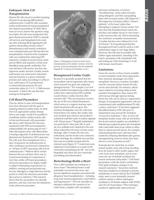
Embryonic Stem CellTransplantationHuman ES cells served as another jumping<strong>of</strong>fpoint for producing differentiatedcardiomyocytes. Could ES cells normalizecardiac performance by being transplantedinto injured myocardium? In 2002, Min’steam set out to answer this question usingrat models. ES cells were transfected withgreen fluorescent protein (GFP) to identifycell survival, and transplanted into malerats after inducing MI by ligating the leftanterior descending coronary artery. 18Hemodynamics and muscle contractionwere evaluated both post-MI and after thetransplantation. Survival <strong>of</strong> the transplantcells was confirmed by observing GFPexpression. Cardiac α-myosin heavy chainand (α-MHC) and troponin I (cTnI) wereidentified using specific antibodies. Notonly did these cells survive, but they alsoimproved cardiac function. Cardiac musclewall tension was measured to determinemyocyte function at a given ventricularpressure and radius (according to LaPlace’sLaw, wall tension (T) is proportionalto intraventricular pressure (P) andventricular radius (r): T ∝ P ∙ r). Wall tensionincreased ~2-fold in the rats that hadundergone transplants. 18Cell-Based PacemakersThus far, efforts in stem cell transplantationhave been discussed with the goal <strong>of</strong>repairing infarcted cardiac tissue. In 2004,Xue et al. approached cardiac therapyfrom a new angle: would it be possible tocoordinate inactive cardiac muscle cellsto beat synchronously with pacemakerlikedonor cells? Human ES cells weretransfected with GFP and transplantedsubepicardially into guinea pigs in vivo. 19After allowing the stem cells differentiate,a beating outgrowth <strong>of</strong> cardiomyocytes wasdissected and checked for GFP expression.The cells were then transplanted onto alayer <strong>of</strong> quiescent rat cardiomyocytes invitro, resulting in synchronous contractionsat ~49 bpm <strong>of</strong> GFP-expressing cells and ratcardiomyocytes. 19 Without direct contactbetween the two cell lines, there was nosynchronous beating. It is important tonote that, unlike cell-based pacemakers,electronic pacemakers are largely unableto adapt to fluctuating requirements. Inaddition, sensing and pacing leads maybecome dislodged or malpositioned, and thepocket in which the electronic pacemakersits is prone to infection. 20 This researchshows that bio-pacemakers are clinicallyattractive, and may overcome the limitations<strong>of</strong> the electronic pacemaker.Figure 3. Photograph <strong>of</strong> a heart at week 9 posttransplantation<strong>of</strong> the cellular construct. Note thepresence <strong>of</strong> neovascularization into the implantedbiograft (B). Courtesy <strong>of</strong> Circulation: Leor, J.Bioengineered Cardiac GraftsBecause it is generally accepted that themyocardium cannot regenerate after injury,much research has gone into replacingdamaged muscle. 21 For example, Leor et al.tested whether bioengineering cardiac tissuewithin three-dimensional (3D) scaffoldswould enhance cardiac function afterextensive MI. 21 This novel practice involvesthe use <strong>of</strong> 3D cross-linked biopolymer,which serves as a support structure uponwhich functional cells can grow. Thestructure biodegrades once the cells haveformed their own matrix. Rat cardiac cellswere isolated and cultured, and seeded incylindrical scaffolds made <strong>of</strong> sodium alginatewith 100 μm pores. 21 Biograft implantationwas performed 7 days post-MI: in eachrat, two scaffolds were attached to the scartissue induced by left main coronary arteryblockage. After 9 weeks, the rats wereeuthanized, and the hearts were examined.Under histology, the scaffolds showed thatthey had successfully merged with theinfarcted area (Figure 3). 21 Control rats weresubjected to heart failure as a result <strong>of</strong> leftventricular remodeling post-MI – but in thebiografted rats, there was less ventricularremodeling and deterioration.Biotechnology Builds a HeartOver 1,000 Canadians are waiting for adonor heart. 22 Bioartificial hearts couldpotentially circumvent this issue, andprevent significant sequelae associated withallogeneic heart transplantation – includinglong-term immunosuppression, renal failure,and hypertension. 23 Ott et al. describes anattempt to fabricate the construct <strong>of</strong> anentire heart, complete with vasculatureand inner architecture: it involves“decellularizing” whole adult rat heartsusing detergents, and then repopulatingthem with neonatal cardiac cells (Figure 4). 23This ingenious technique utilizes “nature’splatform” <strong>of</strong> the heart, rather thanattempting to engineer it from scratch. Notonly does this bioartifical heart mimic thestructure and cellular layout <strong>of</strong> a true heart –it also functions like one. When stimulatingthe constructs, acceptable measurements<strong>of</strong> cardiac function were obtained in 5 out<strong>of</strong> 8 hearts. 23 It is hypothesized that thebioengineered heart could be used as a fullreplacement organ in end-stage failure.However, this will only be possible withfurther organ maturation, reseeding <strong>of</strong> theheart’s vasculature with endothelial cells,and scaling up <strong>of</strong> the technology to workwith human-sized hearts.LimitationsGiven the scarcity <strong>of</strong> donor hearts availableto meet transplant needs, these approacheshave immense advantages over hearttransplants. However, a number <strong>of</strong> hurdlesmust be overcome before human ES cellscan be used clinically. For instance, ethicalissues related to accessing embryos limitscientists’ investigations. Also, human EScells must go through rigorous testingbefore the cells can be used as a regenerativetherapy. If transplanted regenerative cells arecontaminated with undifferentiated ES cells,a tumor could form. 24 It has been suggestedthat differentiation <strong>of</strong> ES cells prior toimplantation may prevent the formation<strong>of</strong> cancerous teratomas. 25 Also, in most celltransplant studies, many cells are lost beforeblood and nutrient supplies are established. 7Introducing exogenous genes into transplantcells could make them more robust or allowthem to release growth factors. For example,induced pluripotent stem cells have beenstudied by obtaining fibroblasts after geneticreprogramming; however, these have beenshown to form teratomas as well. 26It should also be noted that, in certainanimal models, stem cells end up travellingfrom the heart to other nearby organs onlya few hours post-transplant, and in somecases, the use <strong>of</strong> skeletal myoblasts hascaused ventricular tachycardia. 27 Cell-basedpacemakers could also lead to arrhythmiasif the graft undergoes changes in ionchannel expression. 19 Innovative methodshave been studied in an attempt to preventarrhythmias. A recent clinical, the CAuSMICstudy, used a novel minimally-invasivecatheter system to deliver autologousmyoblasts to 23 human subjects with NYHAclass II to IV heart failure. 28 This techniqueresulted in significantly improved heartREVIEW<strong>University</strong> <strong>of</strong> <strong>Alberta</strong> <strong>Health</strong> <strong>Sciences</strong> <strong>Journal</strong> • April 2012 • Volume 7 • <strong>Issue</strong> 1 15