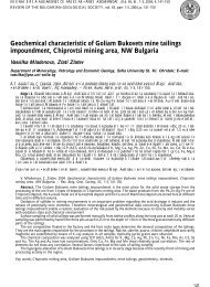
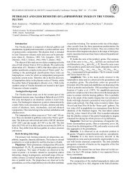
Fig. 1. Location map <strong>and</strong> lithostratigraphic column of the studied boreholes. Sample positions are indicated to the right of thecolumnsÔèã. 1. Ãåîãðàôñêî ïîëîæåíèå è ëèòîñòðàòèãðàôèÿ íà èçñëåäâàíèòå ñîíäàæè. Ìåñòàòà íà ïðîáèòå ñà îçíà÷åíè îòäÿñíîíà êîëîíêèòå108
109dinocyst assemblages; (2) to assess the age <strong>and</strong> correlatethe succession to the biostratigraphically well controlledframework established for the western Tethyanrealm; (3) to provide a direct calibration of the dinocystassociations to the calpionellid <strong>and</strong> calcareous dino<strong>cysts</strong>uccessions <strong>and</strong> zones, recognized in the same boreholes.Material <strong>and</strong> methodsThe present study is based on eleven core samples,containing relatively rich <strong>and</strong> well-preserved <strong>dinoflagellate</strong>cyst assemblages. The position of the samples isindicated in Fig. 1. They were collected <strong>from</strong> the marlylimestones in the sampled interval.The samples were prepared for palynological analysisfollowing st<strong>and</strong>ard preparation techniques, includingHCl, HF treatment <strong>and</strong> separation with heavy liquid.Strew mounts were prepared in Elvacit, a commercialmounting medium on the basis of resin. The slides containingthe illustrated specimens are stored in the collectionsof the Geological Institute, Bulgarian Academyof Sciences.The morphological analysis of the species is primarilybased on the results in transmitted light microscopy,since this is the main tool used in routine palynologicalstudies. Photomicrographs in transmitted light havebeen taken using Differential Interference contrast(DIC) on a BH2 Olympus microscope at the NaturalHistory Museum, London.In addition to conventional transmitted light microscopy,a selection of species has been studied in confocallaser scanning microscopy. Only recently, CLSMhas been successfully applied for the first time to themorphological analysis of fossil <strong>dinoflagellate</strong> <strong>cysts</strong> byFeist-Burkhardt, Pross (1999) <strong>and</strong> Feist-Burkhardt,Monteil (2001).The novel method of CLSM is now applied for thefirst time to the morphological analysis <strong>and</strong> final identificationof Bulgarian fossil <strong>dinoflagellate</strong> cyst species.The present contribution aims also to describe thismethod, thus introducing it to the Bulgarian palynologicalliterature.Confocal Laser Scanning Microscopy(CLSM)In confocal laser scanning microscopy, a fine laserbeam is used to excite fluorescence in the study object.To obtain a full image, the image point is moved acrossthe specimen by mirror scanners. The emitted fluorescencelight passing through the detector pinhole istransformed into electrical signals by a photomultiplier<strong>and</strong> displayed on a computer monitor. Because of thetwo conjugated pinholes in the light path, the so-calledconfocal principle, only the light <strong>from</strong> the focal plane isdetected, <strong>and</strong> stray light is minimized. By imaging serialsections through a microscopic object, an image stackof very thin optical sections is obtained, preserving thetrue 3D information of the specimen. Subsequently, theconfocal imaging software is used to superimpose theslices, giving an extended focus image with high depthof focus. Stereoscopic images <strong>and</strong> computer animationscan be generated facilitating visualization of 3Dstructures <strong>and</strong> showing the microscopic specimen<strong>from</strong> all directions.In the present study, palynomorphs have been studiedusing the Leica TCS SP confocal microscope at theNatural History Museum, London. The unstained <strong>dinoflagellate</strong><strong>cysts</strong> in conventional palynological slideswere analysed using a 40×1.0 NA oil PL Fluotar objective.TRITC_wide filter settings of the confocal microscopewere used to obtain fluorescence image stacks.This means that excitation takes place at a wavelengthof 568 nm <strong>and</strong> the emitted fluorescence light with awavelength longer than 570 nm is detected. Imageswere captured with a lateral resolution of 1024×1024pixels <strong>and</strong> a thickness of generally less than 500 nm.The resulting image stacks were then processed usingthe Leica 3D rendering software to obtain maximumprojections extended focus images <strong>and</strong> red/green anaglyphs.Confocal microscopy has been used in many biological<strong>and</strong> medical studies, in which a wide range offluorescent dyes is applied to stain specific cell areas orcell organelles. It is only recently, that Feist-Burkhardt,Pross (1998) <strong>and</strong> Feist-Burkhardt, Monteil (2001) appliedfluorescence confocal laser scanning microscopyfor morphological analysis <strong>and</strong> imaging purposes to <strong>dinoflagellate</strong><strong>cysts</strong> <strong>from</strong> the Jurassic <strong>and</strong> the Cretaceous.A fairly detailed description <strong>and</strong> explanation ofconfocal microscopy in the application to palynomorphscan be found on the Internet under the followingaddress: http://www.nhm.ac.uk/palaeontology/micro/clsm/clsm.html.Modern <strong>and</strong> fossil palynomorphsgenerally show strong autofluorescence <strong>and</strong> can thereforebe easily analysed in confocal microscopy in theconventional palynological strew mounts without specialsample preparation. So far, only some few exceptionsare known. Mature organic residues with palynomorphshaving suffered thermal alteration may havelost their autofluorescence. Even chemical oxidation,which is part of many st<strong>and</strong>ard palynological processingtechniques, in most cases does not negatively affectthe analysis of palynomorphs in fluorescence confocalmicroscopy.Confocal microscopy of palynomorphs providesadditional information to conventional transmitted lightmicroscopy <strong>and</strong> scanning electron microscopy (SEM).In transmitted light microscopy the depth of field is limited<strong>and</strong> illustration of palynomorphs, especially, whenwell preserved in <strong>three</strong> dimensions, needs several focuslevels to appropriately document the specimens morphology.In confocal microscopy, very thin optical sectionsare imaged, allowing detailed inspection, e.g. ofthe wall structure in cross-section. By using the 3Drenderingalgorithms provided with the confocal software,the image information of the image stack is combinedforming projection images with an extendeddepth of focus, resembling those obtained in SEM. Theadvantage of confocal microscopy compared to SEMis, even though resolution in SEM is for obvious reasonsmuch higher, that individual, specific grains firstselected in a slide in transmitted light, can be analysed.