Chemistry & Biology<strong>Hammerhead</strong> <strong>Ribozyme</strong> Metal Ion <strong>and</strong> <strong>Solvent</strong> <strong>Structure</strong>crystal structure under conditions of (nonphysiological) highmonovalent salt (1 M NH 4 + ) <strong>and</strong> physiological divalent (1 mM)Mg 2+ concentration. Under physiological conditions, a divalentmetal ion may be required to bind to <strong>and</strong> possibly to bridge thetwo closely approaching phosphates (Nelson <strong>and</strong> Uhlenbeck,2006) in a manner similar to that suggested (Wang et al., 1999)based on phosphorothioate substitutions. On the other h<strong>and</strong>,the potential bridging metal-binding site, when comprised oftwo phosphorothioates, may simply recruit thiophilic divalentmetal ions opportunistically.To test these <strong>and</strong> related hypotheses, we have solved thestructure of a full-length hammerhead ribozyme to 2.0 Å resolutionin the presence of 10 mM Mn 2+ (in the presence of 1 MNH 4 + ). We chose Mn 2+ due to the facts that (a) Mn 2+ stimulatescatalysis in the hammerhead ribozyme to an even greater extent(Dahm et al., 1993; Dahm <strong>and</strong> Uhlenbeck, 1991; Kim et al., 2005;Kisseleva et al., 2005; Osborne et al., 2005; Zhou et al., 2002)than Mg 2+ , (b) Mn 2+ binds more tightly (Schiemann et al., 2003)to the hammerhead than does Mg 2+ , <strong>and</strong> (c) Mn 2+ is more electron-richthan Mg 2+ <strong>and</strong> possesses a distinct <strong>and</strong> unambiguousX-ray absorption K edge at 1.896 Å, permitting positive crystallographicidentification of Mn 2+ -ion-binding sites. The 2.0 Å resolutiondata allowed for the identification of five Mn 2+ -ion-bindingsites, including one Mn 2+ ion at the active site, in additionto 200 bound water molecules. The crystal structure, togetherwith molecular dynamics simulations, provides detailed structuralinsight into the possible role of divalent metal ions <strong>and</strong> othersolvent components in hammerhead ribozyme catalysis <strong>and</strong>establishes a structural basis from which further experimental<strong>and</strong> theoretical investigations into the mechanism can beinitiated.RESULTSCrystallographyAn F o (+) F o ( ) or anomalous difference Fourier map was calculatedby using sA-weighted phases generated in Refmac (CCP4,1994; Murshudov et al., 1997) from the previously determinedpositions of the RNA atoms in the full-length hammerhead ribozymecrystal structure. Six prominent peaks above 7s (1s thermsd of the map) were detected. One of these (with a peak heightof 9.67s) corresponded to residual absorption from a single bromineatom in 5Br-U1.5 incorporated in Chain B of stem I as a positivecontrol. The other five peaks, labeled C, D, E, F, <strong>and</strong> G inFigure 1B, correspond to Mn 2+ -ion-binding sites. These rangein prominence from 21s to 7.8s. The single strongest site, D,is a Mn 2+ ion bound to the 5 0 -terminal GTP of the enzyme str<strong>and</strong><strong>and</strong> is thus most likely an artificial Mn 2+ -ion-binding site. Almostas strong is a peak near the active site of the hammerhead, C, inwhich a Mn 2+ ion is observed to coordinate the N7 of G10.1 <strong>and</strong>the adjacent A9 phosphate, in a manner very similar to that observedin structures of the minimal hammerhead RNA (Pleyet al., 1994; Scott et al., 1996; Figure 1C). No other Mn 2+ ionsare bound near the cleavage site; E, F, <strong>and</strong> G are all more distantlylocated. Over 200 ordered water (or similar solvent) moleculesbound to the hammerhead ribozyme (Figure 1B) were alsoidentified, tested, <strong>and</strong> confirmed, based upon difference Fourieranalyses <strong>and</strong> stereochemical criteria implemented within COOT(Emsley <strong>and</strong> Cowtan, 2004). These include water molecules thatcomplete the octahedral coordination of the Mn 2+ ions, alongwith several well-ordered solvent molecules, such as W73, thatparticipate in extensive hydrogen-bonding interactions with thefunctional groups implicated in hammerhead catalysis (Figure1D). Data collection <strong>and</strong> refinement statistics are reportedin Table 1, <strong>and</strong> key features of the Mn 2+ -ion-binding sites aresummarized in Table 2.Molecular Dynamics CalculationsMolecular simulations, together with X-ray crystallography, providea powerful means by which to characterize solvent structure<strong>and</strong> hydrogen-bond networks <strong>and</strong> to probe protonation states<strong>and</strong> ion occupancies. In the present work, molecular dynamicscrystal simulations were performed under conditions of the experimentto probe the solvent identity, protonation propensity,<strong>and</strong> nature of the hydrogen-bonding network in the hammerheadactive site (see Experimental Procedures). Three sets of simulationswere carried out in which the active-site solvent moleculeidentified at position 73 was assumed to be an uncharged watermolecule (designated W73), a positively charged hydronium ion(designated WH + 73), or an ammonium ion (designated NH 4 + );the latter was considered because 1 M NH 4 + is present in thecrystal. As a control, simulations of all three systems were carriedout both with <strong>and</strong> without harmonic B factor restraints thatensure that the RNA simulation structure did not significantly driftfrom the crystal structure. Hence, a total of four crystal simulationswere performed, each containing four copies of the fulllengthhammerhead RNA, <strong>and</strong> were carried out to 10 ns (postequilibration).The rms positional deviation for heavy atoms stablyfluctuates around values of 0.35 <strong>and</strong> 0.70 Å from the startingstructure in the restrained <strong>and</strong> unrestrained simulations, respectively.There is only one monomer per asymmetric unit in thecrystal; hence, each of the four crystallographic monomers areidentical. In the crystal simulations, each of the hammerheadmonomers move independently (i.e., without explicit symmetryconstraints). Thus, for finite simulations, each monomer willsample slightly different regions of configurational space, <strong>and</strong>consequently exhibit correspondingly variable statistics. Thesestatistics are useful in ascertaining the degree of variation ofstructural properties derived from the simulations. For the unrestrainedsimulations, the unit cell average structure, createdfrom averaging all four hammerhead monomers over the last10 ns of simulation, has an overall heavy-atom rmsd of 0.8 Åfrom the crystal structure. The rmsd values for the individualtime-averaged monomers with respect to the crystal structurerange from 1.0 to 1.2 Å <strong>and</strong>, with respect to one another, rangefrom 0.6 to 0.8 Å (see Figure 2; Table S1 in the SupplementalData available with this article online). A comparison of the simulationresults with the crystal structure are shown in Table 3, <strong>and</strong>a detailed analysis of the hydrogen bonding in the active site arepresented in Table 4 (<strong>and</strong> Table S2). The general features in thesetables are quite similar between unrestrained <strong>and</strong> restrained(control) simulations, but somewhat significant differences arisebetween simulations with water, hydronium ion, <strong>and</strong> ammoniumion; the hydronium ion simulations are, in general, most consistentwith the crystallographic data. The sugar pucker of the2 0 methylated C17 is C2 0 -endo in the crystal as in B-formDNA, <strong>and</strong> remains predominantly in this puckering state in the334 Chemistry & Biology 15, 332–342, April 2008 ª2008 Elsevier Ltd All rights reserved
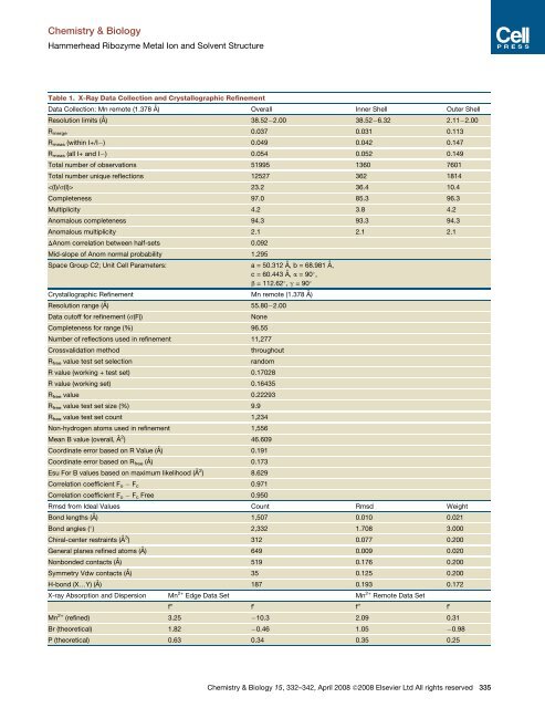
Chemistry & Biology<strong>Hammerhead</strong> <strong>Ribozyme</strong> Metal Ion <strong>and</strong> <strong>Solvent</strong> <strong>Structure</strong>Table 1. X-Ray Data Collection <strong>and</strong> Crystallographic RefinementData Collection: Mn remote (1.378 Å) Overall Inner Shell Outer ShellResolution limits (Å) 38.52 2.00 38.52 6.32 2.11 2.00R merge 0.037 0.031 0.113R meas (within I+/I ) 0.049 0.042 0.147R meas (all I+ <strong>and</strong> I ) 0.054 0.052 0.149Total number of observations 51995 1360 7601Total number unique reflections 12527 362 1814 23.2 36.4 10.4Completeness 97.0 85.3 96.3Multiplicity 4.2 3.8 4.2Anomalous completeness 94.3 93.3 94.3Anomalous multiplicity 2.1 2.1 2.1DAnom correlation between half-sets 0.092Mid-slope of Anom normal probability 1.295Space Group C2; Unit Cell Parameters: a = 50.312 Å, b = 68.981 Å,c = 60.443 Å, a = 90 ,b = 112.62 , g = 90 Crystallographic Refinement Mn remote (1.378 Å)Resolution range (Å) 55.80 2.00Data cutoff for refinement (s(F))NoneCompleteness for range (%) 96.55Number of reflections used in refinement 11,277Crossvalidation methodR free value test set selectionthroughoutr<strong>and</strong>omR value (working + test set) 0.17028R value (working set) 0.16435R free value 0.22293R free value test set size (%) 9.9R free value test set count 1,234Non-hydrogen atoms used in refinement 1,556Mean B value (overall, Å 2 ) 46.609Coordinate error based on R Value (Å) 0.191Coordinate error based on R free (Å) 0.173Esu For B values based on maximum likelihood (Å 2 ) 8.629Correlation coefficient F o F c 0.971Correlation coefficient F o F c Free 0.950Rmsd from Ideal Values Count Rmsd WeightBond lengths (Å) 1,507 0.010 0.021Bond angles ( ) 2,332 1.708 3.000Chiral-center restraints (Å 3 ) 312 0.077 0.200General planes refined atoms (Å) 649 0.009 0.020Nonbonded contacts (Å) 519 0.176 0.200Symmetry Vdw contacts (Å) 35 0.125 0.200H-bond (X.Y) (Å) 187 0.193 0.172X-ray Absorption <strong>and</strong> Dispersion Mn 2+ Edge Data Set Mn 2+ Remote Data Setf 00 f 0 f 00 f 0Mn 2+ (refined) 3.25 10.3 2.09 0.31Br (theoretical) 1.82 0.46 1.05 0.98P (theoretical) 0.63 0.34 0.35 0.25Chemistry & Biology 15, 332–342, April 2008 ª2008 Elsevier Ltd All rights reserved 335