ESSENTIALS OF ECHOCARDIOGRAPHY #4 - Echo in Context
ESSENTIALS OF ECHOCARDIOGRAPHY #4 - Echo in Context
ESSENTIALS OF ECHOCARDIOGRAPHY #4 - Echo in Context

You also want an ePaper? Increase the reach of your titles
YUMPU automatically turns print PDFs into web optimized ePapers that Google loves.
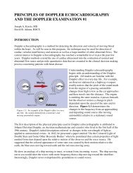
Similarly, the position of the defects guide the surgeon’s approach. Fig. 17 (left) shows ashort-axis view of a subarterial outlet defect enter<strong>in</strong>g the right ventricle just below thepulmonic valve. Such a defect would be very difficult to approach through the tricuspidvalve as it is a great distance away and requires a ventriculotomy for proper closure. Inaddition, these types of defects may, over time, allow for prolapse of the right coronary cuspof the aortic valve <strong>in</strong>to the defect result<strong>in</strong>g <strong>in</strong> aortic <strong>in</strong>sufficiency. Fig. 17 (right), <strong>in</strong>contrast, shows a short axis view of a perimembranous defect. Its proximity just under thetricuspid valve <strong>in</strong>dicates its easy surgical closure through the tricuspid valve and obviates anunwarranted ventriculotomy.Atrioventricular septal defectsFormerly known as “canal defects” or “endocardial cushion defects” atrioventricular septaldefects (AVSD) are present when any abnormality exists of the shared septum between theleft ventricle and the right atrium (the atrioventricular septum). As previously discussed,ostium primum atrial septal defects extend <strong>in</strong>to this common septal area and are really a mildform of an atrioventricular septal defect.In its most severe form (Fig. 18) the primum septum is absent with the defect extend<strong>in</strong>g tothe muscular septum leav<strong>in</strong>g the entire central portion of the heart without divid<strong>in</strong>g septaltissue, either atrial or ventricular. In this sett<strong>in</strong>g, a common atrioventricular valve orifice ispresent with a double-committed s<strong>in</strong>gle atrioventricular valve (i.e., the s<strong>in</strong>gle atrioventricularvalve enters <strong>in</strong>to both ventricles). In this case the AVSD is referred to as a complete defect.Those <strong>in</strong>experienced with congenital heart disease frequently have difficulty understand<strong>in</strong>gthese defects. This is pr<strong>in</strong>cipally brought about because of the failure to recognize thatAVSDs are comprised of a family of defects with ostium primum as a mild form and acomplete AVSDs as the most severe. Many possibilities exist between the two extremes.Fig. 19 shows a parasternal diastolic four-chamber view of an <strong>in</strong>fant with a complete AVSD.Only a small portion of the <strong>in</strong>teratrial septum is present. There is total communication andmix<strong>in</strong>g through the primum defect, ventricular septal defect, and common atrioventricularorifice. Fig. 7 demonstrated the other extreme of a primum defect alone.Fig. 20 shows the diastolic and systolic appearance of a patient with a complete AVSD andcommon atrium. Note the <strong>in</strong>sertion of some of the chordal structures onto the crest of theventricular septum. AVSDs <strong>in</strong>clude a broad spectrum of atrioventricular junctionabnormalities, all of which have two common features: an absent atrioventricular septum andabnormally formed atrioventricular valves. Fig. 21 shows an unusual subcostal short-axisview through the common atrioventricular valve orifice from a patient with a total AVSD. Itis important to identify all the leaflets present and trace their <strong>in</strong>sertion <strong>in</strong>to the left or rightventricle. Occasionally, an atrioventricular leaflet may bridge the central defect and havechordal <strong>in</strong>sertion <strong>in</strong>to both ventricles. In such cases, the leaflet is known as a “bridg<strong>in</strong>gleaflet” and must be surgically divided <strong>in</strong>to left and right portions. It then is resuspendedfrom a central patch to create separate atrioventricular orifices at the time of correction.Certa<strong>in</strong> “transitional” forms of AVSD exist. In these, a primum defect is seen <strong>in</strong> the atrialseptum and a small ventricular septal defect is noted near the atrioventricular junction. Twoseparate atrioventricular valve orifices may be seen. Notably, patients thought to havemerely a primum defect may, <strong>in</strong>deed, have a ventricular component.