Patellar Instability (West and Colvin) - The Patellofemoral Foundation
Patellar Instability (West and Colvin) - The Patellofemoral Foundation
Patellar Instability (West and Colvin) - The Patellofemoral Foundation

You also want an ePaper? Increase the reach of your titles
YUMPU automatically turns print PDFs into web optimized ePapers that Google loves.
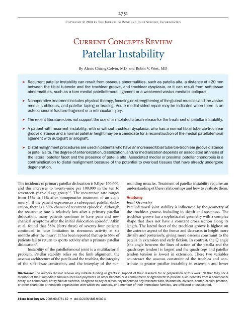
2751COPYRIGHT Ó 2008 BY THE JOURNAL OF BONE AND JOINT SURGERY, INCORPORATEDCurrent Concepts Review<strong>Patellar</strong> <strong>Instability</strong>By Alexis Chiang <strong>Colvin</strong>, MD, <strong>and</strong> Robin V. <strong>West</strong>, MDä Recurrent patellar instability can result from osseous abnormalities, such as patella alta, a distance of >20 mmbetween the tibial tubercle <strong>and</strong> the trochlear groove, <strong>and</strong> trochlear dysplasia, or it can result from soft-tissueabnormalities, such as a torn medial patellofemoral ligament or a weakened vastus medialis obliquus.ä Nonoperative treatment includes physical therapy, focusing on strengthening of the gluteal muscles <strong>and</strong> the vastusmedialis obliquus, <strong>and</strong> patellar taping or bracing. Acute medial-sided repair may be indicated when there is anosteochondral fracture fragment or a retinacular injury.ä <strong>The</strong> recent literature does not support the use of an isolated lateral release for the treatment of patellar instability.ä A patient with recurrent instability, with or without trochlear dysplasia, who has a normal tibial tubercle-trochleargroove distance <strong>and</strong> a normal patellar height may be a c<strong>and</strong>idate for a reconstruction of the medial patellofemoralligament with autograft or allograft.ä Distal realignment procedures are used in patients who have an increased tibial tubercle-trochlear groove distanceor patella alta. <strong>The</strong> degree of anteriorization, distalization, <strong>and</strong>/or medialization depends on associated arthrosis ofthe lateral patellar facet <strong>and</strong> the presence of patella alta. Associated medial or proximal patellar chondrosis is acontraindication to distal realignment because of the potential to overload tissues that have already undergonedegeneration.<strong>The</strong> incidence of primary patellar dislocation is 5.8 per 100,000,<strong>and</strong> this increases to twenty-nine per 100,000 in the ten toseventeen-year-old age group 1,2 . <strong>The</strong> recurrence rate rangesfrom 15% to 44% after nonoperative treatment of an acuteinjury 2 . If the patient experiences a subsequent patellar dislocation,there is a 50% chance of recurrent episodes 1 . Althoughthe recurrence rate is relatively low after a primary patellardislocation, many patients continue to have pain <strong>and</strong> mechanicalsymptoms after the initial dislocation episode 3 . Atkinet al. found that 58% (forty-three) of seventy-four patientscontinued to have limitation in strenuous activity at sixmonths after the injury 4 . It has been reported that up to 55% ofpatients fail to return to sports activity after a primary patellardislocation 4 .<strong>Instability</strong> of the patellofemoral joint is a multifactorialproblem. <strong>Patellar</strong> stability relies on the limb alignment, theosseous architecture of the patella <strong>and</strong> the trochlea, the integrityof the soft-tissue constraints, <strong>and</strong> the interplay of the surroundingmuscles. Treatment of patellar instability requires anunderst<strong>and</strong>ing of these relationships <strong>and</strong> how to evaluate them.AnatomyJoint Geometry<strong>Patellofemoral</strong> joint stability is influenced by the geometry ofthe trochlear groove, including its depth <strong>and</strong> steepness. <strong>The</strong>trochlear groove has a sophisticated geometry with a complexshape that does not have a constant cross section along itslength. <strong>The</strong> lateral facet of the trochlear groove is highest onthe anterior aspect of the femur <strong>and</strong> decreases in height moredistally <strong>and</strong> posteriorly, giving more osseous constraint to thepatella in extension <strong>and</strong> early flexion. In contrast, the Q angle(the angle between the lines of action of the patella <strong>and</strong> thequadriceps tendon) is largest <strong>and</strong> the quadriceps <strong>and</strong> patellartendon tension is lowest in extension. <strong>The</strong>se two variablescounteract the osseous constraint of the trochlea <strong>and</strong> contributeto greater patellar instability in extension <strong>and</strong> lowerDisclosure: <strong>The</strong> authors did not receive any outside funding or grants in support of their research for or preparation of this work. Neither they nor amember of their immediate families received payments or other benefits or a commitment or agreement to provide such benefits from a commercialentity. No commercial entity paid or directed, or agreed to pay or direct, any benefits to any research fund, foundation, division, center, clinical practice,or other charitable or nonprofit organization with which the authors, or a member of their immediate families, are affiliated or associated.J Bone Joint Surg Am. 2008;90:2751-62 d doi:10.2106/JBJS.H.00211
NUMBERD2752dTHE J OURNAL OF B ONE &JOINT S URGERY JBJS. ORGd dVOLUME 90-A 12 ECEMBER 2008PATELLAR I NSTABILITYdegrees of flexion. <strong>The</strong> quadriceps <strong>and</strong> patellar tendons providea strong posterior force vector during knee flexion, contributingto increased patellar stability with knee flexion. Asthe knee flexes <strong>and</strong> extends, the contact area moves across thepatella. <strong>The</strong> patella leaves its engagement with the groove asthe knee reaches full extension. When the knee starts to bend,the initial contact is at the distal <strong>and</strong> lateral edge of the patellararticular surface, which does not extend to the inferior facet.As the patella moves distally with knee flexion, the contact areaon the patella moves proximally. In deep knee flexion (120°),the medial facet, or so-called odd facet, contacts the lateralmargin of the medial femoral condyle 5 .Patella alta has been associated with recurrent dislocations6,7 . Patella alta results in less osseous stability because thedegree of flexion at which the patella engages in the trochlea ishigher than that in a normal knee. Under normal conditions,the patella usually engages by 20° of flexion. Furthermore,knees with patella alta have reduced patellar contact areaswhen compared with knees with normal patellar height, <strong>and</strong>these reduced patellar contact areas lead to greater patellofemoralstress during fast walking 8,9 .Limb AlignmentFemoral <strong>and</strong> tibial torsion can play an important role in patellarinstability. A more widely recognized aspect of osseous alignmentis the Q angle. <strong>The</strong> Q angle is largest in full extensionbecause the tibia rotates externally in terminal knee extension(the so-called screw-home mechanism), moving the tibial tuberositymore laterally 10 . Because the Q angle is greatest in fullextension, this is the position in which the patella is at greatestrisk for dislocation. In this position, the patella disengagesfrom the trochlea <strong>and</strong> the posteriorly directed force from theextensor mechanism that holds the patella in the groove is thelowest.<strong>The</strong> Q angle is difficult to measure because of the mobilityof the patella. Quadriceps tension pulls the patella in a proximallateraldirection in full extension. If the patella is unstable, itsubluxates laterally, resulting in a falsely low Q-angle measurement.<strong>The</strong>refore, it is important to keep the patella locatedin the trochlear groove manually during the measurement.Limb rotation should also be controlled during measurementsince external tibial torsion can increase the apparent Q angle.Retinacula<strong>The</strong> iliotibial b<strong>and</strong> attaches to the Gerdy tubercle distally but alsohas attachments to the patellar <strong>and</strong> quadriceps tendons. It hasbeen found that tension in the iliotibial b<strong>and</strong> causes the patella totrack in a more lateral position. <strong>The</strong>re are three layers that makeup the lateral side of the patellar attachments. <strong>The</strong> superficiallayer is confluent with the iliotibial b<strong>and</strong>. <strong>The</strong> intermediate layeris the lateral patellofemoral b<strong>and</strong>, or the iliotibial patellar b<strong>and</strong>.This b<strong>and</strong> extends from the deep layer of the iliotibial b<strong>and</strong> to themidlateral aspect of the patella. <strong>The</strong> deep layer is confluent withthe knee capsule 11 .<strong>The</strong> medial patellofemoral ligament is the primary passivesoft-tissue restraint to lateral patellar displacement. It provides50% to 60% of lateral restraint from 0° to 30° of knee flexion 12 .<strong>The</strong> medial patellofemoral ligament runs transversely from theproximal half of the medial patellar border to the femur near themedial epicondyle. <strong>The</strong> superficial fibers of the medial patellofemoralligament pass over the saddle between the epicondyle<strong>and</strong> the adductor tubercle <strong>and</strong> insert 1.9 mm anterior <strong>and</strong>3.8 mm distal to the adductor tubercle 13 . <strong>The</strong> medial patellofemoralligament provides an important stabilizing force onthe medial side of the knee. A study of cadavers showed thatcutting the medial structures results in a 50% decrease in theforce required to move the patella 10 mm laterally 14 .Muscles<strong>The</strong> vastus medialis obliquus <strong>and</strong> vastus lateralis obliquus originatefrom septa alongside the femur <strong>and</strong> approach the patellafrom directions that deviate from the anatomic axis of the femur.<strong>The</strong>se muscles can pull the patella medially or laterally. <strong>The</strong> vastusmedialis obliquus has a mean orientation that deviates 47° ± 5°medially from the femoral axis, <strong>and</strong> the vastus lateralis obliquushas a mean orientation that deviates 35° ± 4° laterally from theaxis 15 . An imbalance of strength may lead to instability. <strong>The</strong>vastus medialis obliquus is the first part of the quadriceps toweaken <strong>and</strong> the last to strengthen when function is inhibited 16 .It has been shown that, if the muscle force vectors areadded together in the coronal plane, their resultant force isalmost exactly parallel to the femoral anatomic axis. If the forceproducingcapacity of each muscle head is in proportion to itsphysiologic cross-sectional area, the vastus medialis obliquuscould contribute 10% of the total quadriceps tension 15 .If the vastus medialis obliquus is completely relaxed,lateral patellar stability is reduced at all angles of knee flexionfrom 0° to 90°. Goh et al. found lateral stability to be reducedby 30% when the vastus medialis obliquus was relaxed at 20° ofknee flexion 17 <strong>and</strong> that relaxation of the vastus medialis obliquuscaused the patella to displace laterally 4 mm <strong>and</strong> alsoincreased the load on the lateral facet 17 .Radiographic EvaluationSt<strong>and</strong>ard radiographs for assessment of patellar instability includeposteroanterior weight-bearing views of both knees in 45°of flexion, lateral views, <strong>and</strong> Merchant views. For the Merchantview, the knee is flexed 45° over the end of the table <strong>and</strong> the x-raybeam is inclined 30° downward 18 . This view is used to assess forpatellar tilt, patellar subluxation, <strong>and</strong> trochlear dysplasia. <strong>Patellar</strong>subluxation is assessed by measuring the congruenceangle, which reflects the relationship of the patellar articularridge to the intercondylar sulcus <strong>and</strong> averages approximately6° ± 11° in the medial direction 18 . <strong>The</strong> sulcus angle is formedby the highest points of the medial <strong>and</strong> lateral femoral condyles<strong>and</strong> the lowest point of the intercondylar sulcus <strong>and</strong> is approximately138° ± 6° 18 . A sulcus angle of >145° is indicative oftrochlear dysplasia 19 . <strong>The</strong> lateral patellofemoral angle, as describedby Laurin et al., is used to assess patellar tilt <strong>and</strong> is bestevaluated on an axial radiograph of the patella with the kneeflexed 20° 20 . Further flexion can result in a falsely normalangle 20 .
NUMBERD2754dTHE J OURNAL OF B ONE &JOINT S URGERY JBJS. ORGd dVOLUME 90-A 12 ECEMBER 2008PATELLAR I NSTABILITYFig. 2Classification of trochlear dysplasia. Type A: crossing sign, with trochlear morphology preserved (fairly shallow trochlea [>145°]).Type B: crossing sign, supratrochlear spur, <strong>and</strong> flat or convex trochlea. Type C: crossing sign, with double contour. Type D:crossing sign, supratrochlear spur, double contour, asymmetry of trochlear facets, <strong>and</strong> vertical link between medial <strong>and</strong> lateralfacets (cliff pattern). (Reprinted, with permission, from: Dejour D, Le Coultre B. Osteotomies in patello-femoral instabilities.Sports Med Arthrosc. 2007;15:40.)Escamilla et al. also found that open-chain exercises promotedmore rectus femoris activity <strong>and</strong> that closed-chain exercisesproduced more vastus activity 38 . Closed kinetic trainingallows training of the vastus muscles simultaneously with gluteal<strong>and</strong> trunk-muscle strengthening to control limb position.Operative TreatmentMore than 100 different operations have been described for thetreatment of patellar instability, <strong>and</strong> these procedures typicallyinvolve a combination of lateral release, medial imbrication,distal realignment, <strong>and</strong> anteromedialization of the tibial tubercle39 . <strong>The</strong> so-called gold-st<strong>and</strong>ard treatment for patellarinstability has yet to be defined. <strong>The</strong> literature reflects this inthat no two studies have used the same operative procedure,inclusion <strong>and</strong> exclusion criteria, or outcome measures. Furthermore,there is a lack of prospective r<strong>and</strong>omized trials.Lateral ReleaseAn isolated lateral release is the only procedure that has beenshown to be ineffective for the treatment of patellar instability.While a lateral release can be useful in the treatment of lateralpatellar compression syndrome, it does not yield acceptableresults in patients with patellar instability 40 . In fact, all twentyeightpatients in one series who underwent lateral release forpatellar dislocation continued to experience dislocations 40 .Lattermann et al. reviewed the results from fourteen studies onlateral release for the treatment of patellar instability 41 . Althoughthere was an average 80% patient-satisfaction rating inthe short term, this rating had dropped to 63.5% after morethan four years of follow-up 41 . <strong>The</strong> poor results after lateralrelease can be attributed to the inability of the procedure toalign the patella more medially 42 . Furthermore, lateral releasecan be complicated by medial patellar instability if the releaseextends into, <strong>and</strong> detaches, the vastus lateralis obliquus 41 .If the tibial tubercle-to-trochlear groove distance is
NUMBERD2755dTHE J OURNAL OF B ONE &JOINT S URGERY JBJS. ORGd dVOLUME 90-A 12 ECEMBER 2008PATELLAR I NSTABILITYof 208 N 52 , <strong>and</strong> a hamstring graft used to reconstruct themedial patellofemoral ligament can generate up to 1600 N 43 .However, because medial imbrication is a nonanatomic procedure,it can result in excessive medialization of the patella orabnormal tracking. In a biomechanical study, Ostermeier et al.found that the combination of a lateral release <strong>and</strong> a medialimbrication tensioned with the knee at 45° resulted in significantlymedialized (p < 0.01) <strong>and</strong> internally tilted (p < 0.01)patellar movement when compared with the intact knee condition53 . Furthermore, medial imbrication fails to addressproblems with the medial patellofemoral ligament at the femoralattachment 54 .Fig. 3<strong>The</strong> tibial tubercle-to-trochlear groove (TT-TG) distance is measured bysuperimposing axial computed tomography images of the tibial tubercle<strong>and</strong> the trochlear groove with the knee in extension. <strong>The</strong> normaldistance ranges from 10 to 15 mm. (Reprinted, with permission, from:Dejour D, Le Coultre B. Osteotomies in patello-femoral instabilities.Sports Med Arthrosc. 2007;15:41.)operative <strong>and</strong> nonoperative treatment with respect to scoresdetermined with the systems of Kujala et al. 45 (p = 0.6), Fl<strong>and</strong>ryet al. 46 (p = 0.1), <strong>and</strong> Tegner <strong>and</strong> Lysholm 47 (p = 0.7); they alsofound no difference in the rate of recurrence of subluxations ordislocations. Palmu et al. found that the rates of redislocation(approximately 70%) were similar in patients who had beentreated with repair of the medial structures <strong>and</strong> those who hadhad nonoperative treatment 34 .Atfourteenyears,thetwogroups had similar good-to-excellent subjective outcomescores 34 . Both groups of authors concluded that there was noadvantage to primary repair of the medial structures after afirst-time dislocation. On the other h<strong>and</strong>, several authors havereported good or excellent functional outcome scores <strong>and</strong> fewredislocations after arthroscopic medial plication for treatmentof recurrent patellar instability 48,49 .Acute medial-sided repair does have its proponents inclinical practice. In a recent survey of the National Football LeaguePhysician’s Society (NFLPS), 6% (two) of thirty-one surgeonsindicated that they would perform an early repair to treat an acutepatellar dislocation without a loose body in a high-school, college,or professional athlete 50 . Early operative repair to treat an acutepatellar dislocation without a loose body was not recommendedfor athletes at any level by 58% of the surgeons. Ahmad et al.repaired the medial patellofemoral ligament in addition to thevastus medialis obliquus, if it was torn, because of the importanceof the vastus medialis obliquus as a dynamic medial stabilizer51 , <strong>and</strong> there were no recurrent dislocations in their series.Advocates for medial imbrication, as opposed to reconstructionof the medial patellofemoral ligament, cite the potentialfor overload of the patella with a graft reconstruction 43 .<strong>The</strong> native medial patellofemoral ligament has a load to failureReconstruction of the Medial <strong>Patellofemoral</strong> LigamentReconstruction of the medial patellofemoral ligament has theadvantage of addressing damage at the adductor tubercle 54 .Comparing studies is difficult, as a review of the Englishlanguageliterature identified only eight papers describing avariety of medial patellofemoral ligament reconstruction procedures<strong>and</strong> outcome scales 55 . <strong>The</strong>re was no consensus withregard to the choice of graft, graft positioning, graft tension, orstatic versus dynamic reconstruction.Adductor magnus autografts, semitendinosus autografts<strong>and</strong> allografts, <strong>and</strong> tibialis anterior allografts have all been proposedas possible graft choices 53,56-61 . Steiner et al. recommendedthe use of bone-quadriceps tendon autograft or bone-patellartendon allograft for severely dysplastic knees in which morestrength was thought to be warranted 60 . Farr <strong>and</strong> Schepsis advocatedthe use of a doubled semitendinosus allograft, not for itsstrength but rather to reproduce the broad attachment site onthe patella 57 .Use of a doubled hamstring tendon graft could beproblematic if it is malpositioned, since it is stronger <strong>and</strong> stifferthan the native medial patellofemoral ligament 62 . Elias <strong>and</strong>Cosgarea performed a biomechanical study <strong>and</strong> found a significantincrease in force on the medial patellar facet witheither 5 mm of proximal malpositioning (p < 0.01) or a graftthat was 3 mm shorter than the native medial patellofemoralligament (p < 0.01) 61 . Furthermore, a combination of the twoerrors led to a medial tilt moment from full extension through90° of flexion. Increased pressures could theoretically lead todegeneration of the cartilage <strong>and</strong> arthrosis, while undertighteningcould lead to recurrent instability 60 . Thus, Elias <strong>and</strong>Cosgarea recommended placing the femoral attachment of thegraft 1 cm distal to the adductor tubercle to avoid overloadingthe medial patellofemoral cartilage. A biomechanical study byBeck et al. demonstrated that, when >2 N of tension was usedto secure the reconstruction of the medial patellofemoral ligament,there was a significant increase in medial patellofemoralcontact pressures (p < 0.05) 62 . <strong>The</strong>re is also a risk ofapplying a net posteromedial force on the patella as the reconstructionresults in a posterior force as well 62 .<strong>The</strong> appropriate knee flexion angle at which to tensionthe graft is also controversial. While some believe the medialpatellofemoral ligament to be isometric 60,63 , others have shownthat it is not 52,54 . Tensioning the graft at between 60° <strong>and</strong> 90° of
NUMBERD2756dTHE J OURNAL OF B ONE &JOINT S URGERY JBJS. ORGd dVOLUME 90-A 12 ECEMBER 2008PATELLAR I NSTABILITYflexion 54,59,60 , instead of at the lower flexion angles (30° to 45°)that have been recommended by other authors 53,57 , has beenadvocated to avoid overtightening of the graft <strong>and</strong> to ensurethat the patella has engaged the trochlea. LeGr<strong>and</strong> et al. recommendedapplying tension at 45° to 60° of flexion <strong>and</strong> alsochecking that there is symmetric medial <strong>and</strong> lateral translationof the patella at 20° of flexion 64 . Farr <strong>and</strong> Schepsis described an‘‘anatomometric’’ placement of the graft: tensioning the graftwith the knee in 30° of flexion so that it becomes more lax withfurther flexion <strong>and</strong> tighter in terminal extension 57 .A dynamic reconstruction of the medial patellofemoralligament has been proposed as an alternative that is better than astatic reconstruction. Ostermeier et al. performed a dynamicreconstruction by transferring the distal end of the semitendinosusbehind the proximal aspect of the medial collateral ligamentto the medial margin of the patella 53 . <strong>The</strong> authors foundthat a static reconstruction medialized the patella significantlymore than the dynamic reconstruction did (p < 0.01). Thus, adynamic reconstruction could theoretically protect againstovertensioning of the graft. Deie et al. 56 found that dynamicreconstruction provided a significant improvement (p 0.05).Fracture of the patella after fixation of the graft through abone tunnel has been described 54,58 . In a study of twenty-fourknees treated with reconstruction of the medial patellofemoralligament, Mikashima et al. reported two patellar fractures,both of which occurred through bone tunnels in the patella 58 .<strong>The</strong> authors recommended suturing the graft to the patellarperiosteum in all patients except those with a thin periosteum.However, we are not aware of any biomechanical studies comparingtunnel with suture-anchor fixation.Reconstruction of the medial patellofemoral ligament hashad good results in terms of preventing future subluxations ordislocations 54,58,59 . However, not all patients with recurrentpatellar instability may benefit from this reconstruction. Nomura<strong>and</strong> Inoue evaluated twelve knees in twelve patients at anaverage of 4.2 years (range, 3.1 to 5.6 years) after reconstructionof the medial patellofemoral ligament 59 . Using the Insallscale, they found only fair results in patients with preexistingchondromalacia patella. Thus, they recommended reconstructionof the medial patellofemoral ligament for patients withoutadvanced changes in the patellar cartilage.Biomechanically, reconstruction of the medial patellofemoralligament provides more stability than a medial tibialtubercle transfer does. Ostermeier et al. evaluated patellar kinematicsin cadaver knees after either a medial transfer of thetibial tubercle or a reconstruction of the medial patellofemoralligament with a semitendinosus autograft 67 . <strong>Patellar</strong> movement<strong>and</strong> strain in the medial patellofemoral ligament were measuredwith <strong>and</strong> without a 100-N lateral subluxation forceunder both testing conditions. While loading of the nativemedial patellofemoral ligament was greatest in full extension,the reconstruction of the medial patellofemoral ligament reducedthe ligament load <strong>and</strong> lateral patellar displacementcompared with those parameters after the medial transfer ofthe tibial tubercle, regardless of the knee flexion angle. On thebasis of their results, the authors concluded that reconstructionof the medial patellofemoral ligament was better thanmedial transfer of the tibial tuberosity for stabilizing patellarmovement under a laterally directed force. However, reconstructionof the medial patellofemoral ligament does notaddress potential osseous problems <strong>and</strong> can also result in overloadof the medial patellofemoral cartilage 60,61 .TrochleoplastyTrochleoplasty has been used with equivocal results, as reportedin the European literature. Concerns about possibleserious <strong>and</strong> irreversible articular <strong>and</strong> subchondral injury to thetrochlea have limited its use in the United States.Indications for a sulcus-deepening trochleoplasty includeabnormal patellar tracking with a J-sign, usually manifested by atibial tubercle-trochlear groove distance of greater than 10 to20 mm 23 , <strong>and</strong>/or a dome-shaped trochlea noted on a perfectlateral radiograph with overlap of the posterior condyles in apatient with recurrent patellar instability 68 . In a trochleoplasty,cancellous bone is exposed in the trochlea by elevating a stripof cortical bone around the edge of the trochlea. <strong>The</strong> newtrochlear sulcus is created proximal <strong>and</strong> 3° to 6° lateral to theprevious sulcus by removing cancellous bone. <strong>The</strong> trochlearbone shell is then impacted into the new sulcus <strong>and</strong> fixed withtwo small staples (Fig. 4). <strong>The</strong> bone can also be secured withresorbable sutures 69,70 .Verdonk et al. reported equivocal results at eighteenmonths (range, eight to thirty-four months) after trochleoplastyin thirteen knees in twelve patients 71 . <strong>The</strong>ir indicationfor the operation was patellar pain with or without recurrentpatellar instability. According to the Larsen-Lauridsen scoring
NUMBERD2758dTHE J OURNAL OF B ONE &JOINT S URGERY JBJS. ORGd dVOLUME 90-A 12 ECEMBER 2008PATELLAR I NSTABILITYFig. 5Tibial tubercle realignment. A: A flat (no-angle) osteotomy allows medialization of the tibial tubercle. <strong>The</strong> elevator protects theneurovascular bundle. B: A steeper cut for equal anteriorization <strong>and</strong> medialization of the tibial tubercle. C: A very steep cut providesmaximum anteriorization of the tibial tubercle with less medialization. (Reprinted, with permission, from: Buuck DA, Fulkerson JP.Anteromedialization of the tibial tubercle: a 4- to 12-year follow-up. Op Tech Sports Med. 2000;8:136-7.)Carney et al. reviewed the results of the Elmslie-Trillatprocedure for the treatment of recurrent subluxation <strong>and</strong>dislocation of the patella in fifteen knees in fourteen patients 89 .<strong>The</strong> authors compared the outcomes at a mean of three yearswith those at a mean of twenty-six years <strong>and</strong> found no differencein instability between the two time-periods. Althoughnot significant, there was a trend toward a worsening Coxscore 90 with time, which occurred even though the articularcartilage was grossly intact.Koeter et al. reported the results of medial realignment ofthe tibial tubercle for patients with either painful lateral maltracking(with no instability) or patellar instability for longerthan one year <strong>and</strong> a tibial tubercle-trochlear groove distanceof >15 mm 86 . <strong>The</strong>re was no difference in the distance of themedial displacement between the groups. A mean of 5.7 mm ofdistalization of the tibial tubercle was achieved in nine patientswith lateral maltracking <strong>and</strong> twenty-two patients with patellarinstability. At two years postoperatively, although the patientswith patellar instability had more variable outcomes, bothgroups had improvement in all scores, with no significantdifference noted between the groups. Thus, the authors advocateda medial transfer, with or without distalization, for patientswith either patellar maltracking or patellar instability.Diks et al. found that a tibial tubercle transfer providedbetter results for patients with patellar maltracking <strong>and</strong> no instabilitythan for patients with isolated patellar instability 83 .Tibialtubercle transfer was performed in forty-three knees, twentysevenwith objective evidence of patellar instability <strong>and</strong> sixteenwith a laterally tracking patella. <strong>The</strong> mean duration of follow-upwas thirty-seven months. <strong>The</strong> transfers in the patients with patellarinstability were, overall, more effective in improving stability,doing so in 96% (twenty-six) of the twenty-seven patients,than they were in improving pain (63% of the patients). Onthe other h<strong>and</strong>, a higher percentage of patients (81%, thirteenof sixteen) with patellar maltracking had good pain relief.Barber <strong>and</strong> McGarry advocated the use of the modifiedElmslie-Trillat procedure for treatment of patellofemoral instabilitywithout evidence of arthritis 87 .<strong>The</strong>seauthorsperformeda medial translation of the tibial tubercle hinged on adistal periosteal flap along with an arthroscopic lateral retinacularrelease <strong>and</strong> medial capsular reefing for patients with atleast three recurrent patellar dislocations or subluxations thathad been resistant to a minimum of three months of physicaltherapy or bracing. Of thirty-five patients followed for a meanof ninety-eight months, 91% (thirty-two) had no additionalsubluxations or dislocations. Furthermore, there was improvementin the IKDC 65 ,Fulkersonknee 79 , <strong>and</strong> Lysholm <strong>and</strong>Gillquist 77 scores.Fulkerson et al. described anteromedialization of the tibialtubercle to address degenerative changes of the articular cartilage79 (Fig. 5). In a cadaver model of this procedure, pressure wasshown to be decreased on the lateral patellar facet at lowerangles of flexion (up to 30°) whereas pressure was equalizedbetween the medial <strong>and</strong> lateral patellar facets at greater angles offlexion 85 . <strong>The</strong>re was slight superior migration of the contact areaof the patellofemoral joint with distalization 85 . Cadaver studieshave also demonstrated that anteromedialization decreases thecontact pressures on the trochlear side overall, primarily on the
NUMBERD2759dTHE J OURNAL OF B ONE &JOINT S URGERY JBJS. ORGd dVOLUME 90-A 12 ECEMBER 2008PATELLAR I NSTABILITYTABLE I Grades of Recommendation for Summaries or Review ofOrthopaedic Surgical StudiesGradeABCIDescriptionGood evidence (Level-I studies with consistentfindings) for or against recommending intervention.Fair evidence (Level-II or III studies with consistentfindings) for or against recommending intervention.Poor-quality evidence (Level-IV or V studies withconsistent findings) for or against recommendingintervention.<strong>The</strong>re is insufficient or conflicting evidence notallowing a recommendation for or againstintervention.lateral <strong>and</strong> central areas of the trochlea 88 . However, pressures areelevated on the medial aspect of the trochlea <strong>and</strong> the proximalmedialaspect of the patella at all flexion angles, <strong>and</strong> thus cautionshould be used when performing an anteromedialization procedurein patients with medial-sided defects 88 .Buuck <strong>and</strong> Fulkerson reviewed their results with anteromedializationat an average of 8.2 years postoperatively 81 .<strong>The</strong>ir indications for the procedure were painful patellofemoralmaltracking (subluxation or tilt) with degenerativechanges on the distal <strong>and</strong> lateral aspects of the patella. Poorerresults were associated with Outerbridge 91 grade-3 or 4 lesionsin the central or medial aspects of the trochlea. Notably, threeof the six fair or poor results were in patients who had compensationclaims or were involved in litigation. Overall, 74%(thirty-one) of the forty-two patients had a good or excellentresult at an average of 8.2 years postoperatively.Pritsch et al. reported their results of tibial tubercle transferfor treatment of recurrent patellar instability, anterior knee pain,<strong>and</strong> evidence of maltracking on a dynamic computed tomographyscan 85 . Sixty-nine knees followed for a median of 6.2 yearshad a significant improvement between the preoperative <strong>and</strong>postoperative Lysholm <strong>and</strong> Gillquist 77 <strong>and</strong> Karlsson 92 score categoriesof instability, pain, <strong>and</strong> stair-climbing (p < 0.001).However, patients who had had only instability preoperativelydid better than patients with pain or both pain <strong>and</strong> instabilitypreoperatively. A better outcome was correlated with male sex,intact patellar articular cartilage, <strong>and</strong> symptoms of patellar instability.Physical examination findings that correlated with aworse prognosis included a positive patellar grind test, retinacularpain, a positive patellar tilt test, <strong>and</strong> a positive J-sign. <strong>The</strong>duration of follow-up was also positively correlated with betterLysholm <strong>and</strong> Gillquist 77 <strong>and</strong> Karlsson 92 scores, which the authorsattributed to the need for quadriceps recovery in the short term.Furthermore, there was no deterioration of the results with time.Pidoriano et al. found that the location of articular cartilagedamage in the patellofemoral joint correlated with the outcomeafter anteromedialization 80 . Of thirty-six patients (thirty-sevenknees), 56% (twenty) had the procedure performed because ofpatellar instability. All twenty-three patients with distal <strong>and</strong>lateral patellar lesions were extremely satisfied with the result ofthe procedure. <strong>The</strong>re was a 95% rate of good-to-excellent results(if no Workers’ Compensation claim was involved), <strong>and</strong> 87%had good-to-excellent pain relief. <strong>The</strong> authors recommendednot proceeding with the operation if medial, proximal, or diffuselesions were present on the patella or if central lesions werepresent on the trochlea. Interestingly, the Outerbridge 91 classificationof the lesion had no effect on the outcome.Palmer et al. also reported satisfactory results after anteromedializationfor treatment of both instability <strong>and</strong> painfulpatellar maltracking 84 . In a study of eighty-four patients followedfor a mean of 5.6 years, the result was good to excellent in 80% ofpatients with dislocation <strong>and</strong> 71% of patients with pain frommaltracking. <strong>The</strong>re was no significant difference in outcomebetween the groups, leading the authors to recommend anteromedializationof the tibial tubercle with distalization as aneffective procedure for both instability <strong>and</strong> pain due to maltracking.Predictors of poor postoperative results were postoperativeanterior knee pain <strong>and</strong> a previous realignment procedure.Fracture of the proximal part of the tibia or of the tibialtubercle after tibial tubercle transfer has been reported by severalauthors 86,93,94 . All cases occurred within three months after theoperation <strong>and</strong> were attributed to early weight-bearing. Suggestedpreventive measures have included avoidance of step cuts 86 ,anosteotomy of at least 5 cm in length <strong>and</strong> 0.75 cm in thickness toavoid fracture of the tuberosity 86 , protected weight-bearing oversix to eight weeks in a hinged knee brace, <strong>and</strong> advancement tofull weight-bearing once the osteotomy site is fully healed 93-95 .Overview<strong>The</strong> evaluation <strong>and</strong> treatment of patellar instability continue toevolve. <strong>The</strong> importance of a thorough physical examination <strong>and</strong>an accurate diagnosis cannot be stressed enough. We typicallyTABLE II Grades of Recommendation for the Treatment of Acute <strong>Patellar</strong> Dislocation <strong>and</strong> Chronic <strong>Patellar</strong> <strong>Instability</strong> with Associated FactorsDisorder Treatment Grade of RecommendationAcute patellar dislocation Early medial-sided repair or nonoperative treatment AChronic patellar instability with associated factorsPatella alta Tibial tubercle realignment CMedial patellofemoral ligament injury Medial patellofemoral ligament reconstruction CTrochlear dysplasia Trochleoplasty CElevated tibial tubercle-to-trochlear groove distance Tibial tubercle realignment C
NUMBERD2760dTHE J OURNAL OF B ONE &JOINT S URGERY JBJS. ORGd dVOLUME 90-A 12 ECEMBER 2008PATELLAR I NSTABILITYrecommend nonoperative treatment with patellar bracing <strong>and</strong>therapy for primary patellar dislocations. We aspirate the effusionacutely to allow the patient to regain quadriceps strength<strong>and</strong> control. However, if a patient has a loose body after a dislocation,we recommend arthroscopy for removal or possiblyfixation of the fracture fragment, in which case a medial repairwill usually be performed simultaneously. When there is anextensive medial-sided injury, such as a femoral avulsion of themedial patellofemoral ligament in association with an extensiveretinacular injury or avulsion of the vastus medialis obliquus,repair is usually recommended as well.When physical therapy <strong>and</strong> bracing have failed, the surgicaloptions should be tailored to the underlying pathological condition.<strong>The</strong> literature provides little support for the performanceof an isolated lateral release for the treatment of patellar instability.Recurrent patellar instability can be addressed with either areconstruction of the medial patellofemoral ligament or a distalpatellar realignment. Reconstruction of the medial patellofemoralligament can be performed in patients with recurrent instability,with or without trochlear dysplasia, who have a normaltibial tubercle-trochlear groove distance <strong>and</strong> a normal patellarheight. Distal realignment procedures can be used in patientswho have an increased tibial tubercle-trochlear groove distanceor patella alta. A st<strong>and</strong>ard medialization of the tibial tubercle canbe performed if there is a normal patellar height <strong>and</strong> trochlearanatomy <strong>and</strong> an increased tibial tubercle-trochlear groove distance.Distalization of the tubercle can be added if there is concomitantpatella alta, <strong>and</strong> anteromedialization of the tubercle isperformed if there is lateral <strong>and</strong>/or distal patellar facet chondrosis.To avoid overloading the patella, a tubercle osteotomyshould not be performed if there is associated medial or proximalpatellar chondrosis.While there is good evidence (Table I) for the nonoperativetreatment of an acute patellar dislocation, most of thecurrent surgical treatments for chronic patellar instability arebased on Level-IV evidence (Table II). Prospective r<strong>and</strong>omizedtrials are necessary to determine the optimal surgical treatmentfor chronic patellar instability. nAlexis Chiang <strong>Colvin</strong>, MDMount Sinai School of Medicine, 5 East 98th Street,Box 1188, New York, NY 10029.E-mail address: alexis.colvin@gmail.comRobin V. <strong>West</strong>, MDCenter for Sports Medicine,University of Pittsburgh Medical Center,3200 South Water Street, Pittsburgh, PA 15203.E-mail address: westrv@upmc.eduReferences1. Fithian DC, Paxton EW, Stone ML, Silva P, Davis DK, Elias DA, White LM.Epidemiology <strong>and</strong> natural history of acute patellar dislocation. Am J Sports Med.2004;32:1114-21.2. Hawkins RJ, Bell RH, Anisette G. Acute patellar dislocations. <strong>The</strong> natural history.Am J Sports Med. 1986;14:117-20.3. Cofield RH, Bryan RS. Acute dislocation of the patella: results of conservativetreatment. J Trauma. 1977;17:526-31.4. Atkin DM, Fithian DC, Marangi KS, Stone ML, Dobson BE, Mendelsohn C.Characteristics of patients with primary acute lateral patellar dislocation <strong>and</strong> theirrecovery within the first 6 months of injury. Am J Sports Med. 2000;28:472-9.5. Goodfellow J, Hungerford DS, Zindel M. Patello-femoral joint mechanics <strong>and</strong>pathology. 1. Functional anatomy of the patello-femoral joint. J Bone Joint Surg Br.1976;58:287-90.6. Insall J, Goldberg V, Salvati E. Recurrent dislocation <strong>and</strong> the high-riding patella.Clin Orthop Relat Res. 1972;88:67-9.7. Kannus PA. Long patellar tendon: radiographic sign of patellofemoral painsyndrome—a prospective study. Radiology. 1992;185:859-63.8. Ward SR, Powers CM. <strong>The</strong> influence of patella alta on patellofemoral joint stressduring normal <strong>and</strong> fast walking. Clin Biomech (Bristol, Avon). 2004;19:1040-7.9. Ward SR, Terk MR, Powers CM. Patella alta: association with patellofemoralalignment <strong>and</strong> changes in contact area during weight-bearing. J Bone Joint SurgAm. 2007;89:1749-55.10. Hallén LG, Lindahl O. <strong>The</strong> ‘‘screw-home’’ movement in the knee-joint. ActaOrthop Sc<strong>and</strong>. 1966;37:97-106.11. Terry GC, Hughston JC, Norwood LA. <strong>The</strong> anatomy of the iliopatellar b<strong>and</strong> <strong>and</strong>iliotibial tract. Am J Sports Med. 1986;14:39-45.12. Desio SM, Burks RT, Bachus KN. Soft tissue restraints to lateral patellartranslation in the human knee. Am J Sports Med. 1998;26:59-65.13. LaPrade RF, Engebretsen AH, Ly TV, Johansen S, Wentorf FA, Engebretsen L. <strong>The</strong>anatomy of the medial part of the knee. J Bone Joint Surg Am. 2007;89:2000-10.14. Senavongse W, Amis AA. <strong>The</strong> effects of articular, retinacular, or musculardeficiencies on patellofemoral joint stability. J Bone Joint Surg Br. 2005;87:577-82.15. Farahm<strong>and</strong> F, Senavongse W, Amis AA. Quantitative study of the quadricepsmuscles <strong>and</strong> trochlear groove geometry related to instability of the patellofemoraljoint. J Orthop Res. 1998;16:136-43.16. Stokes M, Young A. Investigations of quadriceps inhibition: implications forclinical practice. Physiotherapy. 1984;70:425-8.17. Goh JC, Lee PY, Bose K. A cadaver study of the function of the oblique part ofvastus medialis. J Bone Joint Surg Br. 1995;77:225-31.18. Merchant AC, Mercer RL, Jacobsen RH, Cool CR. Roentgenographic analysis ofpatellofemoral congruence. J Bone Joint Surg Am. 1974;56:1391-6.19. Dejour H, Walch G, Nove-Josser<strong>and</strong> L, Guier C. Factors of patellar instability: ananatomic radiographic study. Knee Surg Sports Traumatol Arthrosc. 1994;2:19-26.20. Laurin CA, Dussault R, Levesque HP. <strong>The</strong> tangential x-ray investigation of thepatellofemoral joint: x-ray technique, diagnostic criteria <strong>and</strong> their interpretation.Clin Orthop Relat Res. 1979;144:16-26.21. Seil R, Muller B, Georg T, Kohn D, Rupp S. Reliability <strong>and</strong> interobserver variabilityin radiological patellar height ratios. Knee Surg Sports Traumatol Arthrosc.2000;8:231-6.22. Berg EE, Mason SL, Lucas MJ. <strong>Patellar</strong> height ratios. A comparison of fourmeasurement methods. Am J Sports Med. 1996;24:218-21.23. Dejour D, Le Coultre B. Osteotomies in patello-femoral instabilities. SportsMed Arthrosc. 2007;15:39-46.24. Remy F, Chantelot C, Fontaine C, Demondion X, Migaud H, Gougeon F. Inter<strong>and</strong>intraobserver reproducibility in radiographic diagnosis <strong>and</strong> classification offemoral trochlear dysplasia. Surg Radiol Anat. 1998;20:285-9.25. S<strong>and</strong>ers TG, Morrison WB, Singleton BA, Miller MD, Cornum KG. Medialpatellofemoral ligament injury following acute transient dislocation of the patella:MR findings with surgical correlation in 14 patients. J Comput Assist Tomogr.2001;25:957-62.26. Kirsch MD, Fitzgerald SW, Friedman H, Rogers LF. Transient lateral patellardislocation: diagnosis with MR imaging. AJR Am J Roentgenol. 1993;161:109-13.27. Elias DA, White LM, Fithian DC. Acute lateral patellar dislocation at MRimaging: injury patterns of medial patellar soft-tissue restraints <strong>and</strong> osteochondralinjuries of the inferomedial patella. Radiology. 2002;225:736-43.
NUMBERD2761dTHE J OURNAL OF B ONE &JOINT S URGERY JBJS. ORGd dVOLUME 90-A 12 ECEMBER 2008PATELLAR I NSTABILITY28. Nomura E, Horiuchi Y, Inoue M. Correlation of MR imaging findings <strong>and</strong> openexploration of medial patellofemoral ligament injuries in acute patellar dislocations.Knee. 2002;9:139-43.29. Maenpaa H, Lehto MU. <strong>Patellar</strong> dislocation. <strong>The</strong> long-term results of nonoperativemanagement in 100 patients. Am J Sports Med. 1997;25:213-7.30. Nikku R, Nietosvaara Y, Kallio PE, Aalto K, Michelsson JE. Operative versusclosed treatment of primary dislocation of the patella. Similar 2-year results in 125r<strong>and</strong>omized patients. Acta Orthop Sc<strong>and</strong>. 1997;68:419-23.31. Nikku R, Nietosvaara Y, Aalto K, Kallio PE. Operative treatment of primarilypatellar dislocation does not improve medium-term outcome: a 7-year follow-upreport <strong>and</strong> risk analysis of 127 r<strong>and</strong>omized patients. Acta Orthop. 2005;76:699-704.32. Christiansen SE, Lind M, Maul M, Hansen MS, Lund B, Jakobsen BW. Repair ofthe medial patellofemoral ligament in primary dislocation of the patella: a prospectiver<strong>and</strong>omized study. Presented at the Sixth Biennial Congress of the InternationalSociety of Arthroscopy, Knee Surgery, <strong>and</strong> Orthopaedic Sports Medicine;2007 May 27-31; Florence, Italy.33. Stefancin JJ, Parker RD. First-time traumatic patellar dislocation: a systematicreview. Clin Orthop Relat Res. 2007;455:93-101.34. Palmu S, Kallio PE, Donell ST, Helenius I, Nietosvaara Y. Acute patellar dislocationin children <strong>and</strong> adolescents: a r<strong>and</strong>omized clinical trial. J Bone Joint SurgAm. 2008;90:463-70.35. Cowan SM, Bennell KL, Hodges PW. <strong>The</strong>rapeutic patellar taping changes thetiming of vasti muscle activation in people with patellofemoral pain syndrome. ClinJ Sport Med. 2002;12:339-47.36. McConnell J. Rehabilitation <strong>and</strong> nonoperative treatment of patellar instability.Sports Med Arthrosc. 2007;15:95-104.37. Stensdotter AK, Hodges PW, Mellor R, Sundelin G, Häger-Ross C. Quadricepsactivation in closed <strong>and</strong> in open kinetic chain exercise. Med Sci Sports Exerc.2003;35:2043-7.38. Escamilla RF, Fleisig GS, Zheng N, Barrentine SW, Wilk KE, Andrews JR.Biomechanics of the knee during closed kinetic chain <strong>and</strong> open kinetic chainexercises. Med Sci Sports Exerc. 1998;30:556-69.39. Vahasarja V, Kinnunen P, Lanning P, Serlo W. Operative realignment of patellarmalalignment in children. J Pediatr Orthop. 1995;15:281-5.40. Kolowich PA, Paulos LE, Rosenberg TD, Farnsworth S. Lateral releaseof the patella: indications <strong>and</strong> contraindications. Am J Sports Med. 1990;18:359-65.41. Lattermann C, Toth J, Bach BR Jr. <strong>The</strong> role of lateral retinacular release in thetreatment of patellar instability. Sports Med Arthrosc. 2007;15:57-60.42. Fulkerson JP. Diagnosis <strong>and</strong> treatment of patients with patellofemoral pain.Am J Sports Med. 2002;30:447-56.43. Tom A, Fulkerson JP. Restoration of native medial patellofemoral ligamentsupport after patella dislocation. Sports Med Arthrosc. 2007;15:68-71.44. Mulford JS, Wakeley CJ, Eldridge JD. Assessment <strong>and</strong> management of chronicpatellofemoral instability. J Bone Joint Surg Br. 2007;89:709-16.45. Kujala UM, Jaakkola LH, Koskinen SK, Taimela S, Hurme M, Nelimarkka O.Scoring of patellofemoral disorders. Arthroscopy. 1993;9:159-63.46. Fl<strong>and</strong>ry F, Hunt JP, Terry GC, Hughston JC. Analysis of subjective knee complaintsusing visual analog scales. Am J Sports Med. 1991;19:112-8.47. Tegner Y, Lysholm J. Rating systems in the evaluation of knee ligament injuries.Clin Orthop Relat Res. 1985;198:43-9.48. Nam EK, Karzel RP. Mini-open medial reefing <strong>and</strong> arthroscopic lateral releasefor the treatment of recurrent patellar dislocation: a medium-term follow-up. AmJ Sports Med. 2005;33:220-30.49. Ali S, Bhatti A. Arthroscopic proximal realignment of the patella for recurrentinstability: report of a new surgical technique with 1 to 7 years of follow-up.Arthroscopy. 2007;23:305-11.50. <strong>West</strong> RV. NFL Physician’s Society Survey. Unpublished data; 2008.51. Ahmad CS, Stein BE, Matuz D, Henry JH. Immediate surgical repair of themedial patellar stabilizers for acute patellar dislocation. A review of eight cases.Am J Sports Med. 2000;28:804-10.52. Amis AA, Firer P, Mountney J, Senavongse W, Thomas NP. Anatomy <strong>and</strong> biomechanicsof the medial patellofemoral ligament. Knee. 2003;10:215-20. Erratumin: Knee. 2004;11:73.53. Ostermeier S, Holst M, Bohnsack M, Hurschler C, Stukenborg-Colsman C,Wirth CJ. In vitro measurement of patellar kinematics following reconstruction ofthe medial patellofemoral ligament. Knee Surg Sports Traumatol Arthrosc.2007;15:276-85.54. Panagopoulos A, van Niekerk L, Triantafillopoulos IK. MPFL reconstruction forrecurrent patella dislocation: a new surgical technique <strong>and</strong> review of the literature.Int J Sports Med. 2008;29:359-65.55. Smith TO, Walker J, Russell N. Outcomes of medial patellofemoral ligamentreconstruction for patellar instability: a systematic review. Knee Surg SportsTraumatol Arthrosc. 2007;15:1301-14.56. Deie M, Ochi M, Sumen Y, Adachi N, Kobayashi K, Yasumoto M. A long-termfollow-up study after medial patellofemoral ligament reconstruction using thetransferred semitendinosus tendon for patellar dislocation. Knee Surg SportsTraumatol Arthrosc. 2005;13:522-8.57. Farr J, Schepsis AA. Reconstruction of the medial patellofemoral ligament forrecurrent patellar instability. J Knee Surg. 2006;19:307-16.58. Mikashima Y, Kimura M, Kobayashi Y, Miyawaki M, Tomatsu T. Clinical resultsof isolated reconstruction of the medial patellofemoral ligament for recurrent dislocation<strong>and</strong> subluxation of the patella. Acta Orthop Belg. 2006;72:65-71.59. Nomura E, Inoue M. Hybrid medial patellofemoral ligament reconstructionusing the semitendinosus tendon for recurrent patellar dislocation: minimum 3years’ follow-up. Arthroscopy. 2006;22:787-93.60. Steiner TM, Torga-Spak R, Teitge RA. Medial patellofemoral ligament reconstructionin patients with lateral patellar instability <strong>and</strong> trochlear dysplasia. AmJ Sports Med. 2006;34:1254-61.61. Elias JJ, Cosgarea AJ. Technical errors during medial patellofemoral ligamentreconstruction could overload medial patellofemoral cartilage: a computationalanalysis. Am J Sports Med. 2006;34:1478-85.62. Beck P, Brown NA, Greis PE, Burks RT. <strong>Patellofemoral</strong> contact pressures <strong>and</strong>lateral patellar translation after medial patellofemoral ligament reconstruction. AmJ Sports Med. 2007;35:1557-63.63. Steensen RN, Dopirak RM, McDonald WG 3rd. <strong>The</strong> anatomy <strong>and</strong> isometry ofthe medial patellofemoral ligament: implications for reconstruction. Am J SportsMed. 2004;32:1509-13.64. LeGr<strong>and</strong> AB, Greis PE, Dobbs RE, Burks RT. MPFL reconstruction. Sports MedArthrosc. 2007;15:72-7.65. Hefti F, Muller W, Jakob RP, Staubli HU. Evaluation of knee ligament injurieswith the IKDC form. Knee Surg Sports Traumatol Arthrosc. 1993;1:226-34.66. Mountney J, Senavongse W, Amis AA, Thomas NP. Tensile strength of themedial patellofemoral ligament before <strong>and</strong> after repair or reconstruction. J BoneJoint Surg Br. 2005;87:36-40.67. Ostermeier S, Stukenborg-Colsman C, Hurschler C, Wirth CJ. In vitro investigationof the effect of medial patellofemoral ligament reconstruction <strong>and</strong> medialtibial tuberosity transfer on lateral patellar stability. Arthroscopy. 2006;22:308-19.68. Donell ST, Joseph G, Hing CB, Marshall TJ. Modified Dejour trochleoplasty forsevere dysplasia: operative technique <strong>and</strong> early clinical results. Knee. 2006;13:266-73.69. Schottle PB, Fucentese SF, Pfirrmann C, Bereiter H, Romero J. Trochleaplastyfor patellar instability due to trochlear dysplasia: a minimum 2-year clinical <strong>and</strong>radiological follow-up of 19 knees. Acta Orthopaedica. 2005;75:693-8.70. von Knoch F, Bohm T, Burgi ML, von Knoch M, Bereiter H. Trochleaplasty forrecurrent patellar dislocation in association with trochlear dysplasia. A 4- to 14-yearfollow-up study. J Bone Joint Surg Br. 2006;88:1331-5.71. Verdonk R, Jansegers E, Stuyts B. Trochleoplasty in dysplastic knee trochlea.Knee Surg Sports Traumatol Arthrosc. 2005;13:529-33.72. Koeter S, Pakvis D, van Loon CJ, van Kampen A. Trochlear osteotomy forpatellar instability: satisfactory minimum 2-year results in patients with dysplasia ofthe trochlea. Knee Surg Sports Traumatol Arthrosc. 2007;15:228-32.73. Utting MR, Mulford JS, Eldridge JD. A prospective evaluation of trochleoplastyfor the treatment of patellofemoral dislocation <strong>and</strong> instability. J Bone Joint Surg Br.2008;90:180-5.74. Iwano T, Kurosawa H, Tokuyama H, Hoshikawa Y. Roentgenographic <strong>and</strong>clinical findings of patellofemoral osteoarthrosis. With special reference to its relationshipto femorotibial osteoarthrosis <strong>and</strong> etiologic factors. Clin Orthop RelatRes. 1990;252:190-7.75. Schottle PB, Schell H, Duda G, Weiler A. Cartilage viability after trochleoplasty.Knee Surg Sports Traumatol Arthrosc. 2007;15:161-7.76. Mainil-Varlet P, Aigner T, Brittberg M, Bullough P, Holl<strong>and</strong>er A, Hunziker E,K<strong>and</strong>el R, Nehrer S, Pritzker K, Roberts S, Stauffer E; International Cartilage RepairSociety. Histological assessment of cartilage repair: a report of the Histology
NUMBERD2762dTHE J OURNAL OF B ONE &JOINT S URGERY JBJS. ORGd dVOLUME 90-A 12 ECEMBER 2008PATELLAR I NSTABILITYEndpoint Committee of the International Cartilage Repair Society (ICRS). J BoneJoint Surg Am. 2003;85(Suppl 2):45-57.77. Lysholm J, Gillquist J. Evaluation of knee ligament surgery results with specialemphasis on use of a scoring scale. Am J Sports Med. 1982;10:150-4.78. Trillat A, Dejour H, Couette A. [Diagnosis <strong>and</strong> treatment of recurrentdislocations of the patella]. Rev Chir Orthop Reparatrice Appar Mot. 1964;50:813-24. French79. Fulkerson JP, Becker GJ, Meaney JA, Mir<strong>and</strong>a M, Folcik MA. Anteromedialtibial tubercle transfer without bone graft. Am J Sports Med. 1990;18:490-7.80. Pidoriano AJ, Weinstein RN, Buuck DA, Fulkerson JP. Correlation of patellararticular lesions with results from anteromedial tibial tubercle transfer. Am J SportsMed. 1997;25:533-7.81. Buuck DA, Fulkerson JP. Anteromedialization of the tibial tubercle: a4- to 12-year follow-up. Op Tech Sports Med. 2000;8:131-7.82. Nakagawa K, Wada Y, Minamide M, Tsuchiya A, Moriya H. Deterioration oflong-term clinical results after the Elmslie-Trillat procedure for dislocation of thepatella. J Bone Joint Surg Br. 2002;84:861-4.83. Diks MJ, Wymenga AB, Anderson PG. Patients with lateral trackingpatella have better pain relief following CT-guided tibial tuberosity transferthan patients with unstable patella. Knee Surg Sports Traumatol Arthrosc.2003;11:384-8.84. Palmer SH, Servant CT, Maguire J, Machan S, Parish EN, Cross MJ. Surgicalreconstruction of severe patellofemoral maltracking. Clin Orthop Relat Res.2004;419:144-8.85. Pritsch T, Haim A, Arbel R, Snir N, Shasha N, Dekel S. Tailored tibial tubercletransfer for patellofemoral malalignment: analysis of clinical outcomes. Knee SurgSports Traumatol Arthrosc. 2007;15:994-1002.86. Koeter S, Diks MJ, Anderson PG, Wymenga AB. A modified tibial tubercleosteotomy for patellar maltracking: results at two years. J Bone Joint Surg Br.2007;89:180-5.87. Barber FA, McGarry JE. Elmslie-Trillat procedure for the treatment of recurrentpatellar instability. Arthroscopy. 2008;24:77-81.88. Kuroda R, Kambic H, Valdevit A, Andrish JT. Articular cartilage contact pressureafter tibial tuberosity transfer. A cadaveric study. Am J Sports Med.2001;29:403-9.89. Carney JR, Mologne TS, Muldoon M, Cox JS. Long-term evaluation of theRoux-Elmslie-Trillat procedure for patellar instability: a 26-year follow-up.Am J Sports Med. 2005;33:1220-3.90. Cox JS. An evaluation of the Elmslie-Trillat procedure for management of patellardislocations <strong>and</strong> subluxations: a preliminary report. Am J Sports Med.1976;4:72-7.91. Outerbridge RE. <strong>The</strong> etiology of chondromalacia patellae. J Bone Joint Surg Br.1961;43:752-7.92. Karlsson J, Thomee R, Sward L. Eleven year follow-up of patello-femoral painsyndrome. Clin J Sport Med. 1996;6:22-6.93. Stetson WB, Friedman MJ, Fulkerson JP, Cheng M, Buuck D. Fracture of theproximal tibia with immediate weightbearing after a Fulkerson osteotomy. AmJ Sports Med. 1997;25:570-4.94. Bellemans J, Cauwenberghs F, Brys P, Victor J, Fabry G. Fracture of the proximaltibia after Fulkerson anteromedial tibial tubercle transfer. A report of fourcases. Am J Sports Med. 1998;26:300-2.95. Cosgarea AJ, Schatzke MD, Seth AK, Litsky AS. Biomechanical analysis of flat<strong>and</strong> oblique tibial tubercle osteotomy for recurrent patellar instability. Am J SportsMed. 1999;27:507-12.