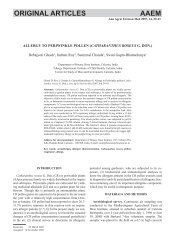
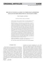
26 Sroka Jbelieved that the most important factors in theepidemiology of T. gondii are cats, pigs, sheep, cattle,fowl and wild game [4]. Serologic examination fortoxoplasmosis in Poland showed a high percent ofpositive reactions in cattle (55%), pigs (21.2%-53%) andin sheep (up to 80%) [28].There is much evidence that the habit of consumptionof undercooked or raw meat is related to hightoxoplasmosis morbidity. In the case of bad sanitary andconsumption habits toxoplasmosis may concern moremembers of the family and thus have the familyenvironmentalcharacter [28].Toxoplasmosis may result from consumption ofundercooked or raw meat containing parasite cysts, orfrom contact with food, water or sand contaminated withoocysts spread by infected cats. There is also thepossibility of transplacental transmission of the parasite tothe foetus during pregnancy. The disease may alsodevelop in the laboratory workers handling infectiousmaterial, after transplantation of organ containing cysts,and through transfusion of infected blood. In people witha well-functioning immunologic system the infectionusually has no symptoms except for development ofspecific antibodies. In people with immunologic disorderprimary invasion may develop or recurrence of chronicinvasion with clinical signs. Most common is thelymphonodular form of toxoplasmosis involving mainlylymph glands in the neck and nape area. Changes mayconcern also liver, spleen, lungs and cardiac muscle,central nervous system and eyes. In the case ofimmunologic disorder generalized toxoplasmosis maydevelop, as in AIDS patients. Especially dangerous is thecongenital form of toxoplasmosis; it develops in abouthalf of pregnant women who were primarily infectedduring pregnancy. The parasites may cause intracranialcalcifications, hydrocephalus and chorioretinitis [28].In animals T. gondii rarely ignites symptoms. The mostfrequent is the latent form characterized by the presenceof tissue cysts in muscles and organs of animals forprolonged periods.The aim of the present study was an evaluation of thefrequency of anti-Toxoplasma gondii antibodies inforestry workers and farmers in the Lublin region.Detection of infected animals as the potential sources oftoxoplasmosis for humans and characterization of the fociof family-environmental toxoplasmosis were the secondarygoals.MATERIALS AND METHODSExamined subjectsSeroepidemiologic examinations of humans. Examinationswere carried out in 1,327 people employed inforestry from five districts and in 86 farmers from twodistricts of the Lublin region (Fig. 1). The administrativedivision before the reform introduced in 1998 is takeninto consideration throughout the paper. 34 office workers=GXY`G]G3XáDZ\/8%/,1-DQyZ/XEHOVNL7$512%5=(*%,$à$32'/$6.$6RVQRZLFD&+(à0=$02û:áRGDZD6RELEyUFigure 1. Map showing area of study: five districts in theLublin region.employed in the forestry headquarters in Lublin and 50blood donors (healthy inhabitants of Lublin) wereexamined as the control group.The majority of forestry workers were people actuallyworking in the forest. Age, gender, working time, contactwith free living and domestic animals, and past diseaseswere taken into account. The mean age of 1,086 men and241 women from the forestry group was 40 years, and themean period of employment was 13 years. Epidemiologicanamnesis has shown that almost everyone from thisgroup had contact with domestic and wild animals. Themean age of 54 women and 32 men from farmers groupwas 41 years and the mean age of 75 men and 9 women inthe control group was 35 years.Seroepidemiologic examination of animals. Sera from494 animals were examined: 262 from cows, 120 frompigs, 34 from geese, 65 from 38-week old chickens from abreeding farm. Three roe deer and 10 sheep were alsoexamined from areas where high percentages of positivereactions in people were found. All animals originatedfrom the Lublin region.Examination of the family-environmental case oftoxoplasmosis. During the study a case of familyenvironmentalcharacter was evidenced in Potok Wielkivillage (Tarnobrzeg district). Detailed epidemiologicanamnesis was performed. Data about family membersconcerning age, gender, place and time of employment,past diseases, contact with domestic and wild animals,information about diet, origin of the food, type of foodand method of cooking, and the presence of raw meat inthe diet were collected. Data about animal diseases werealso collected. The sera collected from men and animalswere examined for toxoplasmosis.
Seroepidemiology of toxoplasmosis in the Lublin region 27Serological testsThe basic test used in serological examination ofhumans and animals was direct agglutination test with 2-mercaptoethanol (DA). Other tests, such as indirectfluorescence test (IFT), immunosorbent agglutinationassay for IgM (ISAGA IgM, bioMérieux, France),enzyme linked fluorescent assay (ELFA, Vidas Toxo IgGand Vidas Toxo IgM, bioMérieux, France), were usedadditionally in the examination of clinical cases.Most of the subjects: 1,327 forestry workers, 86farmers, 84 persons of the control group, 5 persons fromthe case of family-environmental toxoplasmosis, a forestworker from Sosnowica with primary infection and all502 animals were examined with DA. Other tests (IFT,ISAGA IgM, ELFA IgG and ELFA IgM) were usedadditionally for the examination of the case of familyenvironmentaltoxoplasmosis (5 persons), 10 clinicalFDVHV IURP %LDáD 3RGODVND DQG &KHáP GLVWULFWV DQG RQHforestry worker with primary toxoplasmosis.Direct agglutination test with modifications suggestedby Desmonts and Remington was used to detectantibodies in IgG class [1]. Sera were treated with 2-mercaptoethanol to reduce IgM antibodies and thenincubated with formalin-treated Toxoplasma antigen. Theantigen was prepared in the Department of OccupationalBiohazards of the Institute of Agricultural Medicine inLublin. Tachyzoites from 3 day peritoneal exudate ofmice inoculated with RH strain of Toxoplasma gondiiwere centrifugated at 3,000 rpm, washed with PBS (pH7.6) twice and then mixed with trypsin for 20 min in anelectromagnetic mixer. After washing with PBS twice,tachyzoites were formalinized (2%) for 24 hrs and aftercentrifugation and washing with PBS were dyed with0.7% methylene blue for 20 min. After centrifugation andfinal washing, tachyzoites were suspended in 0.2%formalin. Antigen was added to examined sera and thetest was incubated for 24 hrs at 37ºC. Agglutination ofparasites took place if the serum contained antibodies.This test is easy to perform, specific and sensitive and canbe used for testing human or animal sera [12, 25, 30].Indirect fluorescence test (IFT) detects specificantibodies of IgG and IgM classes. The fresh antigen wasprepared in our laboratory. Tachyzoites from 3 dayperitoneal exudate of mice inoculated with RH strain ofToxoplasma gondii were centrifugated at 3,000 rpm andformalinized (1%) for 90 min. After centrifugation,sediment was suspended in PBS (pH 7.6). Then antigenwas diluted with PBS (until 10-30 tachyzoites werevisible in the field of view of the microscope at 400 ×magnification) and dripped on glass slides, air-dried andstored at –20ºC until needed. Before use, antigen-coatedslides were rinsed with water and repeatedly air-dried.7HVWHG VHUDZHUHSODFHGRQDQWLJHQVSRWVOVSRWLQtwo dilutions (1:32 and 1:128) and incubated at 37ºC for30 min. The slides were then rinsed in water, washed inPBS (pH 7.6) three times for 5 min each, and dried. Adrop of fluorescein isothiocyanate labelled goat antihumanimmunoglobulin diluted in PBS (in proportionestablished by titration) was added to each spot. Anaddition of equal volume of 0.01% Evans blue toimmunoglobulin helped in later visualisation ofToxoplasma fluorescence. Slides were incubated at 37ºCfor 30 min, washed, dried and mounted with bufferedglycerol. Slides were examined using a fluorescentmicroscope. Visible fluorescence of cytoplasmicmembrane contrasting with red dyed cytoplasma ofparasite was regarded as positive. Positive titres weregreater or equal to 32. Serum positive in both dilutionswas examined in higher dilutions to establish endpointtitre.Immunosorbent agglutination assay IgM (ISAGAIgM, bioMérieux, France) detects immunoglobulins ofIgM class. All human IgM immunoglobulins in examinedsera are captured in the wells coated with mousemonoclonal antibodies. Specific IgM antibodies are thendetected with the suspension of Toxoplasma gondiiantigen in formalin. Every serum is examined with anincreasing amount of antigen. Positive reaction evokesfull agglutination at the bottom of the well (no sediment -4 points); if antigen remains unbound it elicitesintermediate reactions with visible sedimentation (1–3points). Whole antigen sediments at the bottom in the caseof negative reaction (0 points). The result of this test isexpressed as an index (0–12 points) being the sum of theresults from three consecutive wells filled with differentamounts of antigen. The result of index from 9–12 pointsis regarded as positive, from 6–8 points as equivocal, andfrom 0–5 points as negative [10].Enzyme linked fluorescent assay (ELFA, VidasToxo IgG, bioMérieux, France) with the use ofMiniVidas. Toxoplasma antigens from membrane andcytoplasm with high content of P30 protein were placedon solid phase and covered with examined sera. Anti-T.gondii IgG antibodies were detected by mousemonoclonal anti-human IgG antibodies conjugated withalkaline phosphatase. Final reading was based on thefluorescence measurement, automatically compared tocalibration curve and calculated to IU/ml. The resultswere considered as positive from 10 IU per ml and 8-9IU/ml were regarded as equivocal.Enzyme linked fluorescent assay (ELFA, VidasToxo IgM, bioMérieux, France) with the use ofMiniVidas. This is an “immunocapture” method. Mouseantibodies anti-KXPDQ FKDLQV ZHUH SODFHG RQ VROLGphase. Human IgM immunoglobulins from tested serawere captured and their presence was detected withimmunologic complex (Toxoplasma antigen + monoclonalantibodies anti-P30 conjugated with alkaline phosphatase).Final reading was based on the fluorescence measurement.The results were expressed as a ratio of examined serum