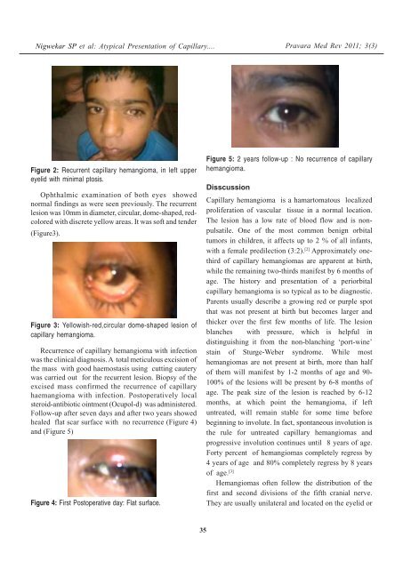
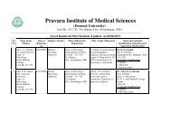
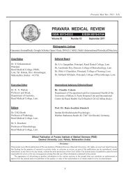
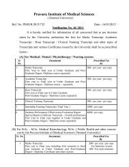
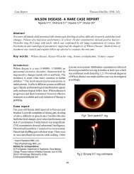
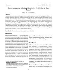
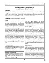
Nigwekar SP et al: <strong>Atypical</strong> Presentation <strong>of</strong> Capillary....Pravara Med Rev 2011; 3(3)Figure 2: Recurrent <strong>capillary</strong> <strong>hemangioma</strong>, in left <strong>upper</strong><strong>eyelid</strong> with minimal ptosis.Ophthalmic examination <strong>of</strong> both eyes showednormal findings as were seen previously. The recurrentlesion was 10mm in diameter, circular, dome-shaped, redcoloredwith discrete yellow areas. It was s<strong>of</strong>t and tender(Figure3).Figure 3: Yellowish-red,circular dome-shaped lesion <strong>of</strong><strong>capillary</strong> <strong>hemangioma</strong>.Recurrence <strong>of</strong> <strong>capillary</strong> <strong>hemangioma</strong> with infectionwas the clinical diagnosis. A total meticulous excision <strong>of</strong>the mass with good haemostasis using cutting cauterywas carried out for the recurrent lesion. Biopsy <strong>of</strong> theexcised mass confirmed the recurrence <strong>of</strong> <strong>capillary</strong>haemangioma with infection. Postoperatively localsteroid-antibiotic ointment (Ocupol-d) was administered.Follow-up after seven days and after two years showedhealed flat scar surface with no recurrence (Figure 4)and (Figure 5)Figure 4: First Postoperative day: Flat surface.Figure 5: 2 years follow-up : No recurrence <strong>of</strong> <strong>capillary</strong><strong>hemangioma</strong>.DisscussionCapillary <strong>hemangioma</strong> is a hamartomatous localizedproliferation <strong>of</strong> vascular tissue in a normal location.The lesion has a low rate <strong>of</strong> blood flow and is nonpulsatile.One <strong>of</strong> the most common benign orbitaltumors in children, it affects up to 2 % <strong>of</strong> all infants,with a female predilection (3:2). [2] Approximately onethird<strong>of</strong> <strong>capillary</strong> <strong>hemangioma</strong>s are apparent at birth,while the remaining two-thirds manifest by 6 months <strong>of</strong>age. The history and <strong>presentation</strong> <strong>of</strong> a periorbital<strong>capillary</strong> <strong>hemangioma</strong> is so typical as to be diagnostic.Parents usually describe a growing red or purple spotthat was not present at birth but becomes larger andthicker over the first few months <strong>of</strong> life. The lesionblanches with pressure, which is helpful indistinguishing it from the non-blanching ‘port-wine’stain <strong>of</strong> Sturge-Weber syndrome. While most<strong>hemangioma</strong>s are not present at birth, more than half<strong>of</strong> them will manifest by 1-2 months <strong>of</strong> age and 90-100% <strong>of</strong> the lesions will be present by 6-8 months <strong>of</strong>age. The peak size <strong>of</strong> the lesion is reached by 6-12months, at which point the <strong>hemangioma</strong>, if leftuntreated, will remain stable for some time beforebeginning to involute. In fact, spontaneous involution isthe rule for untreated <strong>capillary</strong> <strong>hemangioma</strong>s andprogressive involution continues until 8 years <strong>of</strong> age.Forty percent <strong>of</strong> <strong>hemangioma</strong>s completely regress by4 years <strong>of</strong> age and 80% completely regress by 8 years<strong>of</strong> age. [3]Hemangiomas <strong>of</strong>ten follow the distribution <strong>of</strong> thefirst and second divisions <strong>of</strong> the fifth cranial nerve.They are usually unilateral and located on the <strong>eyelid</strong> or35
Nigwekar SP et al: <strong>Atypical</strong> Presentation <strong>of</strong> Capillary....Pravara Med Rev 2011; 3(3)brow and may result in significant ptosis <strong>of</strong> an involved<strong>eyelid</strong>. Reduced visual acuity may be noted due toptosis or astigmatism. Amblyopia is seen inapproximately 50% <strong>of</strong> patients with <strong>eyelid</strong><strong>hemangioma</strong>. (4) The lesion may be cutaneous or mayextend to subcutaneous tissue. The subcutaneousportion may extend into the orbit causing painless, nonpulsatingproptosis <strong>of</strong> the involved eye. [5]The ophthalmologist should be aware that manychildren with periorbital <strong>hemangioma</strong> may also haveintraorbital extension <strong>of</strong> the lesion and/or systemicangiogenic lesions (including pulmonary, s<strong>of</strong>t-tissueand/or skin lesions). Orbital imaging with B-scanultrasonography is helpful in determining the posteriorextent <strong>of</strong> suspicious lesions, while chest or abdominalimaging may uncover other <strong>hemangioma</strong>s. Somepatients with large visceral <strong>capillary</strong> <strong>hemangioma</strong>s maydevelop thrombocytopenia, a condition known asKasabach-Merritt syndrome. Capillary haemangiomawith skin haemorrhages, enchondromata <strong>of</strong> hands andfeet and bowing <strong>of</strong> long bones is known as Maffuci’ssyndrome. [6]Adviced Treatment OptionsSpontaneous involution is the rule and only observationmay be appropriate. In cases <strong>of</strong> <strong>eyelid</strong> occlusionleading to amblyopia or significant astigmatism, prompttreatment in the form <strong>of</strong> occlusion and refractivecorrection should be initiated.1. Use <strong>of</strong> Corticosteroids:Intralesional, systemic and topical corticosteroidsmay be used as treatment.2. Surgical excision:Surgery carries the risks associated with generalanesthesia. As the lesion is not encapsulated,surgical excision is <strong>of</strong>ten difficult and there is a risk<strong>of</strong> recurrence. One has to anticipate intraoperativebleeding and control it with application <strong>of</strong>cautery. [7]3. Laser photocoagulation:It is effective in attenuating proliferation andfacilitating regression <strong>of</strong> superficial cutaneous<strong>hemangioma</strong>s <strong>of</strong> thickness more than 2mm.4. Immunomodulator:Vincristine, Cyclophosphamide and Interferon alfa-2a has been used as monotherapy or in combinationwith systemic corticosteroids. [7] Howevermyelosuppression, hepatotoxicity and neurotoxicityare the side effects.DiscussionThe present case had a short two month history andlate <strong>presentation</strong> at seven years <strong>of</strong> age. This is notclassic for <strong>capillary</strong> <strong>hemangioma</strong>. Clinically thedifferential diagnosis consists <strong>of</strong> Pyogenic granuloma.The typical solitary pyogenic granuloma (lobular<strong>capillary</strong> <strong>hemangioma</strong>) is a bright red, friable, polypoidpapule or nodule ranging from a few millimeters toseveral centimeters (average size is 6.5mm) indiameter. Bleeding, erosion, ulceration, and crusting arefrequently noted. Regressing lesions appear as a s<strong>of</strong>tfibroma. The head, neck and face; distal extremities,especially the fingers (notably in pregnant woman) arethe sites <strong>of</strong> predilection, but lesions can occuranywhere on the integument. [8] Recurrences aftersurgical excision can occur in <strong>capillary</strong> <strong>hemangioma</strong> aswas a feature in our case.ConclusionsThough <strong>capillary</strong> <strong>hemangioma</strong> presents usually at birthand upto 1 year <strong>of</strong> age, it may present as late as 7years. Meticulous surgical excision gives good resultseven in recurrent cases.References1. Sihota-Tandon. ‘Parson’s diseases <strong>of</strong> eye’. 21 stedition 2011:456.2. Haik BG. Capillary <strong>hemangioma</strong>. Surv Ophthalmol1994;38(5):399-426.3. Review <strong>of</strong> Capillary Hemangioma, Peralta RJ,Glavas. New York University and the ManhattanEye, Ear and Throat Hospital, New York. Eye NetMagazine February 2009.4. Basak SK. ‘Essenials <strong>of</strong> Ophthalmology’, 4 thedition 2007: 127.5 Capillary Hemangioma right <strong>upper</strong> <strong>eyelid</strong>: Casereport. JM Graff, Adam Garza, KD Carter, January31, 2006.6 Kanski JJ. ‘Clinical Ophthalmology A SystemicApproach’, 6 th edition 2007; 105,189.7 Yan<strong>of</strong>f M. ‘Ophthalmology’ 3 rd edition 2009: 1462.8. Dermatologic Manifestations <strong>of</strong> PyogenicGranuloma (Lobular Capillary Hemangioma) JCPierson, DM Elston. Medscape 29 th April 2010.36