Partial Purification and Characterization of the mRNA for a-Amylase ...
Partial Purification and Characterization of the mRNA for a-Amylase ...
Partial Purification and Characterization of the mRNA for a-Amylase ...

Create successful ePaper yourself
Turn your PDF publications into a flip-book with our unique Google optimized e-Paper software.
Plant Physiol. Vol. 65, 1980 BARLEY a-AMYLASE <strong>mRNA</strong><br />
835<br />
DMSO-Formamide-Sucrose Density Gradient Centrifugation.<br />
Using methods described by R. Beachy (personal communication),<br />
<strong>the</strong> poly(A)-selected RNA was centrifuged, dried, <strong>and</strong> resuspended<br />
into 100 pl deionized H20. To this solution, 400 ul <strong>of</strong> a<br />
solution containing 95% DMS0, 4% deionized <strong>for</strong>mamide, <strong>and</strong><br />
1% 1 MTns-HCl (pH 7.4) containing I M LiCl <strong>and</strong> 100 mm EDTA<br />
(v/v/v) was added. This was heated to 60 C <strong>for</strong> 5 min, cooled,<br />
<strong>and</strong> applied to a sucrose density gradient. This gradient was<br />
prepared <strong>the</strong> previous day <strong>and</strong> consisted <strong>of</strong> successive layers <strong>of</strong> 5,<br />
10, 15, <strong>and</strong> 20% sucrose in 95% DMSO, 4% <strong>for</strong>mamide, <strong>and</strong> 1%<br />
buffer. The gradient was centrifuged in an SW 40 rotor at 40,000<br />
rpm at 28 C <strong>for</strong> 48 h. The gradients were fractionated using an<br />
Isco automatic fractionator coupled with an UV monitor to determine<br />
A at 280 nm. Each fraction collected was ethanol precipitated<br />
overnight, centrifuged at l0,OOOg <strong>for</strong> 30 min, washed three times<br />
with 70o ethanol <strong>and</strong> 0.3 M ammonium acetate, <strong>and</strong> finally dried.<br />
In Vitro Protein Translation Reactions. The reticulocyte lysate<br />
system was prepared <strong>and</strong> used essentially as described by Pelham<br />
<strong>and</strong> Jackson (15). The final assay mixture <strong>of</strong> 25 ,il contained 40<br />
mm Hepes (pH 7.6), 80 mm K-acetate, 1 mm Mg-acetate, 0.5 mM<br />
spermidine, 2 mm DTT, 20 ,ug wheat germ tRNA, 60 tLM amino<br />
acids minus leucine, 6.7 tLM leucine, 50 ,Ci [3H]leucine, <strong>and</strong> an<br />
energy source consisting <strong>of</strong> I mM ATP, 0.4 mm GTP, 0.4 mm CTP,<br />
3 mM phosphocreatine, <strong>and</strong> 20 ,ug/ml phosphocreatine kinase. The<br />
wheat germ extract was prepared as described by Bruening et al.<br />
(4). For <strong>the</strong> wheat germ in vitro translation system, <strong>the</strong> following<br />
concentrations <strong>of</strong> reagents were used: 41 mM Hepes (pH 7.6), 149<br />
mm K-acetate, 10 mM KCI, 2.2 iM DTT, 2.24 mm Mg-acetate,<br />
0.39 mM spermidine, 50 pM CaC12, 10 ,m EDTA, 60 ^lm amino<br />
acids minus leucine, 6.7 .tM leucine, 50 pCi [3H]leucine, <strong>and</strong> <strong>the</strong><br />
same energy source as above. All reactions were initiated by <strong>the</strong><br />
addition <strong>of</strong> between 50 <strong>and</strong> 200 ng <strong>of</strong> RNA <strong>and</strong> were continued<br />
<strong>for</strong> 90 min. Trichloroacetic acid precipitable cpm <strong>for</strong> each assay<br />
were determined as described by Bruening et al. (4).<br />
Each assay was electrophoresed on an SDS-polyacrylamide slab<br />
gel system which separates proteins on <strong>the</strong> basis <strong>of</strong> mol wt (13).<br />
An acrylamide to bis ratio <strong>of</strong> 30:0.174 was used, <strong>and</strong> <strong>the</strong> acrylamide<br />
concentrations in <strong>the</strong> separating gel <strong>and</strong> stacking gel were<br />
12.5 <strong>and</strong> 5%, respectively. After electrophoresis, <strong>the</strong> gels were<br />
prepared <strong>for</strong> fluorography <strong>and</strong> exposed to Kodak X-omat R fim<br />
<strong>for</strong> 1-2 days be<strong>for</strong>e development (3).<br />
Methyl Mercury Agarose Gel Electrophoresis. Agarose gel<br />
electrophoresis using methylmercuric hydroxide as <strong>the</strong> denaturing<br />
agent was used (1). The agarose concentration in <strong>the</strong> st<strong>and</strong>ard slab<br />
gel was 2%. Electrophoresis <strong>of</strong> <strong>the</strong> RNA was carried out <strong>for</strong> 4 h at<br />
80 v at room temperature. After electrophoresis, <strong>the</strong> gel was placed<br />
in a tray <strong>of</strong> H20 <strong>and</strong> two to three drops <strong>of</strong> ethidium bromide (1<br />
mg/ml) were added. After 1 h, <strong>the</strong> gel was rinsed with H20 <strong>and</strong><br />
photographed over an UV light box.<br />
RESULTS<br />
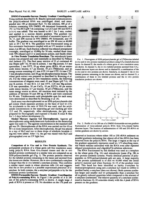
Comparison <strong>of</strong> in Vivo <strong>and</strong> in Vitro Protein Syn<strong>the</strong>sis. The<br />
polypeptide products <strong>of</strong> a wheat germ cell free translation assay<br />
using poly(A) RNA from GA3-treated tissues <strong>and</strong> <strong>the</strong> in vivo<br />
labeled proteins syn<strong>the</strong>sized at a similar time are compared on an<br />
SDS-polyacrylamide gel (Fig. 1). The protein patterns observed<br />
<strong>for</strong> <strong>the</strong> labeled proteins remaining in <strong>the</strong> tissue <strong>and</strong> secreted from<br />
<strong>the</strong> tissues are similar. However, <strong>the</strong> in vitro-syn<strong>the</strong>sized a-amylase<br />
is larger than <strong>the</strong> in vivo-labeled a-amylase protein. This confirms<br />
a similar report made in wheat aleurone cells (14) <strong>and</strong> suggests<br />
that <strong>the</strong> in vitro product is a possible unprocessed precursor <strong>of</strong> aamylase.<br />
In both systems, <strong>the</strong> a-amylase polypeptide is by far <strong>the</strong><br />
dominant protein syn<strong>the</strong>sized.<br />
DMSO-Formamide-Sucrose Density Gradient Centrifugation<br />
<strong>of</strong> Poly(A)-containing RNA. In Figure 2 <strong>the</strong> pr<strong>of</strong>ile <strong>of</strong> A at 280<br />
am <strong>of</strong> <strong>the</strong> poly(A)-containing RNA is shown. A single large A<br />
peak <strong>of</strong> RNA is present at 17S. No shoulders on <strong>the</strong> 17S peak are<br />
A B C D E<br />
-~~<br />
FIG. 1. Fluorogram <strong>of</strong> SDS-polyacrylamide gel <strong>of</strong> [3H]leucine-labeled<br />
in vivo <strong>and</strong> in vitro protein translation products using GA3-treated aleurone<br />
layers. In channel E, <strong>the</strong> results <strong>of</strong> a wheat germ in vitro translation assay<br />
are shown. In channel A, <strong>the</strong> in vivo labeled proteins secreted from GA3treated<br />
aleurone layers are shown <strong>and</strong> in channel B, a combination <strong>of</strong> <strong>the</strong><br />
in vivo <strong>and</strong> in vitro proteins are shown. Similarly, in channel C <strong>the</strong> in vivo<br />
labeled proteins remaining in <strong>the</strong> tissues are shown, <strong>and</strong> in channel D, a<br />
combination <strong>of</strong> <strong>the</strong>se in vivo labeled proteins <strong>and</strong> <strong>the</strong> in vitro protein<br />
translation products are shown.<br />
0.5-<br />
E<br />
09.4 > 18S 25$<br />
u" 0.3<br />
z<br />
202<br />
4 8 12 16 20 24<br />
FRACTION NUMBER<br />
FIG. 2. Pr<strong>of</strong>ile <strong>of</strong> A at 280 nm <strong>of</strong> a DMSO-<strong>for</strong>mamide-sucrose gradient<br />
fractionation <strong>of</strong> twice-selected poly(A) RNA from GA3-treated barley<br />
aleurone layers. The location <strong>of</strong> sedimentation <strong>of</strong> 18S <strong>and</strong> 25S rRNA on<br />
similar gradients are shown by arrows.<br />
observed at locations where ei<strong>the</strong>r 18S or 25S rRNA sediment on<br />
parallel gradients indicating that almost all <strong>of</strong> <strong>the</strong> rRNA has been<br />
removed from <strong>the</strong> poly(A) RNA. The large A region at <strong>the</strong> top <strong>of</strong><br />
<strong>the</strong> gradient apparently represents small wt, UV-absorbing material<br />
which remains associated with <strong>the</strong> RNA even after repeated<br />
ethanol precipitations <strong>and</strong> oligo[d(pT)J-cellulose selections.<br />
The RNA fractionated by this method was used as a template<br />
in <strong>the</strong> wheat germ (Fig. 3A) <strong>and</strong> reticulocyte lysate (Fig. 3B) in<br />
vitro protein syn<strong>the</strong>sis systems. Nearly identical patterns <strong>of</strong> polypeptides<br />
on SDS-polyacrylamide gels are seen. A large majority<br />
<strong>of</strong> <strong>the</strong> protein syn<strong>the</strong>sized is <strong>of</strong> mol wt 45,000 when <strong>the</strong> initial<br />
poly(A) material <strong>and</strong> when fractions 16-18 <strong>of</strong> <strong>the</strong> gradient were<br />
used. This b<strong>and</strong> has been identified as barley a-amylase by<br />
immunoprecipitation using anti a-amylase IgG made in rabbits to<br />
purified barley a-amylase (data not shown). Both systems syn<strong>the</strong>size<br />
larger <strong>and</strong> smaller mol wt polypeptides than a-amylase but<br />
all in greatly reduced quantities when compared to <strong>the</strong> amount <strong>of</strong><br />
a-amylase syn<strong>the</strong>sized. The relative quantity <strong>of</strong> a-amylase syn<strong>the</strong>sized<br />
in fractions 16-18 coincides closely with <strong>the</strong> A peak <strong>of</strong> RNA<br />
present on <strong>the</strong> gradient.