77TH ANNIVERSARY ADDRESS - Indian National Science Academy
77TH ANNIVERSARY ADDRESS - Indian National Science Academy
77TH ANNIVERSARY ADDRESS - Indian National Science Academy

Create successful ePaper yourself
Turn your PDF publications into a flip-book with our unique Google optimized e-Paper software.
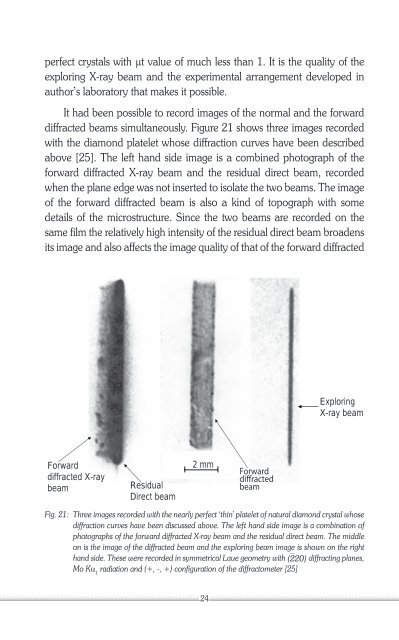
perfect crystals with mt value of much less than 1. It is the quality of theexploring X-ray beam and the experimental arrangement developed inauthor’s laboratory that makes it possible.It had been possible to record images of the normal and the forwarddiffracted beams simultaneously. Figure 21 shows three images recordedwith the diamond platelet whose diffraction curves have been describedabove [25]. The left hand side image is a combined photograph of theforward diffracted X-ray beam and the residual direct beam, recordedwhen the plane edge was not inserted to isolate the two beams. The imageof the forward diffracted beam is also a kind of topograph with somedetails of the microstructure. Since the two beams are recorded on thesame film the relatively high intensity of the residual direct beam broadensits image and also affects the image quality of that of the forward diffractedExploringX-ray beamForwarddiffracted X-raybeamResidualDirect beam2 mmForwarddiffractedbeamFig. 21: Three images recorded with the nearly perfect ‘thin’ platelet of natural diamond crystal whosediffraction curves have been discussed above. The left hand side image is a combination ofphotographs of the forward diffracted X-ray beam and the residual direct beam. The middleon is the image of the diffracted beam and the exploring beam image is shown on the righthand side. These were recorded in symmetrical Laue geometry with ⎺(220) diffracting planes,Mo Ka 1radiation and (+, -, +) configuration of the diffractometer [25]24