Biochemistry of exercise-induced metabolic acidosis
Biochemistry of exercise-induced metabolic acidosis
Biochemistry of exercise-induced metabolic acidosis

You also want an ePaper? Increase the reach of your titles
YUMPU automatically turns print PDFs into web optimized ePapers that Google loves.
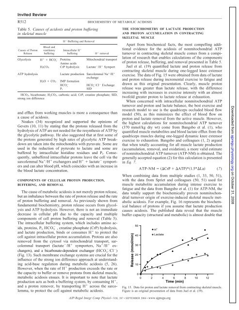
Invited ReviewR512Table 5. Causes <strong>of</strong> <strong>acidosis</strong> and proton bufferingin skeletal muscleCauses <strong>of</strong> ProtonReleaseBlood andventilatorybufferingH Buffering and RemovalIntracellular H bufferingH removalGlycolysis H HCO 3Proteins Mitochondrial transportAmino acidsH 2CO 3 CrP hydrolysis Lactate /H SymportATP hydrolysisH 2O CO 2Lactate production Sarcolemmal Na /H exchangeIMP formationHCO 3HCO 3 /Cl ExchangeSIDP iHCO 3, bicarbonate; H 2CO 3, carbonic acid; CrP, creatine phosphate; SID,strong ion differenceand efflux from working muscles is more a consequence thana cause <strong>of</strong> <strong>acidosis</strong>.Noakes (34) recognized and supported the opinions <strong>of</strong>Gevers (10, 11) by stating that the protons released from thehydrolysis <strong>of</strong> ATP are not needed for the resynthesis <strong>of</strong> ATP bythe glycolytic pathway. He also suggested that at first some <strong>of</strong>the protons generated by high rates <strong>of</strong> glycolytic ATP breakdownare taken into the mitochondria with pyruvate. Some areused in the reduction <strong>of</strong> pyruvate to lactate and some arebuffered by intracellular histidine residues and P i . Consequently,unbuffered intracellular protons leave the cell via thesarcolemmal Na /H exchangers and H lactate symportersand can alter blood pH, which coincides with an increase inthe blood lactate concentration.COMPONENTS OF CELLULAR PROTON PRODUCTION,BUFFERING, AND REMOVALThe cause <strong>of</strong> <strong>metabolic</strong> <strong>acidosis</strong> is not merely proton release,but an imbalance between the rate <strong>of</strong> proton release and the rate<strong>of</strong> proton buffering and removal. As previously shown fromfundamental biochemistry, proton release occurs from glycolysisand ATP hydrolysis. However, there is not an immediatedecrease in cellular pH due to the capacity and multiplecomponents <strong>of</strong> cell proton buffering and removal (Table 5).The intracellular buffering system, which includes amino acids,proteins, P i , HCO 3 , creatine phosphate (CrP) hydrolysis,and lactate production, binds or consumes H to protect thecell against intracellular proton accumulation. Protons are alsoremoved from the cytosol via mitochondrial transport, sarcolemmaltransport (lactate /H symporters, Na /H exchangers),and a bicarbonate-dependent exchanger (HCO 3 /Cl )(Fig. 13). Such membrane exchange systems are crucial for theinfluence <strong>of</strong> the strong ion difference approach at understandingacid-base regulation during <strong>metabolic</strong> <strong>acidosis</strong> (5, 26).However, when the rate <strong>of</strong> H production exceeds the rate orthe capacity to buffer or remove protons from skeletal muscle,<strong>metabolic</strong> <strong>acidosis</strong> ensues. It is important to note that lactateproduction acts as both a buffering system, by consuming H ,and a proton remover, by transporting H across the sarcolemma,to protect the cell against <strong>metabolic</strong> <strong>acidosis</strong>.BIOCHEMISTRY OF METABOLIC ACIDOSISTHE STOICHIOMETRY OF LACTATE PRODUCTIONAND PROTON ACCUMULATION IN CONTRACTINGSKELETAL MUSCLEApart from biochemical facts, the most compelling additionalevidence for the <strong>acidosis</strong> <strong>of</strong> nonmitochondrial ATPturnover in contracting skeletal muscle comes from a compilation<strong>of</strong> research that enables calculations <strong>of</strong> the components<strong>of</strong> proton release, buffering, and removal presented in Table 5.Juel et al. (19) quantified lactate and proton release fromcontracting skeletal muscle during one-legged knee extensor<strong>exercise</strong>. The data <strong>of</strong> Fig. 15 were obtained from data <strong>of</strong> lactateand proton release during incremental <strong>exercise</strong> to fatigue anddrawn as this original presentation. Clearly, muscle protonrelease was greater than lactate release, with the differenceincreasing with increases in <strong>exercise</strong> intensity with an almosttw<strong>of</strong>old greater proton to lactate release at exhaustion.When concerned with intracellular nonmitochondrial ATPturnover and proton and lactate balance, the best <strong>exercise</strong> andresearch model to use is the quadriceps occluded blood flowmodel (50), as this minimizes the effect <strong>of</strong> blood flow onproton and lactate removal from the active muscle. However,the highest calculations for nonmitochondrial ATP turnover(370 mmol/kg dry wt) come from Bangsbo et al. (1), whoquantified muscle metabolites and blood lactate efflux from thequadriceps muscles during one-legged dynamic knee extensor<strong>exercise</strong> to exhaustion. Bangsbo and colleagues (1, 2) arguedthat when totally accounting for all muscle lactate production(accumulation, removal, and oxidation), a more valid estimate<strong>of</strong> nonmitochondrial ATP turnover (ATP-NM) is obtained. Thegenerally accepted equation (2) for this calculation is presentedin Eq. 7.ATP-NM CrP ATP*1.5*La (7)When combining data from multiple studies (1, 33, 50, 51),with the data from Spriet and colleagues (50, 51) used formuscle metabolite accumulation during intense <strong>exercise</strong> t<strong>of</strong>atigue and the data from Bangsbo et al. (1) for ATP-NM, thedata totally support the biochemically proven nonmitochondrialturnover origin <strong>of</strong> <strong>exercise</strong>-<strong>induced</strong> skeletal muscle <strong>metabolic</strong><strong>acidosis</strong>. For example, Fig. 16 represents the biochemicalbalance <strong>of</strong> protons if you assume that lactate productioncauses <strong>acidosis</strong>. The published data reveal that the musclebuffer capacity (structural and <strong>metabolic</strong>) is almost double thatFig. 15. Data for proton and lactate removal from contracting skeletal muscle.Figure is an original presentation <strong>of</strong> data from Juel et al. (19).Downloaded from ajpregu.physiology.org on October 31, 2008AJP-Regul Integr Comp Physiol • VOL 287 • SEPTEMBER 2004 • www.ajpregu.org