Autonomic Nervous System - UMK CARNIVORES 3
Autonomic Nervous System - UMK CARNIVORES 3
Autonomic Nervous System - UMK CARNIVORES 3

You also want an ePaper? Increase the reach of your titles
YUMPU automatically turns print PDFs into web optimized ePapers that Google loves.
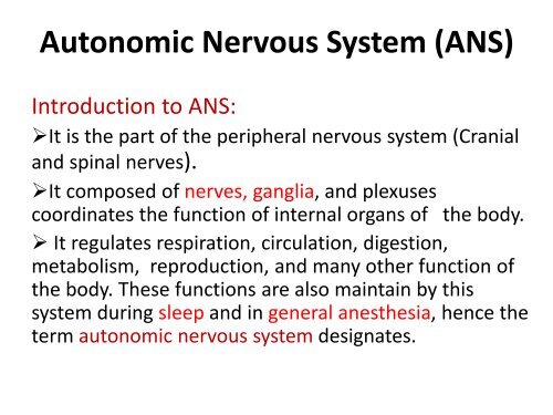
<strong>Autonomic</strong> <strong>Nervous</strong> <strong>System</strong> (ANS)Introduction to ANS:‣It is the part of the peripheral nervous system (Cranialand spinal nerves).‣It composed of nerves, ganglia, and plexusescoordinates the function of internal organs of the body.‣ It regulates respiration, circulation, digestion,metabolism, reproduction, and many other function ofthe body. These functions are also maintain by thissystem during sleep and in general anesthesia, hence theterm autonomic nervous system designates.
<strong>Autonomic</strong> <strong>Nervous</strong> <strong>System</strong> (ANS)(cont.)Introduction to ANS:‣ The life sustaining activities such as visceral (internal softorgan) function and homeostasis (internal physiology)which is out of individuals controlled is maintain by theautonomic nervous system.‣ The visceral function include also the secretion ofendocrine and exocrine function and the function ofblood vessels also.‣ The ganglia include paravertebral chain ganglia andprevertebral ganglia, and intramural ganglia.
Difference between Somatic <strong>Nervous</strong><strong>System</strong> and <strong>Autonomic</strong> <strong>Nervous</strong> <strong>System</strong>(1) Somatic <strong>Nervous</strong> <strong>System</strong> (SNS), is undervoluntary control, and regulates thecontraction of skeletal muscle. In contrast, theautonomic nervous system (ANS) or visceralmotor system, is generally not undervoluntary control and controls smooth muscle,cardiac muscle, glandular tissue and visceralreflexes.
Difference between Somatic Motor<strong>System</strong> and <strong>Autonomic</strong> <strong>Nervous</strong> <strong>System</strong>(cont.)(2) In the SNS, efferent fibers (motor nerve) beginsfrom the cortex of brain, and travel either to thecranial nerve nuclei located in the brain or to thealpha-motor neuron of the dorsal horn of thespinal cord. From here, motor nerves exit theCNS and innervates into the skeletal muscle.Therefore, in somatic nervous system the nervecell bodies of the motor nerves located in theCNS.
Difference between Somatic Motor<strong>System</strong> and <strong>Autonomic</strong> <strong>Nervous</strong> <strong>System</strong>(cont)(2) In the ANS, efferent fibers (motor nerve) beginsfrom the hypothalamus of brain and travel tothe nuclei of the cranial nerves belong the ANSor intermediate horn of the spinal cord. Fromhere the nerves (preganglionic fibers) travelagain and synapse in the 2 nd ganglion(paravertebral, prevertebral, or intramuralganglia) located outside the CNS.
Difference between Somatic Motor<strong>System</strong> and <strong>Autonomic</strong> <strong>Nervous</strong> <strong>System</strong>(cont)3. The fibers of SNS innervates into the skeletalmuscle as neuromuscular spindle. Whereas theANS innervates into the smooth muscle asvaricosities and vesicles.
Division of <strong>Autonomic</strong> <strong>Nervous</strong><strong>System</strong>Based on anatomy, physiology, andpharmacology, the ANS can be divided into:(1) Sympathetic division associated withautonomic nerves, paravertebral chainganglia and prevertebral ganglia.(2) Parasympathetic division, associated withautonomic nerves and intramural ganglia.
Characteristics of the Fibers of<strong>Autonomic</strong> <strong>Nervous</strong> <strong>System</strong> (ANS)‣ The fibers are: Preganglionic & postganglionic fibers.‣ The autonomic fibers from the intermediated horn ofthe spinal cord to the paravertebral or prevertebralganglionic synapse is called preganglionic fibers aremyelinated, therefore, it is white.‣ The postganglionic fibers are the fibers after the pre- orparavertebral ganglia which are little myelinated,therefore, it is gray.
Structural Organization ofSympathetic division• In sympathetic division the multipolar nerve cellbody is located in the intermediate horn of thethoracic and lumbar spinal cord (between 1 stthoracic and 3 rd lumbar segment).• The preganglionic nerve fiber (myelinated axon) isshort and travel from the multipolar nerve cell bodyand synapse within the paravertebral chain ganglia/prevertebral ganglia.• The postganglionic fiber (non-myelinated) is longand terminates on the cells of effector organ.
Nerve cell bodies located in the Paravertebral andPrevertebral ganglionIntermediatehornParavertebralGanglionPrevertebralGanglion
<strong>Autonomic</strong> Nerve FibersSynapse here inThe paravertebralGanglionPreganglionicnerve fiberPostganglionicnerve fiberSynapse here in thePrevertebral ganglionPreganglionicnerve fiberPostganglioninerve fiber
White ramiGray ramiPVGPrevertebralGanglion (PrVG)Review of Ganglia and Nerves ofSympathetic DivisionIntermediatehornMyelinatedPreganglionicfiberNon-myelinatedPostganglionicfiber<strong>Autonomic</strong> fibers fromIntermediate horn ofspinal cordVentral root of thoracicand 1 st few lumbar spinalnerveWhite ramiSynapse in the paravertebralGanglia/prevertebral ganglia(This is the preganglionic fiber)The postganglionic fiber from here innervates into the autonomic organ
Sympathetic TrunkSympathetic trunk consist of paravertebral ganglia oneither side of the vertebral column and are interconn -ected longitudinally and transversely. It can be dividedinto:(1) Cephalic and Cervical Part(2) Thoracic Part(3) Abdominal part and(4) Sacral and Coccygeal part.
Cephalic and Cervical part of SympatheticTrunkIt is the cranial continuation of the thoracic part,without directly contacting the vertebral column. The cervical part starts at the cervico-thoracicganglion, which is connected to the middle cervicalganglion. From here sympathetic trunk passes craniallyin combination with the vagus nerve forming“vagosympathetic trunk”. At the atlas, the sympatheticpart separates from the vagus and terminate intocranial cervical ganglion.
Ganglia and autonomic innervation in theCervical Trunk The cranial cervicalganglion is located near theatlas bone and serves for theautonomic innervation ofthe head and face. Middle cervical ganglionis located close to thesubclavian artery and servesfor the autonomicinnervation of the heart.
Ganglia and autonomic innervation in theCervico-thoracic region Cervicothoracicganglion: The cervico -thoracic ganglion is locatedmedial to the first rib. It isthe results of the fusion ofcaudal cervical and one ortwo thoracic ganglion. Itsend following branches: Vertebral nerveCervical cardiac nerve,&Caudal continuation ofthoracic sympathetic part.
Thoracic part of the sympathetic trunk(See Fig. Slide 14)• The number of paravertebral ganglia is correspond to the numberof thoracic vertebra.• The organs of the thoracic cavity (heart, lungs, trachea receivedautonomic innervations from Postganglionic fibers originatingfrom cervicothoracic ganglion or from postganglionic thoracicnerves.• More caudally 7-8 preganglionic thoracic fibers unite to formgreater splanchnic nerve pass through the crus of diaphragm,entered into the abdominal cavity and synapse in the celiacganglia. The postganglionic fiber from here supply to the liver,stomach, spleen, and pancreas.• Last 2-3 thoracic preganglionic fibers unite to form lessersplanchnic nerve, enter into the abdominal cavity and synapse incranial mesenteric ganglion. The postganglionic fibers from heresupply to the jejunum, ileum, cecum, colon, and kidney.
Abdominal part of the sympathetic trunk (SeeFig. Slide 14)• The lumbar autonomic nerves leaves thelumbar segments of spinal cord, passthrough the paravertebral ganglia andforms lumbar splanchnic nerves, andsynapse with the celiac ganglion, cranialmesenteric ganglion, and caudalmesenteric ganglion. The postganglionicfibers innervates into spleen, kidney,pancreas, cecum, colon, rectum, andgenital organs.
Sacral part of the Sympathetic Trunk (SeeFig. Slide 14)First 5 preganglionic nerve fibers leaves the pelvicpart of the spinal cord, pass through theparavertebral ganglion, unite to form sacral (pelvic)spalnchnic nerves which synapse in the sacralganglion (pelvic ganglion). The postganglionicfibers innervates into the rectum and genitalorgans.
Structural Organization ofParasympathetic division• The multipolar nerve cell body is located in thenuclei of the III, VII, IX, and X cranial nerves andintermediate horn of first 3-5 sacral spinal cord ofdifferent animals.• The preganglionic nerve fiber (myelinated axon) islong and travel from the multipolar nerve cellbody and synapse within the intramural ganglia;ciliary, pterigopalatine, mandibular, otic and distalganglia.• The postganglionic fiber (non-myelinated) is shortand terminates on the cells of effector organ.
Parasympathetic Innervation in the HeadRegionOcculo.nerveIII. Oculomotor nervePreganglionic fiber synapseIn the ciliary ganglionPostganglionic fiber innervatesinto ciliary muscle whichregulates lens curvature andthe pupillary diameter.
Parasympathetic Innervation in theHead RegionFacial n.VII. Facial nervePreganglionic fiber synapsein the Pterygopalatine andmandibular ganglionPostganglionic fiberinnervates into sublingual,submandibular, lacrimal,nasal, and palatine gland.
Parasympathetic Innervation in theHead RegionG.Pharyn.nerveIX. Gloss. Phar.nervePreganglionic fiber synapsein the Otic ganglionPostganglionic fiberinnervates into buccal andparotid gland.
Parasympathetic Innervation of theCervical Vagus n.Vagus n. in theThoracic regionVagus NerveVagus n in theAbdomenVagusnerveVagus n in thePelvic regionVagus NervePreganglionic fibersinnervates into the organof neck, thoracicabdominal and pelvicregion and synapse withthe intramural ganglionlocated in the organs.Postganglionic fiber isshort.
Pelvic Parasympathetic InnervationPelvic ParasympatheticnervesPreganglionic fibers fromintermediate horn of thespinal cord forms pelvicautonomic nerves andinnervates into the pelvicorgans and synapse with theintramural ganglion of theorgans. Postganglionic fibersare short.
Function of <strong>Autonomic</strong> <strong>Nervous</strong><strong>System</strong> in GeneralBoth the system have opposing effects on most function:(1) Sympathetic (Thoraco-lumbar nerves) division generallycauses excitation and results in catabolism. This divisionis acted during period of stress and exertion.Sympathetic nerve ending secretes neurotransmittor(chemicals) “norepinephrine”. Norepinephrine act on α,and β adrenergic receptor of various organ, that is whysympathetic nerves is called Adrenergic nerves.(2) The parasympathetic division is responsible for rest,digestion, and anabolism (i.e., building phase ofmetabolism). Parasympathetic nerve fibers secreteneurotransmittor acetylcholine, that’s why these nervesknown as Cholinergic nerves.
Function of <strong>Autonomic</strong> <strong>Nervous</strong> <strong>System</strong>(Specific)Organ Sympathetic Parasympathetic<strong>Nervous</strong> <strong>System</strong> <strong>Nervous</strong> <strong>System</strong>1.Iris Dilates pupil Constrict pupil2. Ciliary m. Relax Constrict3. Glands Inhibit secretion Stimulates secret.4. Heart m. Increased HR Decreased HR5. U. Bladder Inhibit urine release Cause urination6. Lung Dilates bronchioles Constrict Br.7. Digestive tract Decreased peristalsis Increased