African Type Burkitt's Lymphoma - dishekdergi.hacettepe.edu.tr
African Type Burkitt's Lymphoma - dishekdergi.hacettepe.edu.tr
African Type Burkitt's Lymphoma - dishekdergi.hacettepe.edu.tr

You also want an ePaper? Increase the reach of your titles
YUMPU automatically turns print PDFs into web optimized ePapers that Google loves.
32<br />
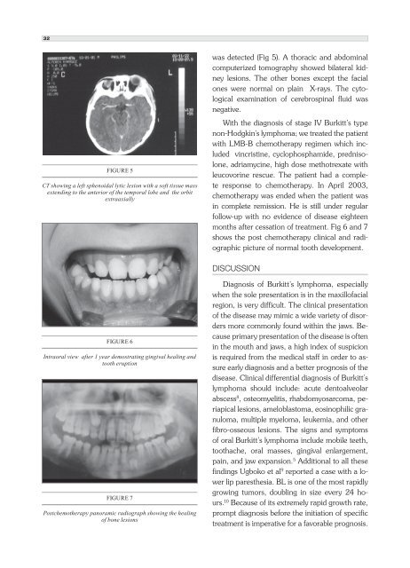
FIGURE 5<br />
CT showing a left sphenoidal lytic lesion with a soft tissue mass<br />
extending to the anterior of the temporal lobe and the orbit<br />
ex<strong>tr</strong>aaxially<br />
FIGURE 6<br />
In<strong>tr</strong>aoral view after 1 year demos<strong>tr</strong>ating gingival healing and<br />
tooth eruption<br />
FIGURE 7<br />
Postchemotherapy panoramic radiograph showing the healing<br />
of bone lesions<br />
was detected (Fig 5). A thoracic and abdominal<br />
computerized tomography showed bilateral kidney<br />
lesions. The other bones except the facial<br />
ones were normal on plain X-rays. The cytological<br />
examination of cerebrospinal fluid was<br />
negative.<br />
With the diagnosis of stage IV Burkitt’s type<br />
non-Hodgkin’s lymphoma; we <strong>tr</strong>eated the patient<br />
with LMB-B chemotherapy regimen which included<br />
vincristine, cyclophosphamide, prednisolone,<br />
adriamycine, high dose metho<strong>tr</strong>exate with<br />
leucovorine rescue. The patient had a complete<br />
response to chemotherapy. In April 2003,<br />
chemotherapy was ended when the patient was<br />
in complete remission. He is still under regular<br />
follow-up with no evidence of disease eighteen<br />
months after cessation of <strong>tr</strong>eatment. Fig 6 and 7<br />
shows the post chemotherapy clinical and radiographic<br />
picture of normal tooth development.<br />
DIsCUssIOn<br />
Diagnosis of Burkitt’s lymphoma, especially<br />
when the sole presentation is in the maxillofacial<br />
region, is very difficult. The clinical presentation<br />
of the disease may mimic a wide variety of disorders<br />
more commonly found within the jaws. Because<br />
primary presentation of the disease is often<br />
in the mouth and jaws, a high index of suspicion<br />
is required from the medical staff in order to assure<br />
early diagnosis and a better prognosis of the<br />
disease. Clinical differential diagnosis of Burkitt’s<br />
lymphoma should include: acute dentoalveolar<br />
abscess 8 , osteomyelitis, rhabdomyosarcoma, periapical<br />
lesions, ameloblastoma, eosinophilic granuloma,<br />
multiple myeloma, leukemia, and other<br />
fibro-osseous lesions. The signs and symptoms<br />
of oral Burkitt’s lymphoma include mobile teeth,<br />
toothache, oral masses, gingival enlargement,<br />
pain, and jaw expansion. 5 Additional to all these<br />
findings Ugboko et al 9 reported a case with a lower<br />
lip paresthesia. BL is one of the most rapidly<br />
growing tumors, doubling in size every 24 hours.<br />
10 Because of its ex<strong>tr</strong>emely rapid growth rate,<br />
prompt diagnosis before the initiation of specific<br />
<strong>tr</strong>eatment is imperative for a favorable prognosis.