Abdominal CT With Model-Based Iterative Reconstruction (MBIR ...
Abdominal CT With Model-Based Iterative Reconstruction (MBIR ...
Abdominal CT With Model-Based Iterative Reconstruction (MBIR ...
You also want an ePaper? Increase the reach of your titles
YUMPU automatically turns print PDFs into web optimized ePapers that Google loves.
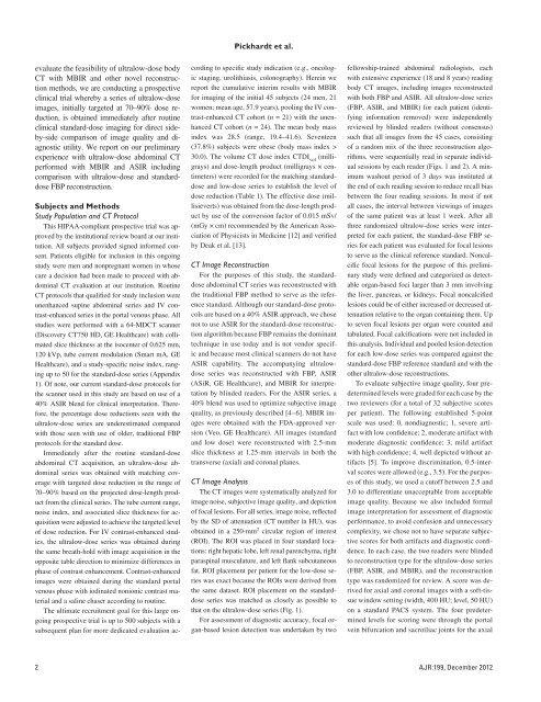
Pickhardt et al.evaluate the feasibility of ultralow-dose body<strong>CT</strong> with <strong>MBIR</strong> and other novel reconstructionmethods, we are conducting a prospectiveclinical trial whereby a series of ultralow-doseimages, initially targeted at 70–90% dose reduction,is obtained immediately after routineclinical standard-dose imaging for direct sideby-sidecomparison of image quality and diagnosticutility. We report on our preliminaryexperience with ultralow-dose abdominal <strong>CT</strong>performed with <strong>MBIR</strong> and ASIR includingcomparison with ultralow-dose and standarddoseFBP reconstruction.Subjects and MethodsStudy Population and <strong>CT</strong> ProtocolThis HIPAA-compliant prospective trial was approvedby the institutional review board at our institution.All subjects provided signed informed consent.Patients eligible for inclusion in this ongoingstudy were men and nonpregnant women in whosecare a decision had been made to proceed with abdominal<strong>CT</strong> evaluation at our institution. Routine<strong>CT</strong> protocols that qualified for study inclusion wereunenhanced supine abdominal series and IV contrast-enhancedseries in the portal venous phase. Allstudies were performed with a 64-MD<strong>CT</strong> scanner(Discovery <strong>CT</strong>750 HD, GE Healthcare) with collimatedslice thickness at the isocenter of 0.625 mm,120 kVp, tube current modulation (Smart mA, GEHealthcare), and a study-specific noise index, rangingup to 50 for the standard-dose series (Appendix1). Of note, our current standard-dose protocols forthe scanner used in this study are based on use of a40% ASIR blend for clinical interpretation. Therefore,the percentage dose reductions seen with theultralow-dose series are underestimated comparedwith those seen with use of older, traditional FBPprotocols for the standard dose.Immediately after the routine standard-doseabdominal <strong>CT</strong> acquisition, an ultralow-dose abdominalseries was obtained with matching coveragewith targeted dose reduction in the range of70–90% based on the projected dose-length productfrom the clinical series. The tube current range,noise index, and associated slice thickness for acquisitionwere adjusted to achieve the targeted levelof dose reduction. For IV contrast-enhanced studies,the ultralow-dose series was obtained duringthe same breath-hold with image acquisition in theopposite table direction to minimize differences inphase of contrast enhancement. Contrast-enhancedimages were obtained during the standard portalvenous phase with iodinated nonionic contrast materialand a saline chaser according to routine.The ultimate recruitment goal for this large ongoingprospective trial is up to 500 subjects with asubsequent plan for more dedicated evaluation accordingto specific study indication (e.g., oncologicstaging, urolithiasis, colonography). Herein wereport the cumulative interim results with <strong>MBIR</strong>for imaging of the initial 45 subjects (24 men, 21women; mean age, 57.9 years), pooling the IV contrast-enhanced<strong>CT</strong> cohort (n = 21) with the unenhanced<strong>CT</strong> cohort (n = 24). The mean body massindex was 28.5 (range, 19.4–41.6). Seventeen(37.8%) subjects were obese (body mass index >30.0). The volume <strong>CT</strong> dose index <strong>CT</strong>DI vol(milligrays)and dose-length product (milligrays × centimeters)were recorded for the matching standarddoseand low-dose series to establish the level ofdose reduction (Table 1). The effective dose (millisieverts)was obtained from the dose-length productby use of the conversion factor of 0.015 mSv/(mGy × cm) recommended by the American Associationof Physicists in Medicine [12] and verifiedby Deak et al. [13].<strong>CT</strong> Image <strong>Reconstruction</strong>For the purposes of this study, the standarddoseabdominal <strong>CT</strong> series was reconstructed withthe traditional FBP method to serve as the referencestandard. Although our standard-dose protocolsare based on a 40% ASIR approach, we chosenot to use ASIR for the standard-dose reconstructionalgorithm because FBP remains the dominanttechnique in use today and is not vendor specificand because most clinical scanners do not haveASIR capability. The accompanying ultralowdoseseries was reconstructed with FBP, ASIR(ASiR, GE Healthcare), and <strong>MBIR</strong> for interpretationby blinded readers. For the ASIR series, a40% blend was used to optimize subjective imagequality, as previously described [4–6]. <strong>MBIR</strong> imageswere obtained with the FDA-approved version(Veo, GE Healthcare). All images (standardand low dose) were reconstructed with 2.5-mmslice thickness at 1.25-mm intervals in both thetransverse (axial) and coronal planes.<strong>CT</strong> Image AnalysisThe <strong>CT</strong> images were systematically analyzed forimage noise, subjective image quality, and depictionof focal lesions. For all series, image noise, reflectedby the SD of attenuation (<strong>CT</strong> number in HU), wasobtained in a 250-mm 2 circular region of interest(ROI). The ROI was placed in four standard locations:right hepatic lobe, left renal parenchyma, rightparaspinal musculature, and left flank subcutaneousfat. ROI placement per patient for the low-dose serieswas exact because the ROIs were derived fromthe same dataset. ROI placement on the standarddoseseries was matched as closely as possible tothat on the ultralow-dose series (Fig. 1).For assessment of diagnostic accuracy, focal organ-basedlesion detection was undertaken by twofellowship-trained abdominal radiologists, eachwith extensive experience (18 and 8 years) readingbody <strong>CT</strong> images, including images reconstructedwith both FBP and ASIR. All ultralow-dose series(FBP, ASIR, and <strong>MBIR</strong>) for each patient (identifyinginformation removed) were independentlyreviewed by blinded readers (without consensus)such that all images from the 45 cases, consistingof a random mix of the three reconstruction algorithms,were sequentially read in separate individualsessions by each reader (Figs. 1 and 2). A minimumwashout period of 3 days was instituted atthe end of each reading session to reduce recall biasbetween the four reading sessions. In most if notall cases, the interval between viewings of imagesof the same patient was at least 1 week. After allthree randomized ultralow-dose series were interpretedfor each patient, the standard-dose FBP seriesfor each patient was evaluated for focal lesionsto serve as the clinical reference standard. Noncalcificfocal lesions for the purpose of this preliminarystudy were defined and categorized as detectableorgan-based foci larger than 3 mm involvingthe liver, pancreas, or kidneys. Focal noncalcifiedlesions could be of either increased or decreased attenuationrelative to the organ containing them. Upto seven focal lesions per organ were counted andtabulated. Focal calcifications were not included inthis analysis. Individual and pooled lesion detectionfor each low-dose series was compared against thestandard-dose FBP reference standard and with theother ultralow-dose reconstructions.To evaluate subjective image quality, four predeterminedlevels were graded for each case by thetwo reviewers (for a total of 32 subjective scoresper patient). The following established 5-pointscale was used: 0, nondiagnostic; 1, severe artifactwith low confidence; 2, moderate artifact withmoderate diagnostic confidence; 3, mild artifactwith high confidence; 4, well depicted without artifacts[5]. To improve discrimination, 0.5-intervalscores were allowed (e.g., 3.5). For the purposesof this study, we used a cutoff between 2.5 and3.0 to differentiate unacceptable from acceptableimage quality. Because we also included formalimage interpretation for assessment of diagnosticperformance, to avoid confusion and unnecessarycomplexity, we chose not to have separate subjectivescores for both artifacts and diagnostic confidence.In each case, the two readers were blindedto reconstruction type for the ultralow-dose series(FBP, ASIR, and <strong>MBIR</strong>), and the reconstructiontype was randomized for review. A score was derivedfor axial and coronal images with a soft-tissuewindow setting (width, 400 HU; level, 50 HU)on a standard PACS system. The four predeterminedlevels for scoring were through the portalvein bifurcation and sacroiliac joints for the axial2 AJR:199, December 2012


