196any study reviewed. Therefore, the evidence does not support thebenign profile that providers and the public have accepted.Another commonly used medication is mirtazapine, an antidepressantfound to have significant sedation effect when used in depressedpatients. It is similar to trazodone, and is used for insomniato capitalize on its side effect profile. It has also become a popularchoice among those who care for dementia patients who suffer fromboth insomnia and anorexia – increased appetite was another sideeffect noted when the drug was used in depressed patients. Ofnote, the side effects of increased appetite and sedation are typicallyseen with the lowest doses (7.5 mg or 15 mg). Case reports havedemonstrated clinical improvement in both insomnia and anorexiawhen the medication is used in Alzheimer’s patients. 9 However, morerigorous studies of mirtazapine’s tolerance and safety profile in elderlypatients are limited. A small study, examining its efficacy, notedthat 11% of patients discontinued use due to adverse events, 18%of which were falls. 10 Caution should be used when prescribing thisagent for insomnia in older adults.The newer drugs that capture the most media attention,and that patients request by name, are the “Z drugs,” whichinclude zaleplon (Sonata), zolpidem, and eszopiclone (Lunesta).One randomized trial compared zaleplon at 5 mg and 10 mgdoses to zolpidem 10mg and to placebo in 549 patients, all overthe age of 65. 11 The results demonstrate better sleep quality inboth groups as compared to placebo. Adverse events were similarin the four groups, with no increased adverse effects as comparedto placebo. However, the follow up time was only twoweeks. Another group of authors reviewed the literature to comparethe effectiveness of the Z drugs to placebo, and included24 trials. 12 Their final conclusions reflected disappointment: moststudies had small sample size, poor methodologic quality, and inmost, pharmaceutical funding. They recommended further studiesin the elderly, with more rigorous methodologic adherencebefore drawing clinical conclusions. 12Finally, ramelteon (Rozerem) warrants discussion. In the limitedbut promising existing studies, this melatonin receptor agonistwas found to produce improvements in all sleep components(latency, efficiency, and duration). 13 The agent has been found tobe effective and safe, with no concerns of dependence or next daysomnolence. It is best used for patients who have difficulty initiatingsleep. However, as a newer agent, more time and study inpost-marketing surveillance must be awaited before recommendationscan be made in vulnerable very old persons. Yet, amongthe options, this one t seems preferable for use in the older adultpopulation, and some studies have demonstrated that elderly patientscan use it safely without increased risk of falls or drowsinessthe following day. 14 Only time and clinical use will tell how trulysafe and effective it is. This agent has recently been added to theformulary at the Lifespan hospitals.In summary, the evidence supports the use of non-pharmacologictreatments as first line due to their proven efficacy,and for the long-term effects. Pharmacologic therapy has animportant role, but only for the short-term, and carries with itsignificant risk of adverse reactions. The newer agent ramelteonis promising, but will require further study and use in practice.Trazodone did not prove to be as safe and harmless as initiallythought, and its efficacy is also in question. The newer Z drugsalso proved efficacious, but had limited data in the older adultMEDICINE & HEALTH/RHODE ISLANDpopulation, are expensive, and are scheduled drugs, makinguse in long-term care settings more troublesome.For the patient presented at the beginning of this article, thefirst assessment the clinician must make is whether the situation isa sleep emergency or not. If the patient can wait, it would bebetter for him to discuss the problem and options with his primaryphysician, who knows him and his history best. This patienthas had three days of symptoms, but is retired and able to napduring the day; he should continue his current regimen untilMonday, when he can call his primary care doctor. Options atthat point include a more detailed review of sleep hygiene andrecommendations to discontinue drinking coffee after breakfast,to decrease caffeine intake and substitute a glass of warm milk orherbal tea after dinner. He should avoid napping during the dayand evaluate the quality of his bedroom for sleep promotion. Thetrazodone dose can remain the same, since there is no evidencebase for increasing the dose to regain initial effect, and strong considerationshould be given to discontinuing it altogether due tothe poor evidence base for its use in primary insomniaREFERENCES1. Alessi, C. Sleep Problems. In: Pompei P, Christmas C, Counsell SR, et al, eds, GeriatricsReview Syllabus, 6 th Edition, NY: American Geriatric <strong>Society</strong>, 2006: 249-57.2. Stanford University patient education webpage “How to Sleep Well” Link:http://www.stanford.edu/~dement/howto.html3. WebMD patient education webpage “How to Sleep Better” Link: http://www.webmd.com/sleep-disorders/guide/sleep-hygiene4. McCurry SM, Gibbons LE, et al. Nighttime insomnia treatment and educationfor Alzheimer’s disease. J Am Geri Soc 2005; 53: 793-802.5. Holbrook A, Crowther R, et al. The role of benzodiazepines in the yreatmentof Insomnia. J Am Geri Soc 2001; 49: 824-6.6. Fick DM, Cooper JW, et al. Updating the Beers criteria for potentially inappropriatemedication use in older adults. Arch Intern Med 2003;163:2716-24.7. IMS intelligence applied pharmaceutical marketing website. Link: http://www.imshealth.com8. Mendelson WB. A review of the evidence for the efficacy and safety oftrazodone in insomnia. J Clin Psychiatry 2005; 66: 469-76.9. Raji MA, Brady SR. Mirtazapine for treatment of depression and comorbiditiesin Alzheimer’s Disease. Ann Pharmacother 2001; 35:1024-7.10. Roose SP, Nelson JC, et al. Open-label study of mirtazapine orally disintegrating tabletsin depressed patients in the nursing home. Curr Med Res Opin 2003; 19: 737-46.11. Ancoli-Israel S, Walsh JK, et al. Zaleplon a novel nonbenzodiazepine hypnoticeffectively treats insomnia in elderly patients without causing reboundeffects. Primar Care Comp J Clin Psyc 1999; 1: 114-12.12, Dundar Y, Dodd S, , et al. Comparative efficacy of newer hypnotic drugs for theshort-term management of insomnia. Hum Psychopharmacol 2004; 19: 305-22.13. Brzezinski A, Vangel MG, et al. Effects of exogenous melatonin on sleep.Sleep Med Rev 2005 ; 9: 41-5.14. CenterWatch clinical trials listing service website. Link: http://www.centerwatch.com/patient/drugs/dru882.htmlAna C. Tuya, MD, is Assistant Professor of Geriatrics, <strong>Rhode</strong> <strong>Island</strong>Hospital, The Warren Alpert <strong>Medical</strong> School of Brown University.8SOW-RI-GERIATRICS -062007THE ANALYSES UPON WHICH THIS PUBLICATION IS BASED wereperformed under Contract Number 500-02-RI02, funded bythe Centers for Medicare & Medicaid Services, an agency ofthe U.S. Department of Health and Human Services. The contentof this publication does not necessarily reflect the viewsor policies of the Department of Health and Human Services,nor does mention of trade names, commercial products, ororganizations imply endorsement by the U.S. Government.The author assumes full responsibility for the accuracy andcompleteness of the ideas presented.
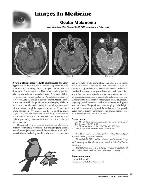
Images In MedicineOcular MelanomaIkue Shimizu, MD, Richard Gold, MD, and Edward Feller, MDFigure 1a. Figure 1b. Figure 1c.A 71 year-old man presented with severe nausea and vomitingfor several days. He had no visual complaints. Physicalexam was normal except for an enlarged, tender liver. AbdominalCT scan revealed a 12cm mass in the right liverlobe, shown to be melanoma by biospy. Skin exam did notreveal a primary cutaneous lesion. An ophthalmologic consultto evaluate for ocular melanoma demonstrated a lesionin the left choroid. Magnetic resonance imaging of the orbitshowed two choroidal masses in the left eye consistentwith melanoma: slightly hyperintense on the T1-weightedimage (Figure 1a), hypointense on the T2-weighted image(Figure 1b), and enhancement on the T1 post-gadoliniumimage with fat saturation (Figure 1c). The patient receivedright hepatic artery chemoembolization, and was dischargedon anti-emetics.The eye and orbit are the most common non-skin sites ofprimary or metastatic melanoma. 1 The most frequent locationis uveal, the majority are choroidal. Presentation of ocular melanomais diverse, including visual disturbance, ocular mass, cranialnerve palsy, orbital myopathy, or uveitis or vitritis. Severepain is uncommon. Some are detected by routine exam or discoveredduring evaluation of known extra-ocular melanoma.Uveal melanomas tend to spread hematogenously, most oftento the liver; as many as 60% of these melanomas have livermetastases at presentation. Diagnosis of uveal melanoma is usuallyestablished by an indirect fundoscopic exam. Fluorosceinangiography and ultrasound studies are also used to diagnoseuveal melanoma. 2 Magnetic resonance imaging can be helpfulin uveal melanoma staging and the evaluation of prognosticfactors such as tumor pigmentation, size, shape, location, retinaldetachment, extrascleral extension. 3REFERENCES1. Grin JM, et al. Ocular melanomas and melanocytic lesions of the eye. J AmAcad Dermatol 1998; 38:716-30.2. Char DH. Ocular melanoma. Surg Clin N Am 2003; 83:253-74.3. Lemke AJ, et al. Uveal melanoma. Radiol 1999;210:775-83.Ikue Shimizu, MD, is a 2007 graduate of The Warren Alpert<strong>Medical</strong> School of Brown University.Richard Gold, MD, is Clinical Associate Professor of DiagnosticImaging at The Warren Alpert <strong>Medical</strong> School of BrownUniversity.Edward Feller, MD, is a Clinical Professor of Medicine atThe Warren Alpert <strong>Medical</strong> School of Brown University.CORRESPONDENCEEdward Feller, MDe-mail: Edward_Feller@brown.eduVOLUME 90 NO. 6 JUNE 2007197