Scientific Session 1 â Breast Imaging: Mammography
Scientific Session 1 â Breast Imaging: Mammography
Scientific Session 1 â Breast Imaging: Mammography
Create successful ePaper yourself
Turn your PDF publications into a flip-book with our unique Google optimized e-Paper software.
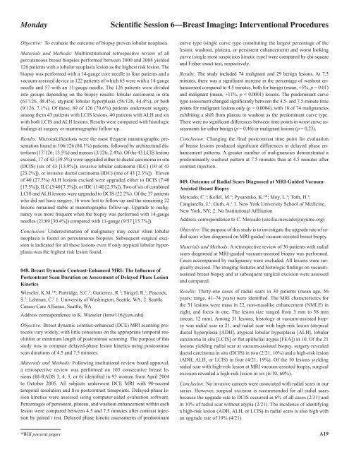
Monday<strong>Scientific</strong> <strong>Session</strong> 6—<strong>Breast</strong> <strong>Imaging</strong>: Interventional ProceduresObjective: To evaluate the outcome of biopsy proven lobular neoplasia.Materials and Methods: Multiinstitutional retrospective review of allpercutaneous breast biopsies performed between 2000 and 2008 yielded126 patients with a lobular neoplasia lesion as the highest risk lesion. Thebiopsy was performed with a 14-gauge core needle in four patients and avacuum-assisted device in 122 patients of which 65 were with a 14-gaugeneedle and 57 with an 11-gauge needle. The 126 patients were dividedinto groups depending on the biopsy results: lobular carcinoma in situ(61/126, 48.4%), atypical lobular hyperplasia (56/126, 44.4%), or both(9/126, 7.1%). Of these, 89 of 126 (70.6%) patients underwent surgery,among them 43 patients with LCIS lesions, 40 patients with ALH and sixwith both LCIS and ALH lesions. Results were compared with histologicfindings at surgery or mammographic follow-up.Results: Microcalcifications were the most frequent mammographic presentationfound in 106/126 (84.1%) patients, followed by architectural distortions(17/126; 13.5%) and masses (3/126; 2.4%). Of the 43 LCIS lesionsexcised, 17 of 43 (39.5%) were upgraded either to ductal carcinoma in situ(DCIS) (six of 43 [13.9%]), invasive lobular carcinoma (ILC) (10 of 43[23.2%]), or invasive ductal carcinoma (IDC) (one of 43 [2.3%]). Elevenof 40 (27.5%) ALH lesions excised were upgraded either to DCIS (7/40[17.5%]), ILC (3/40 [7.5%]), or IDC (1/40 [2.5%]). Two of six of combinedLCIS and ALH lesions were upgraded to DCIS (22.2%). Of the 37 patientswho did not have surgery, 16 were lost to follow-up and the remaining 22lesions remained stable at mammographic follow-up. Upgrade to malignancywas more frequent when the biopsy was performed with 14-gaugeneedles (21/69 [30.4%]) compared with 11-gauge (9/57 [15.7%]).Conclusion: Underestimation of malignancy may occur when lobularneoplasia is found on percutaneous biopsies. Subsequent surgical excisionis indicated for all these lesions even if only atypical lobular hyperplasiawas the highest risk lesion found.048. <strong>Breast</strong> Dynamic Contrast-Enhanced MRI: The Influence ofPostcontrast Scan Duration on Assessment of Delayed Phase LesionKineticsWieseler, K.M. 1 *; Partridge, S.C. 2 ; Gutierrez, R. 2 ; Strigel, R. 1 ; Peacock,S. 2 ; Lehman, C. 2 1. University of Washington, Seattle, WA; 2. SeattleCancer Care Alliance, Seattle, WAAddress correspondence to K. Wieseler (kmw116@uw.edu)Objective: <strong>Breast</strong> dynamic contrast-enhanced (DCE) MRI scanning protocolsvary widely, with little consensus on the appropriate temporal resolutionor minimum length of postcontrast scanning. The purpose of thisstudy was to compare delayed-phase lesion kinetics using postcontrastscan durations of 4.5 and 7.5 minutes.Materials and Methods: Following institutional review board approval,a retrospective review was performed on 103 consecutive breast lesions(BI-RADS 3, 4, 5, or 6) identified in 93 women from April 2004to October 2005. All subjects underwent DCE MRI with 90-secondtemporal resolution and five postcontrast timepoints. Delayed-phase lesionkinetics were assessed using computer-aided evaluation software.Percentages of persistent, plateau, and washout enhancement within eachlesion were compared between 4.5 and 7.5 minutes after contrast injectionby paired t test. Delayed phase kinetic assessments of predominantcurve type (single curve type constituting the largest percentage of thelesion; washout, plateau, or persistent enhancement) and worst lookingcurve (single most suspicious kinetic type) were compared by chi-squareand Fisher exact test, respectively.Results: The study included 74 malignant and 29 benign lesions. At 7.5minutes, there was a significant increase in the percentage of washout enhancementcompared to 4.5 minutes, both for benign (mean, +5%, p = 0.01)and malignant (mean, +11%, p < 0.0001) lesions. The predominant curvetype assessment changed significantly between the 4.5- and 7.5-minute timepoints for malignant lesions only (p = 0.0004), with 18 of 74 malignanciesexhibiting a shift from plateau to washout as the predominant curve type.There were no significant differences between time points in worst curve assessmentsfor either benign (p = 0.46) or malignant lesions (p = 0.23).Conclusion: Changing the final postcontrast time point for evaluationof breast lesions produced significant differences in delayed phase enhancementpatterns. A greater number of malignancies demonstrated apredominantly washout pattern at 7.5 minutes than at 4.5 minutes aftercontrast injection.049. Outcome of Radial Scars Diagnosed at MRI-Guided Vacuum-Assisted <strong>Breast</strong> BiopsyMercado, C. 1 ; Kellel, M. 2 ; Pysarenko, K. 1 *; Moy, L. 1 ; Toth, H. 1 ;Cangiarella, J. 1 ; Guth, A. 1 1. New York University School of Medicine,New York, NY; 2. No Institutional AffiliationAddress correspondence to C. Mercado (cecilia.mercado@nyumc.org)Objective: The purpose of this study is to investigate the upgrade rate of radialscars when diagnosed on MRI-guided vacuum-assisted breast biopsy.Materials and Methods: A retrospective review of 30 patients with radialscars diagnosed at MRI-guided vacuum-assisted biopsy was performed.Cases accompanied by malignancy were excluded. All lesions were surgicallyexcised. The imaging features and histologic findings on vacuumassistedbreast biopsy and at subsequent surgical excision were assessedand compared.Results: Thirty-one cases of radial scars in 30 patients (mean age, 56years; range, 41–74 years) were identified. The MRI characteristics forthe 31 lesions were mass in 22, non-masslike enhancement (NMLE) ineight, and focus in one. The lesion size ranged from 3 mm to 38 mm(mean, 12 mm). Among 31 lesions, histology at vacuum-assisted biopsywas radial scar in 21, and radial scar with high-risk lesion (atypicalductal hyperplasia [ADH], atypical lobular hyperplasia [ALH], lobularcarcinoma in situ [LCIS] or flat epithelial atypia [FEA]) in 10. Of the 21lesions yielding radial scar at vacuum-assisted biopsy, surgery revealedductal carcinoma in situ (DCIS) in two (2/21, 10%) and a high-risk lesion(ADH, ALH, or LCIS) in four (4/21, 19%). Of the 10 lesions yieldingradial scar with high-risk lesion at MRI vacuum-assisted biopsy, surgicalexcision revealed a high-risk lesion in six (6/10, 60%).Conclusion: No invasive cancers were associated with radial scars in ourseries. However, surgical excision is recommended for all radial scarsbecause the upgrade rate to DCIS occurred in 6% of all cases (2/31) andin 10% of radial scar without atypia (2/21). The incidence of identifyinga high-risk lesion (ADH, ALH, or LCIS) in radial scars is also high withan upgrade rate of 19% (4/21).*Will present paperA19


