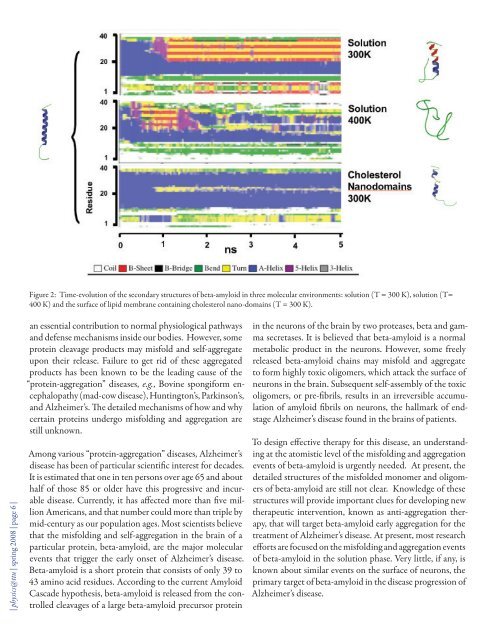
Figure 2: Time-evolution <strong>of</strong> the secondary structures <strong>of</strong> beta-amyloid in three molecular environments: solution (T = 300 K), solution (T=400 K) and the surface <strong>of</strong> lipid membrane containing cholesterol nano-domains (T = 300 K).| physics@ttu | spring <strong>2008</strong> | page 6 |an essential contribution to normal physiological pathwaysand defense mechanisms inside our bodies. However, someprotein cleavage products may misfold and self-aggregateupon their release. Failure to get rid <strong>of</strong> these aggregatedproducts has been known to be the leading cause <strong>of</strong> the“protein-aggregation” diseases, e.g., Bovine spongiform encephalopathy(mad-cow disease), Huntington’s, Parkinson’s,and Alzheimer’s. The detailed mechanisms <strong>of</strong> how and whycertain proteins undergo misfolding and aggregation arestill unknown.Among various “protein-aggregation” diseases, Alzheimer’sdisease has been <strong>of</strong> particular scientific interest for decades.It is estimated that one in ten persons over age 65 and abouthalf <strong>of</strong> those 85 or older have this progressive and incurabledisease. Currently, it has affected more than five millionAmericans, and that number could more than triple bymid-century as our population ages. Most scientists believethat the misfolding and self-aggregation in the brain <strong>of</strong> aparticular protein, beta-amyloid, are the major molecularevents that trigger the early onset <strong>of</strong> Alzheimer’s disease.Beta-amyloid is a short protein that consists <strong>of</strong> only 39 to43 amino acid residues. According to the current AmyloidCascade hypothesis, beta-amyloid is released from the controlledcleavages <strong>of</strong> a large beta-amyloid precursor proteinin the neurons <strong>of</strong> the brain by two proteases, beta and gammasecretases. It is believed that beta-amyloid is a normalmetabolic product in the neurons. However, some freelyreleased beta-amyloid chains may misfold and aggregateto form highly toxic oligomers, which attack the surface <strong>of</strong>neurons in the brain. Subsequent self-assembly <strong>of</strong> the toxicoligomers, or pre-fibrils, results in an irreversible accumulation<strong>of</strong> amyloid fibrils on neurons, the hallmark <strong>of</strong> endstageAlzheimer’s disease found in the brains <strong>of</strong> patients.To design effective therapy for this disease, an understandingat the atomistic level <strong>of</strong> the misfolding and aggregationevents <strong>of</strong> beta-amyloid is urgently needed. At present, thedetailed structures <strong>of</strong> the misfolded monomer and oligomers<strong>of</strong> beta-amyloid are still not clear. Knowledge <strong>of</strong> thesestructures will provide important clues for developing newtherapeutic intervention, known as anti-aggregation therapy,that will target beta-amyloid early aggregation for thetreatment <strong>of</strong> Alzheimer’s disease. At present, most researchefforts are focused on the misfolding and aggregation events<strong>of</strong> beta-amyloid in the solution phase. Very little, if any, isknown about similar events on the surface <strong>of</strong> neurons, theprimary target <strong>of</strong> beta-amyloid in the disease progression <strong>of</strong>Alzheimer’s disease.
From a computational standpoint, thereason for this lack <strong>of</strong> detailed informationabout the misfolding and aggregationevents <strong>of</strong> beta-amyloid and theirstructures on the neuronal membranesurface is the difficulty <strong>of</strong> creating a realisticneuronal membrane surface mimic.Previous computational studies onbeta-amyloid/membrane interactionsfocused on simple one-component andcontinuum lipid membrane systems thatfailed to capture the molecular organizations<strong>of</strong> certain key membrane components(lipids) and atomistic interactionsbetween the lipids and beta-amyloidmolecules.Recently, we have successfully designedand constructed a multi-componentneuronal membrane mimic with nanoscalearchitecture. This model membraneconsists <strong>of</strong> a bilayer made <strong>of</strong> cholesteroland phosphatidylcholine, the major lipidsfound in neurons. In addition, cholesterolnano-domains <strong>of</strong> defined lateralorganization have also been simulated.Recent studies have indicated that thesenano-domains are dynamically stablestructures in lipid membranes. Also,they can regulate the activities <strong>of</strong> certainmembrane surface acting proteins and the amyloid cascadeprocess leading to Alzheimer’s disease. However, the mechanismby which these nano-domains affect the misfoldingand aggregation <strong>of</strong> beta-amyloid on the lipid membranesurface remains unknown and constitutes the major focus<strong>of</strong> our work.To explore the folding and aggregation events <strong>of</strong> beta-amyloidon the neuronal membrane mimic with nano-domains,we plan to utilize a computational approach involving bothcoarse-grained and all-atom molecular dynamics (MD)simulations. This new hybrid MD method will allow usto sample a large configuration space, achieve simulationtimes in milliseconds, and reveal atomistic details <strong>of</strong> conformations-- all necessary to unraveling the complexities<strong>of</strong> protein folding and aggregation events on membranesurfaces.Pr<strong>of</strong>essor Kelvin Cheng (kelvin.cheng@ttu.edu), a biophysicist, hasbeen with our department since1989. One <strong>of</strong> his recent interestsis protein folding and its ramificationsfor our health. He explains hiswork on a computational approachto explore the early misfolding andaggregation events <strong>of</strong> Alzheimer’sbeta-amyloid proteins on the neuronsurface. Pr<strong>of</strong>essor Cheng is teachinggraduate-level Molecular Biophysicsthis spring.Figure 1 summarizes the design framework <strong>of</strong> our computationalstudy. Here, we first constructed the monomeric beta-amyloidin the α-state. We then constructedtwo water/protein/membranemolecular clusters, one with the proteinpartially inserted in the lipid membraneand the other with the protein lying onthe lipid membrane surface (Figure 1B).The lipid membrane surface shown hereconsists <strong>of</strong> 40% cholesterol, a biologicallyrelevant lipid composition found inthe neurons, with stable highly orderedcholesterol nano-domains. Finally, weconstructed a third cluster consisting <strong>of</strong>water/protein only and in the absence <strong>of</strong>the lipid membrane that serves as a control.Some initial results <strong>of</strong> the intermediatestate, or partially folded beta-amyloid(I-state) and completely unfoldedprotein (γ-state) in solution, are shownin Figure 1A. The aggregated beta-sheetstack structure (β-state) <strong>of</strong> beta-amyloidin solution published by another researchgroup is also shown for comparison.Figure 2 shows our 5-ns simulation data<strong>of</strong> beta-amyloid in three different environments:with the solution at T = 300K and 400 K, and on the membrane surfacewith cholesterol nano-domains atT = 300 K. It is clear that the misfoldingpathway <strong>of</strong> the protein in the 3D isotropicsolution phase is distinctly different from that on the2D membrane surface with cholesterol nano-domain structures.The appearance <strong>of</strong> beta sheet (red) and turn (yellow)structures is a signature <strong>of</strong> the misfolding regions <strong>of</strong> theprotein that may lead to the development <strong>of</strong> a beta-sheetstack, the currently hypothesized structure <strong>of</strong> the toxic oligomer<strong>of</strong> beta-amyloid (Figure 1A).Our plan is to compare the structures <strong>of</strong> our aggregated state<strong>of</strong> beta-amyloid on the membrane surface and in solutionwith the published toxic oligomer structures in solution,and to examine the stability <strong>of</strong> the protein that is partiallyinserted in membrane. The latter mimics the structure <strong>of</strong>the protein immediately after its release from the cleavages<strong>of</strong> the large beta-amyloid precursor protein. We anticipatethat our information-rich MD simulation data will provideunique opportunities for data mining, i.e., the search forthe unique conformations <strong>of</strong> the protein that will help usto design drugs that target the misfolding and self-aggregationstructures <strong>of</strong> beta-amyloid. Two Ph.D. students and| physics@ttu | spring <strong>2008</strong> | page 7 |