

Tankyrase inhibition stabilizes axin and antagonizes Wnt signalling
Tankyrase inhibition stabilizes axin and antagonizes Wnt signalling
Tankyrase inhibition stabilizes axin and antagonizes Wnt signalling

Create successful ePaper yourself
Turn your PDF publications into a flip-book with our unique Google optimized e-Paper software.
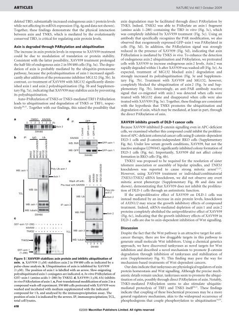
ARTICLES NATURE |Vol 461 |1 October 2009deleted TBD, substantially increased endogenous <strong>axin</strong> 1 protein levelswhile not affecting its mRNA expression (Fig. 4g <strong>and</strong> data not shown).Together, these findings demonstrate that the physical interactionbetween <strong>axin</strong> <strong>and</strong> TNKS, which is mediated by the evolutionarilyconserved TBD, is critical for regulating <strong>axin</strong> protein levels.Axin is degraded through PARsylation <strong>and</strong> ubiquitinationThe increase in <strong>axin</strong> protein levels in response to XAV939 treatmentcould be due to modulation of translation or protein stability.Consistent with the latter possibility, XAV939 treatment prolongedthe half-life of endogenous <strong>axin</strong> 2 in SW480 cells (Fig. 5a). The degradationof <strong>axin</strong> is probably mediated by the ubiquitin-proteasomepathway, because the polyubiquitination of <strong>axin</strong> 1 increased significantlyafter addition of the proteasome inhibitor MG132 (Fig. 5b). Incontrast, co-treatment of XAV939 with MG132 significantly diminished<strong>axin</strong> 1 <strong>and</strong> <strong>axin</strong> 2 polyubiquitination (Fig. 5b <strong>and</strong> SupplementaryFig. 7a), indicating that XAV939 may stabilize <strong>axin</strong> by preventingits polyubiquitination.Auto-PARsylation of TNKS or TNKS-mediated TRF1 PARsylationleads to ubiquitination <strong>and</strong> degradation of TNKS or TRF1, respectively20,21 . Together with our findings, this raised the possibility thatabcWB: <strong>axin</strong> 1TCLDMSO XAV9390 1 2 3 1 2 3 (h)IP: IgG UbDMSOMG132MG132+XAV939DMSOMG132MG132+XAV939WB: UbWB: <strong>axin</strong> 1WB: tubulinSW480GST–<strong>axin</strong> 1(1–280)XAV939TNKS2TNKS2GST–<strong>axin</strong> 1(1–280)SW480––+––+–++–+++–++–+Axin 2Poly-Ub-<strong>axin</strong> 1+++++++––+––deIP:XAV939WB: PARWB: GFPControlGFPGFP–<strong>axin</strong> 1GFP–<strong>axin</strong> 1– – +Axin 1Wash off with:Pre-treat with XAV939 – + + + +IP: <strong>axin</strong> 2TCLWB: UbWB: PARWB: <strong>axin</strong> 2SW480DMSOXAV939MG132MG132+XAV939WB: tubulin1 2 3 4 5SW480GFP–<strong>axin</strong> 1GFP–<strong>axin</strong> 1Poly-Ub<strong>axin</strong>2Axin 2Axin 2Figure 5 | XAV939 <strong>stabilizes</strong> <strong>axin</strong> protein <strong>and</strong> inhibits ubiquitination of<strong>axin</strong>. a, XAV939 (1 mM) <strong>stabilizes</strong> <strong>axin</strong> 2 in SW480 cells as indicated by apulse-chase analysis. b, Ubiquitination of <strong>axin</strong> is inhibited by XAV939(1 mM). The position of <strong>axin</strong> 1 is labelled with an arrow. Slow-migratingpolyubiquitinated <strong>axin</strong> 1 conjugates are indicated. c, In vitro PARsylation ofGST–<strong>axin</strong> 1 (amino acids 1–280) by TNKS2. d, XAV939 (1 mM, 6 h) inhibitsin vivo PARsylation of <strong>axin</strong> 1. e, Post-translational modification of <strong>axin</strong> 2 in acompound wash-off experiment. SW480 cells pretreated with XAV939 werewashed <strong>and</strong> incubated with medium supplemented with the indicatedcompound for 1 h, <strong>and</strong> analysed by the immunoprecipitation assay. Theposition of <strong>axin</strong> 2 is indicated by the arrows. IP, immunoprecipitation; TCL,total cell lysates.618©2009 Macmillan Publishers Limited. All rights reserved<strong>axin</strong> degradation may be facilitated through direct PARsylation byTNKS. Indeed, TNKS2 was able to PARsylate an <strong>axin</strong> 1 fragment(amino acids 1–280) containing the TBD in vitro (Fig. 5c), whichwas completely inhibited by XAV939 treatment (Fig. 5c). Using anantibody that specifically recognizes the PAR modification, we alsoobserved that exogenously expressed GFP–<strong>axin</strong> 1 was PARsylated incells (Fig. 5d). In addition, the PARsylation signal was stronglyreduced in the presence of XAV939 (Fig. 5d), indicating that <strong>axin</strong>PARsylation is mediated by TNKS in vivo. To enhance the detectionof endogenous <strong>axin</strong> 2 ubiquitination <strong>and</strong> PARsylation, we pretreatedcells with XAV939 to increase endogenous <strong>axin</strong> 2 levels. Axin 2 wasrapidly degraded within 1 h after XAV939 was washed off (Fig. 5e). Asexpected, treatment of MG132 blocked <strong>axin</strong> 2 degradation <strong>and</strong>strongly increased its polyubiquitination (Fig. 5e <strong>and</strong> SupplementaryFig. 7b). Treatment with XAV939 <strong>and</strong> MG132, however,completely blocked the ubiquitination of <strong>axin</strong> 2 (Fig. 5e <strong>and</strong> SupplementaryFig. 7b). Interestingly, an anti-PAR antibody reactivesignal that co-migrated with <strong>axin</strong> 2 was detected when cells weretreated with MG132 alone <strong>and</strong> disappeared when cells were alsotreated with XAV939 (Fig. 5e). Together, these findings are consistentwith the hypothesis that TNKS promotes the ubiquitination <strong>and</strong>degradation of <strong>axin</strong>, which may be mediated, at least in part, throughthe direct PARsylation of <strong>axin</strong>.XAV939 inhibits growth of DLD-1 cancer cellsBecause XAV939 inhibited b-catenin <strong>signalling</strong> even in APC-deficientcells, we examined whether this compound could inhibit the proliferationof APC-deficient colorectal cancer cells using b-catenin-dependentDLD-1 cells <strong>and</strong> b-catenin-independent RKO cells (SupplementaryFig. 8a). Under low serum growth conditions, XAV939, but not theinactive analogue LDW643, significantly inhibited colony formation ofDLD-1 cells (Fig. 6a). Importantly, XAV939 did not affect colonyformationinRKOcells(Fig.6b).TNKS1 was proposed to be required for the resolution of sistertelomere association or assembly of bipolar spindles, <strong>and</strong> TNKS1knockdown was reported to cause strong mitotic arrest 22,23 .However, using XAV939 treatment or individual/combinatorialTNKS1/TNKS2 siRNA knockdown, we did not observe any overtmitotic arrest phenotype (Supplementary Fig. 8b <strong>and</strong> data notshown), demonstrating that XAV939 does not inhibit the proliferationof DLD-1 cells through an antimitotic function.If the antiproliferative effect of XAV939 on DLD-1 cells wasinstead mediated by an increase in <strong>axin</strong> protein levels, knockdownof AXIN1/2 may rescue the growth inhibitory effects of compoundtreatment. Indeed, siRNA-mediated depletion of <strong>axin</strong> 1 <strong>and</strong> <strong>axin</strong> 2proteins completely abolished the antiproliferative effect of XAV939(Fig. 6c), indicating that the growth inhibitory effects of XAV939 inDLD-1 cells are due to <strong>axin</strong>-dependent <strong>inhibition</strong> of <strong>Wnt</strong> <strong>signalling</strong>.DiscussionDespite the fact that the <strong>Wnt</strong> pathway is an attractive target for anticancertherapy, there are few druggable targets in this pathway togenerate small molecule <strong>Wnt</strong> inhibitors. Using a chemical geneticsapproach, we have discovered tankyrases as novel targets for <strong>Wnt</strong><strong>inhibition</strong> <strong>and</strong> described a novel mechanism to promote b-catenindegradation through <strong>inhibition</strong> of tankyrases <strong>and</strong> stabilization of<strong>axin</strong> (Supplementary Fig. 9). This finding may pave the way formechanism-based treatments of <strong>Wnt</strong>-dependent cancers.Our data indicate that tankyrases are physiological regulators of <strong>axin</strong>protein homeostasis <strong>and</strong> <strong>Wnt</strong> <strong>signalling</strong>. Although the precise mechanisticdetails remain unclear, tankyrases seem to promote the ubiquitinationof <strong>axin</strong>, possibly through direct PARsylation of <strong>axin</strong>. Notably,TNKS-mediated PARsylation seems to also stimulate ubiquitinmediatedproteolysis of TRF1 <strong>and</strong> TNKS itself 20,21 . These findingsindicate that coupling of these biochemical processes may be a moregeneral regulatory mechanism, akin to the widespread occurrence ofphosphodegrons that couple phosphorylation to ubiquitination 24,25 .